. 1999 Mar 2;96(5):2445-50.
doi: 10.1073/pnas.96.5.2445.

Bilirubin, formed by activation of heme oxygenase-2, protects neurons against oxidative stress injury

Affiliations

Bilirubin, formed by activation of heme oxygenase-2, protects neurons against oxidative stress injury

S Doré et al. Proc Natl Acad Sci U S A. .

Erratum in

  • Proc Natl Acad Sci U S A 1999 Sep 14;96(19):10944

Abstract

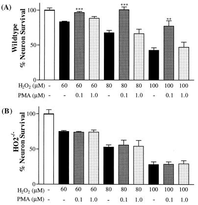
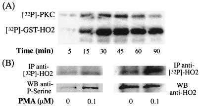
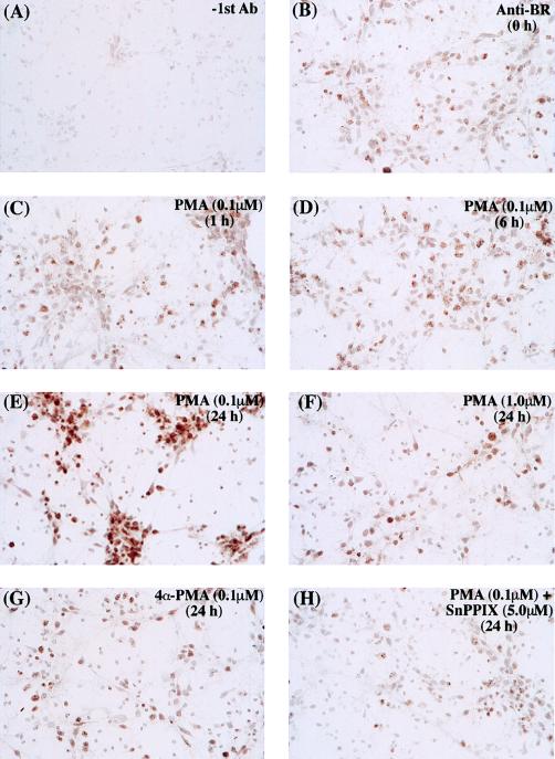
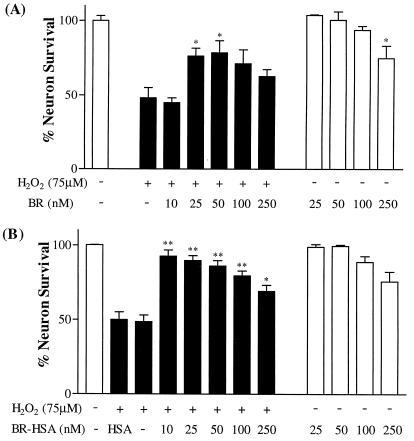
Heme oxygenase (HO) catalyzes the conversion of heme to carbon monoxide, iron, and biliverdin, which is immediately reduced to bilirubin (BR). Two HO active isozymes exist: HO1, an inducible heat shock protein, and HO2, which is constitutive and highly concentrated in neurons. We demonstrate a neuroprotective role for BR formed from HO2. Neurotoxicity elicited by hydrogen peroxide in hippocampal and cortical neuronal cultures is prevented by the phorbol ester, phorbol 12-myristate 13-acetate (PMA) via stimulation of protein kinase C. We observe phosphorylation of HO2 through the protein kinase C pathway with enhancement of HO2 catalytic activity and accumulation of BR in neuronal cultures. The neuroprotective effects of PMA are prevented by the HO inhibitor tin protoporphyrin IX and in cultures from mice with deletion of HO2 gene. Moreover, BR, an antioxidant, is neuroprotective at nanomolar concentrations.


Figures

Figure 1
Figure 1
Neuroprotective effect of PMA against H2O2-induced toxicity on rat primary neurons. Rat primary neurons were exposed to oxidative stress injury induced by H2O2 (75 μM), and neuron survival was estimated 24 h after the beginning of the experiment (A and B). Concentration-dependent biological effects of PMA were observed against H2O2 toxicity when PMA was added 24 h before exposure of H2O2 on hippocampal (A) and cortical (B) neurons. In C, hippocampal neurons were pretreated with PMA (0.1 and 1.0 μM) before the addition of different concentrations of H2O2 (30, 100, and 300 μM). Western blot analysis showed the expression of PKC in hippocampal neuronal cultures after PMA treatment (D). (E) An inactive enantiomer of PMA, 4α-PMA, was added in the same conditions as for PMA. Hippocampal neurons were treated by using identical conditions as described above and 5.0 μM of the HO inhibitor (SnPPIX) was added at the same time as PMA. (F) The effect of a PKC inhibitor, bisindolylmaleimide, on the neuroprotective effect of PMA in hippocampal neuronal cultures.
Figure 2
Figure 2
PMA neuroprotection is lost in hippocampal cultures of HO2−/− mice. Primary hippocampal neurons from wild-type (A) and HO2 knockout (B) mice were cultured similarly as rat neuronal cultures. Neurons were treated with various concentrations of H2O2 (60, 80, and 100 μM). PMA (0.1 and 1.0 μM) was added 24 h before the addition of H2O2. Neuronal survival was assessed 24 h later.
Figure 3
Figure 3
In vitro phosphorylation of hHO2. (A) Time-dependent effect on phosphorylation of GST–hHO2 incubated with [32P]ATP and purified brain PKC. The upper band shows the phosphorylation of PKC itself and the lower band depicts the phosphorylation of GST–hHO2. (B) HO2 phosphorylation in HEK 293 cells stably overexpressing hHO2. The top row shows incorporation of [32P]ATP into HO2 after phosphorylation induced by treatment with PMA (0 and 0.1 μM). HO2 was immunoprecipitated with two different antibodies (Left, SPA297; Right, our antibody), then subjected to electrophoresis and transferred to nitrocellulose membrane. Quantification of the [32P]HO2 with the PhosphorImager reveals an increased of 1.63 (Left) and 1.72 (Right) times after PMA treatment over the control. The Lower row was obtained after Western blot analysis on the same nitrocellulose membrane with an anti-phosphoserine antibody, suggesting that immunoprecipitated HO2 was phosphorylated on serine residues. Right indicates that HO2 was loaded in similar amounts.
Figure 4
Figure 4
Photomicrograph of rat primary hippocampal neurons incubated with an antibody against BR showing BR accumulation after PMA treatment. (A) Control staining performed without the addition of the primary antibody. (BH) Staining with the anti-BR under different stimuli. Cells were treated with 0.1 μM PMA for various time periods (0, 1, 6, or 24 h) (BE), with 1.0 μM PMA for 24 h (F), with 0.1 μM inactive enantiomer 4α-PMA (G), or with 0.1 μM in combination with 5.0 μM HO inhibitor SnPPIX (H).
Figure 5
Figure 5
Neuroprotective effect of bilirubin on H2O2-induced toxicity. (A) Increasing concentrations of free BR were added to rat primary hippocampal neurons before addition of 75 μM H2O2. (B) Increasing concentrations of BR complexed with human serum albumin (BR–HSA was added to neurons). An equivalent concentration HSA of albumin was added. Neuron survival was estimated 24 h after the beginning of the experiment.

References

    1. Maines M D. Annu Rev Pharmacol Toxicol. 1997;37:517–554. - PubMed
    1. Ewing J F, Maines M D. Brain Res Brain Res Protoc. 1997;1:165–174. - PubMed
    1. Verma A, Hirsch D J, Glatt C E, Ronnett G V, Snyder S H. Science. 1993;259:381–384. - PubMed
    1. Zakhary R, Poss K D, Jaffrey S R, Ferris C D, Tonegawa S, Snyder S H. Proc Natl Acad Sci USA. 1997;94:14848–14853. - PMC - PubMed
    1. Stocker R, Yamamoto Y, McDonagh A F, Glazer A N, Ames B N. Science. 1987;235:1043–1046. - PubMed

Publication types

MeSH terms

LinkOut - more resources