. 1999 Mar 15;19(6):1922-31.
doi: 10.1523/JNEUROSCI.19-06-01922.1999.

The synaptophysin-synaptobrevin complex: a hallmark of synaptic vesicle maturation

Affiliations

The synaptophysin-synaptobrevin complex: a hallmark of synaptic vesicle maturation

A Becher et al. J Neurosci. .

Abstract

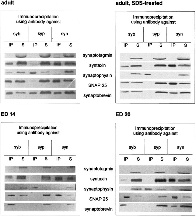
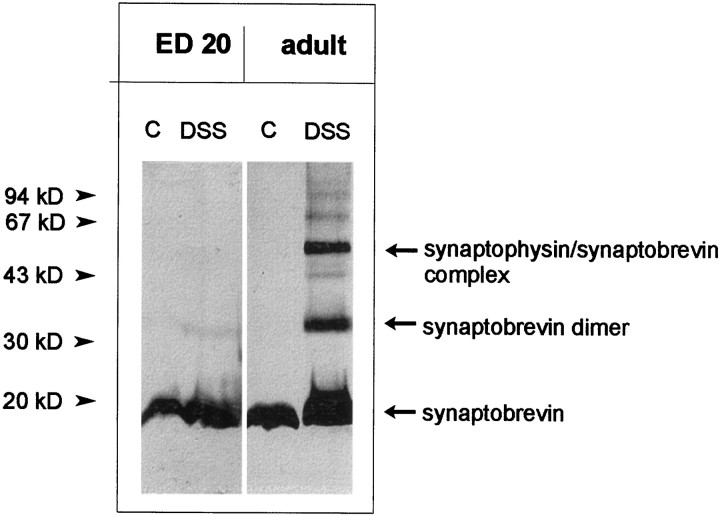
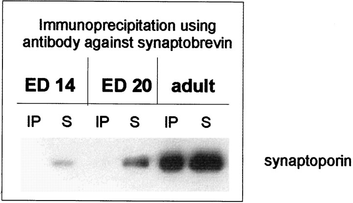
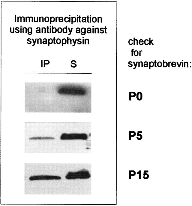
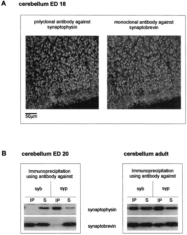
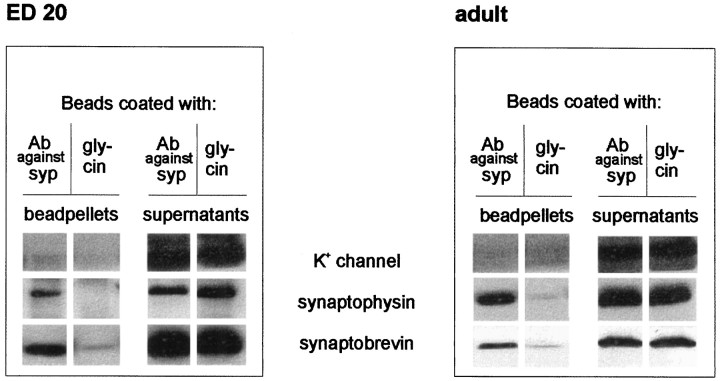
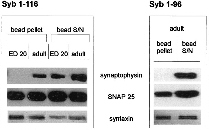
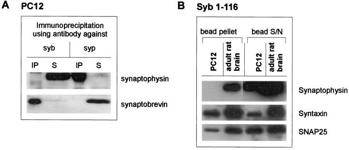
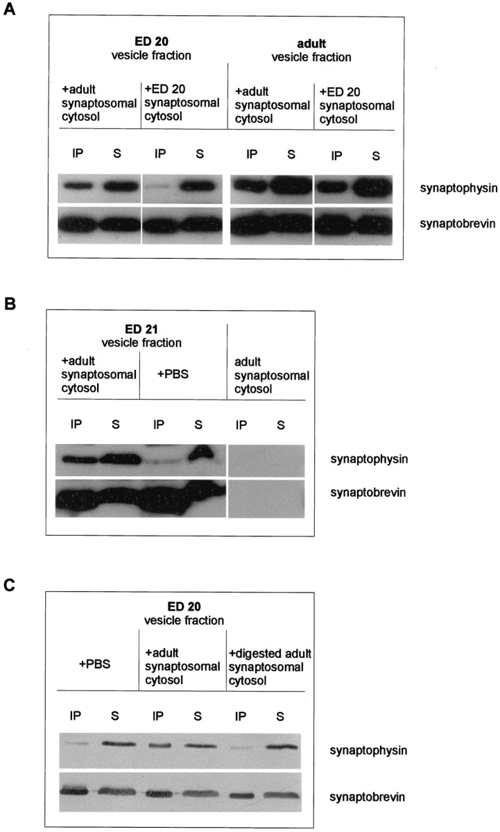
Exocytosis of synaptic vesicles requires the formation of a fusion complex consisting of the synaptic vesicle protein synaptobrevin (vesicle-associated membrane protein, or VAMP) and the plasma membrane proteins syntaxin and soluble synaptosomal-associated protein of 25 kDa (or SNAP 25). In search of mechanisms that regulate the assembly of the fusion complex, it was found that synaptobrevin also binds to the vesicle protein synaptophysin and that synaptophysin-bound synaptobrevin cannot enter the fusion complex. Using a combination of immunoprecipitation, cross-linking, and in vitro interaction experiments, we report here that the synaptophysin-synaptobrevin complex is upregulated during neuronal development. In embryonic rat brain, the complex is not detectable, although synaptophysin and synaptobrevin are expressed and are localized to the same nerve terminals and to the same pool of vesicles. In contrast, the ability of synaptobrevin to participate in the fusion complex is detectable as early as embryonic day 14. The binding of synaptoporin, a closely related homolog of synaptophysin, to synaptobrevin changes in a similar manner during development. Recombinant synaptobrevin binds to synaptophysin derived from adult brain extracts but not to that derived from embryonic brain extracts. Furthermore, the soluble cytosol fraction of adult, but not of embryonic, synaptosomes contains a protein that induces synaptophysin-synaptobrevin complex formation in embryonic vesicle fractions. We conclude that complex formation is regulated during development and is mediated by a posttranslational modification of synaptophysin. Furthermore, we propose that the synaptophysin-synaptobrevin complex is not essential for exocytosis but rather provides a reserve pool of synaptobrevin for exocytosis that can be readily recruited during periods of high synaptic activity.


Figures

Fig. 1.
Fig. 1.
SNARE complex, synaptophysin, and the synaptophysin–synaptobrevin complex in embryonic and adult synaptosomes. Triton X-100 extracts of whole brain crude synaptosomal fractions during different stages of development were immunoprecipitated using monoclonal antibodies against synaptobrevin (syb), synaptophysin (syp), or syntaxin (syn). Immunoprecipitates (IP) and their corresponding supernatants (S) were analyzed using antibodies against the indicated proteins.
Fig. 2.
Fig. 2.
Complexes of synaptophysin and synaptobrevin after chemical cross-linking in embryonic and adult synaptosomes. Crude synaptosomal fractions from embryonic day (ED) 20 whole brain (4 mg/ml protein) or adult whole brain (1.5 mg/ml protein) were treated with disuccinimidyl suberate (DSS) as described in Materials and Methods. After SDS-PAGE and Western blotting, membranes were analyzed using the monoclonal antibody against synaptobrevin. Note that cross-linking reveals the synaptobrevin dimer and the synaptobrevin–synaptophysin complex in the adult synaptosomes, whereas no synaptobrevin–synaptophysin complex and only traces of the synaptobrevin dimer can be detected in embryonic synaptosomes.C, Control.
Fig. 3.
Fig. 3.
Synaptoporin and the synaptoporin–synaptobrevin complex in embryonic and adult synaptosomes. Triton X-100 extracts of whole brain crude synaptosomal fractions during different stages of development were immunoprecipitated using the monoclonal antibody against synaptobrevin. Immunoprecipitates (IP) and their corresponding supernatants (S) were analyzed using an antiserum against the synaptophysin analog synaptoporin.
Fig. 4.
Fig. 4.
Postnatal appearance of the synaptophysin–synaptobrevin complex. Triton X-100 extracts of whole brain crude synaptosomal fractions from different stages of postnatal development were immunoprecipitated using the monoclonal antibody against synaptophysin. Immunoprecipitates (IP) and their corresponding supernatants (S) were analyzed using an antibody against synaptobrevin.
Fig. 5.
Fig. 5.
Synaptophysin, synaptobrevin, and the synaptophysin–synaptobrevin complex in embryonic and adult cerebellum.A, Confocal laser microscopic analysis of synaptophysin and synaptobrevin in embryonic day 18 rat cerebellar cortex. Note the clear presence of synaptophysin (left) and synaptobrevin (right) in the developing molecular and granular layers and the almost complete colocalization of both antigens.B, Triton X-100 extracts of cerebellar crude synaptosomal fraction from embryonic day 18 or adult were immunoprecipitated using antibodies against synaptophysin or synaptobrevin. Immunoprecipitates (IP) and their corresponding supernatants (S) were analyzed using anti-synaptophysin or anti-synaptobrevin antibody. No synaptobrevin–synaptophysin complex could be detected in the embryonic cerebellar synaptosomes.
Fig. 6.
Fig. 6.
Immunoisolation of synaptic vesicles from embryonic and adult whole brain. Eupergit beads coated with either an antibody against synaptophysin or glycine as negative control were incubated with crude synaptic vesicle fractions as described in Materials and Methods. Membranes adsorbed to the beads, as well as the corresponding supernatants, were analyzed using antibodies against synaptobrevin, synaptophysin, or the α subunit of the Kv1.6 channel. Only beads coated with the antibody against synaptophysin pelleted synaptobrevin and synaptophysin from both embryonic and adult vesicles. The potassium channel protein was always detected in the supernatant.
Fig. 7.
Fig. 7.
Binding of recombinant synaptobrevin constructs to embryonic and adult synaptic vesicles. Crude synaptic vesicle extracts from embryonic or adult brain were incubated with either full-length synaptobrevin (1–116) or the N-terminal part of synaptobrevin (1–96) (see Materials and Methods). The bead pellets and the corresponding supernatants were analyzed using antibodies against synaptophysin, SNAP 25, and syntaxin.
Fig. 8.
Fig. 8.
Absence of synaptophysin–synaptobrevin complex in PC 12 cells. A, Triton X-100 extracts of PC 12 cells were immunoprecipitated using monoclonal antibodies against synaptobrevin (syb) and synaptophysin (syp). Immunoprecipitates (IP) and their corresponding supernatants (S) were analyzed using antibodies against synaptophysin and synaptobrevin.B, PC 12 cell extracts and crude synaptic vesicle extracts were incubated with full-length synaptobrevin (1–116). The bead pellets and the corresponding supernatants were analyzed using antibodies against synaptophysin, SNAP 25, and syntaxin.
Fig. 9.
Fig. 9.
Induction of the synaptophysin–synaptobrevin complex in embryonic synaptic vesicles by adult cytosol.A, Crude synaptic vesicle fractions from embryonic day 20 or adult brain were incubated with synaptosomal cytosol fractions (LS2, synaptosomal cytosol) obtained from either adult (left lanes) or embryonic (ED 20,right lanes) brain for 90 min at 37°C before they were subjected to extraction and immunoprecipitation procedures using the anti-synaptobrevin antibody. Immunoprecipitates (IP) and their corresponding supernatants (S) were analyzed using anti-synaptophysin or anti-synaptobrevin antibody.B, Crude synaptic vesicle fractions from embryonic day 21 brain were incubated with either PBS or LS2 (synaptosomal cytosol) obtained from adult brains and processed as described in A. Note that the embryonic cytosol is completely free of both synaptophysin and synaptobrevin (right lanes). C, Crude synaptic vesicle fractions from embryonic day 20 were incubated with either PBS or with undigested or trypsin-digested LS2 (synaptosomal cytosol) obtained from adult brains. Samples were extracted, immunoprecipitated, and analyzed as described in A.

References

    1. Ahnert-Hilger G, Kutay U, Chahoud I, Rapoport TA, Wiedenmann B. Synaptobrevin is essential for secretion but not for the development of synaptic processes. Eur J Cell Biol. 1996;70:1–11. - PubMed
    1. Alder J, Kanki H, Valtorta F, Greengard P, Poo MM. Overexpression of synaptophysin enhances neurotransmitter secretion at Xenopus neuromuscular synapses. J Neurosci. 1995;15:511–519. - PMC - PubMed
    1. Barnstable CJ, Hofstein R, Akagawa K. A marker of early amacrine cell development in rat retina. Brain Res. 1985;352:286–290. - PubMed
    1. Bauerfeind R, Huttner WB (1993) Biogenesis of constitutive secretory vesicles, secretory granules and synaptic vesicles. Curr Opin Cell Biol [Erratum 5:1106] 5:628–635. - PubMed
    1. Baumert M, Maycox PR, Navone F, DeCamilli P, Jahn R. Synaptobrevin: an integral membrane protein of 18,000 daltons in small synaptic vesicles. EMBO J. 1989;8:379–384. - PMC - PubMed

Publication types

LinkOut - more resources