. 1999 Mar 15;19(6):1940-51.
doi: 10.1523/JNEUROSCI.19-06-01940.1999.

Akt-dependent potentiation of L channels by insulin-like growth factor-1 is required for neuronal survival

Affiliations

Akt-dependent potentiation of L channels by insulin-like growth factor-1 is required for neuronal survival

L A Blair et al. J Neurosci. .

Abstract

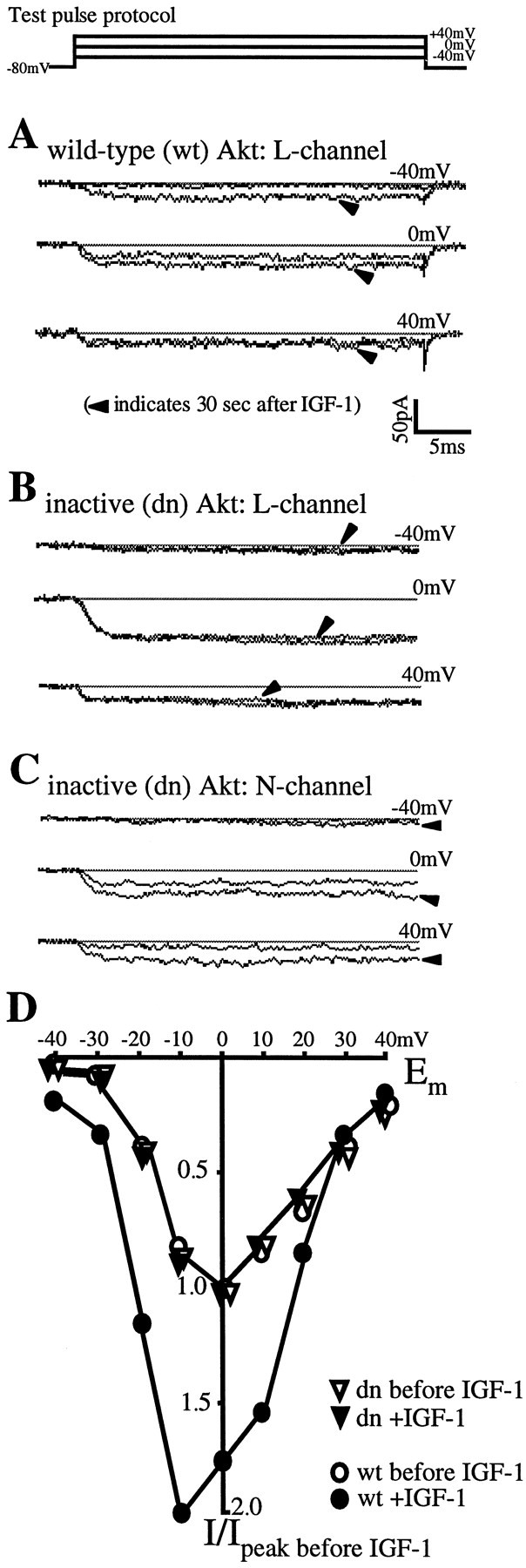
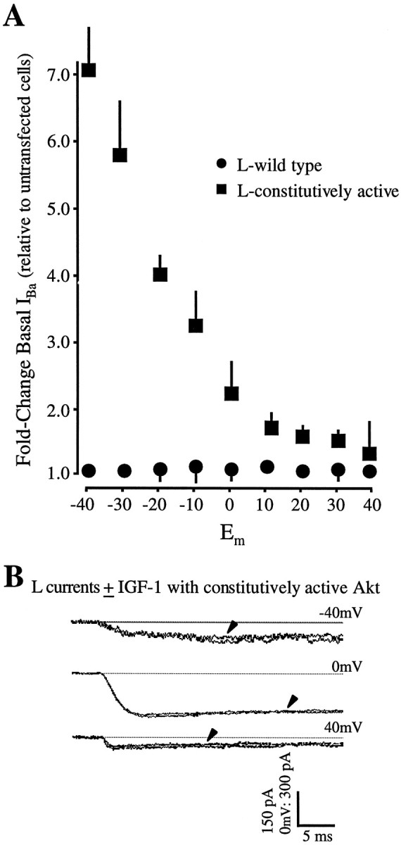
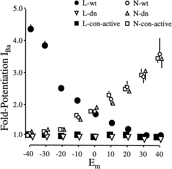
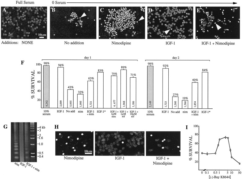
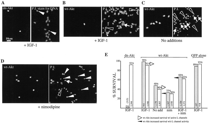
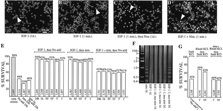
The insulin-like growth factor-1 (IGF-1)/receptor tyrosine kinase recently has been shown to mediate neuronal survival and potentiate the activity of specific calcium channel subtypes; survival requires Akt, a serine/threonine kinase. We demonstrate here that Akt mediates the IGF-1-induced potentiation of L channel currents, but not that of N channels. Transient expression of wild-type, dominant-negative, and constitutively active forms of Akt in cerebellar granule neurons causes, respectively, no change in IGF-1/L channel potentiation, complete inhibition of potentiation, and a dramatic increase in basal L currents accompanied by the loss of ability to induce further increases. In no case is the IGF-1 potentiation of N currents affected. We additionally find that IGF-1 partially mediates granule neuron survival via L channel activity and that Akt-dependent L channel modulation is a necessary component. Interestingly, very brief exposure (1 min) to IGF-1 triggers nearly complete survival and requires L channel activity. These results strongly suggest that neuronal receptor tyrosine kinases can control long-term calcium-dependent processes via the rapid control of voltage-sensitive channels.


Figures

Fig. 1.
Fig. 1.
Rapid IGF-1 potentiation of cerebellar L channels requires the serine/threonine kinase Akt. A, Overexpression of wild-type Akt allows the normal, hyperpolarization-dependent IGF-1 potentiation of L channels.A–C, Each panel shows an individual neuron transfected with either wild-type or kinase-dead Akt. Recordings are of the barium currents evoked at three membrane potentials (−40, 0, and +40 mV) before and 30 sec after IGF-1 (20 ng/ml) addition (arrowheads). The test pulse protocol is shownabove A. Calcium channel subtypes were isolated as described in Materials and Methods. B, A K179M mutation in the ATP-binding site of the kinase renders Akt inactive and blocks IGF-1/L channel potentiation. C, Inactive Akt has no effect on the normal depolarization-dependent potentiation of N channels by IGF-1. D, IGF-1 responses in two neurons, one transfected with wild-type Akt (open circles) and one with inactive dn-Akt (inverted open triangles); shown are membrane L channel current–voltage relationships before (open symbols) and 30 sec after the addition of 20 ng/ml IGF-1 (filled symbols). L currents for each cell were normalized to the peak currents measured before IGF-1. Note that, although the currents are relatively small at hyperpolarized potentials, the fold increase in the wild-type Akt-expressing neuron is much greater at −30 and −40 mV than at potentials ≥0 mV.
Fig. 2.
Fig. 2.
Overexpression of a constitutively active Akt mimics the IGF-1 effect. A, Basal L currents are increased dramatically in neurons transfected with Akt containing a myristoylation sequence that renders it constitutively active. Moreover, superfusion of IGF-1 is incapable of further potentiating L currents. Recordings were performed as in Figure 1; note, however, the change in scale. Arrowheads indicate currents recorded 30 sec after IGF-1 (20 ng/ml) addition. The test pulse protocol is identical to that used in Figure 1. B, Plots of the voltage-dependent changes in basal L currents show that the increase is particularly prominent at hyperpolarized membrane potentials in neurons expressing myristoylated Akt. Current levels before IGF-1 addition in transfected cells were normalized to cell size as estimated by membrane capacitance and were compared with the values obtained in untransfected cells. Values are means ± SEM; where no bars are shown, the errors are smaller than the symbols denoting their means. Errors were calculated as described in Materials and Methods. n= 27 cells transfected with myristoylated Akt; n = 32 cells with wild-type Akt.
Fig. 3.
Fig. 3.
Fold potentiation of N and L currents in wild-type, inactive, and constitutively active Akt-expressing neurons. For each cell the peak current measured after 20 ng/ml IGF-1 was divided by that measured before. Values are means ± SEM; where no bars are shown, the errors are smaller than the symbols denoting their means. Filled circles, inverted open triangles, filled squares, The potentiation of L-currents by 20 ng/ml IGF-1. Transfection conditions: wild-type Akt (L-wt),n = 32 cells; dominant–negative Akt (L-dn), n = 21 cells; constitutively active Akt (L-con-active), n = 27 cells. Open circles, open triangles, open squares, N-current potentiation by IGF-1. Transfection conditions: wild-type Akt (N-wt), n = 11 cells; dominant–negative Akt (N-dn), n = 17 cells; constitutively active Akt (N-con-active),n = 8 cells.
Fig. 4.
Fig. 4.
L channel potentiation by IGF-1 promotes neuronal survival. Granule neurons, cultured for 1 d in the indicated test media, were stained with propidium iodide to reveal DNA; anarrowhead indicates one example/panel of the bright, condensed chromatin characteristic of apoptotic cells.A, In full (10%) serum medium the nuclei are healthy, showing dispersed chromatin. B, At 1 d after serum withdrawal (No addition) most neurons are apoptotic.C, The addition of 10 μm nimodipine, an L channel inhibitor, to block neuronal L channel activity reduces survival over 0 serum alone. D, Conversely, the addition of 50 ng/ml IGF-1 promotes survival almost to the levels observed in full serum. E, Exposure to both IGF-1 and nimodipine partially promotes survival. F, Quantitation of survival: data (mean ± SEM) are from seven (day 1) and four independent (day 2) experiments in which each condition was tested in triplicate. The total number of neurons scored is shownwithin each bar; the percentage of survival is indicatedabove each bar. Shaded bars, Survival in full-serum medium. IGF-1*, Because nimodipine by itself decreases survival beyond that of the basic survival, the effect of nimodipine alone [11% = (survival in No add) − (survival in nim)] was subtracted from the survival in IGF-1 to obtain an estimate (IGF*) that can be compared directly with the survival in the L channel inhibition medium (IGF + nim, nim). IGF + 1 μm nif, IGF + 10 μm nif, IGF + 10 μm nim, Survival in DHP-containing media suggests that 10, but not 1, μm is effective. G, H, DNA laddering (G) and the TUNEL assay (H) for nicked DNA similarly indicate that L channel potentiation by IGF-1 is a component of IGF-1-mediated survival. G, IGF-1 (50 ng/ml) protects chromatin from the cleavage into low molecular weight fragments that occurs in the presence of 10 μm nimodipine. Only partial protection occurs when cells are grown in IGF-1 plus nimodipine. Lane 4, DNA standards. H, Cells with cleaved chromatin are brightly labeled. Unlabeled cells can be identified from the dim autofluorescence. All cells are shown at 1 d in test media. I, The DHP L channel agonist, (−)-Bay K8644, can mimic the IGF-1-dependent and L channel-dependent survival, but only at optimal concentrations. Sister cultures of neurons were grown for 24 hr in the basic survival medium supplemented with the indicated (−)-Bay K8644 concentrations, and survival was assessed by the proportion of cells displaying dispersed chromatin after propidium iodide labeling; the values are expressed as means ± SEM.
Fig. 5.
Fig. 5.
Survival via IGF-1 modulation of L channels is mediated by Akt. Granule neurons were cultured for 1 d in the indicated survival media and transfection conditions.A–D, Left, Akt transfectants as indicated by GFP fluorescence. Right, The same fields showing propidium iodide labeling of all neurons to reveal DNA;arrowheads indicate transfectants with the condensed chromatin characteristic of apoptotic cells, and arrowsindicate healthy transfectants; open arrowheads indicate examples of untransfected apoptotic cells. A, In IGF-1-containing (50 ng/ml) medium, neurons expressing dominant–negative (dn) Akt are apoptotic although all surrounding untransfected neurons are healthy. B–E, Neurons expressing wild-type (wt) Akt show increased, but L channel-dependent, survival. B, In IGF-1-containing test medium, wt-Akt-transfected neurons are healthy.C, In the basic test medium (No addition), wt-Akt-transfected neurons survive much better than their untransfected neighbors. D, In nimodipine-containing (10 μm) medium, the survival of transfected and untransfected neurons is similar. E, Quantitation of survival: data (mean ± SEM) are from three independent experiments (except GFP alone,n = 2), with each condition tested in triplicate. In each bar pair the left bar (shaded) represents transfectants, and the right bar represents untransfected cells from the same cultures. Overexpression of wt-Akt increases survival but to a much lesser degree when L channels are blocked (filled arrowheads) than when they are active (open arrowheads). The total number of neurons scored is shown within each bar; the percentage of survival is indicated above each bar.
Fig. 6.
Fig. 6.
A brief pulse of IGF-1 promotes neuronal survival via L channel potentiation. Granule neurons were kept in survival test media for a total of 24 hr; IGF-1-containing (50 ng/ml) test media (IGF-1, IGF + Nim) were applied for the indicated times, and then the cultures were rinsed three times and placed in either basic survival medium (No add) or in 10 μmnimodipine-containing medium (Nim) for the remainder of the 24 hr period. DNA was labeled as in Figures 4 and 5; anarrowhead indicates one example/panel of apoptotic cells. A, In IGF-1 medium the nuclei are healthy.B, C, A 1 min pulse of IGF-1 strongly promotes survival, even when L channels subsequently are blocked for the remainder of the test duration (C). D, Conversely, the number of apoptotic nuclei increases after a 1 min pulse of IGF-1 in the presence of nimodipine. E, Quantitation of survival: data (mean ± SEM) are from three independent experiments, with each condition tested in triplicate. The total number of neurons scored is shown within each bar; the percentage of survival is indicated above each bar.Shaded bars, Survival in full-serum medium.F, Brief exposure (1 or 10 min) to IGF-1 (50 ng/ml), followed by continuous exposure to nimodipine (10 μm), reduces chromatin cleavage as determined by DNA laddering. Partial protection can be detected for up to 2 d. Lane 1, DNA standards. G, The activation of L channels by depolarization also inhibits apoptosis. Neurons depolarized with 80 mm KCl for the same periods (1 or 10 min) survive much better than those exposed either to the basic survival medium, which contains 5 mm KCl (i.e., the “No add”medium), or to the 80 mm KCl-containing medium in the presence of 10 μm nimodipine (nim + 80 mm KCl). The 80 mm KCl is expected to drive the membrane potential to approximately −15 mV. After the brief application of 80 mm KCl ± nimodipine or basic (5 mm KCl) medium, all neurons were maintained for 1 d in the basic medium; apoptotic versus nonapoptotic cells were scored as above after propidium iodide labeling.

References

    1. Alessi DR, James SR, Downes CP, Holmes AB, Gaffney PR, Reese CB, Cohen P. Characterization of a 3-phosphoinositide-dependent protein kinase which phosphorylates and activates protein kinase Bα. Curr Biol. 1997;7:261–269. - PubMed
    1. Andjelkovic M, Alessi DR, Meier R, Fernandez A, Lamb NJ, Frech M, Cron P, Cohen P, Lucocq JM, Hemmings MA. Role of translocation in the activation and function of protein kinase B. J Biol Chem. 1997;272:31515–31524. - PubMed
    1. Bean BP. Whole-cell recording of calcium channel currents. Methods Enzymol. 1992;207:181–193. - PubMed
    1. Blair LAC, Marshall J. IGF-1 modulates N and L calcium channels in a PI 3-kinase-dependent manner. Neuron. 1997;19:421–429. - PubMed
    1. Blair LAC, Bence KK, Marshall J. The jellyfish green fluorescent protein: a tool for studying ion channels and second messenger signalling in neurons. Methods Enzymol. 1999;302:213–225. - PubMed

Publication types

LinkOut - more resources