Comparative Study
. 1999 Mar 15;19(6):2113-21.
doi: 10.1523/JNEUROSCI.19-06-02113.1999.

Leukemia inhibitory factor and ciliary neurotrophic factor cause dendritic retraction in cultured rat sympathetic neurons

Affiliations
Comparative Study

Leukemia inhibitory factor and ciliary neurotrophic factor cause dendritic retraction in cultured rat sympathetic neurons

X Guo et al. J Neurosci. .

Abstract

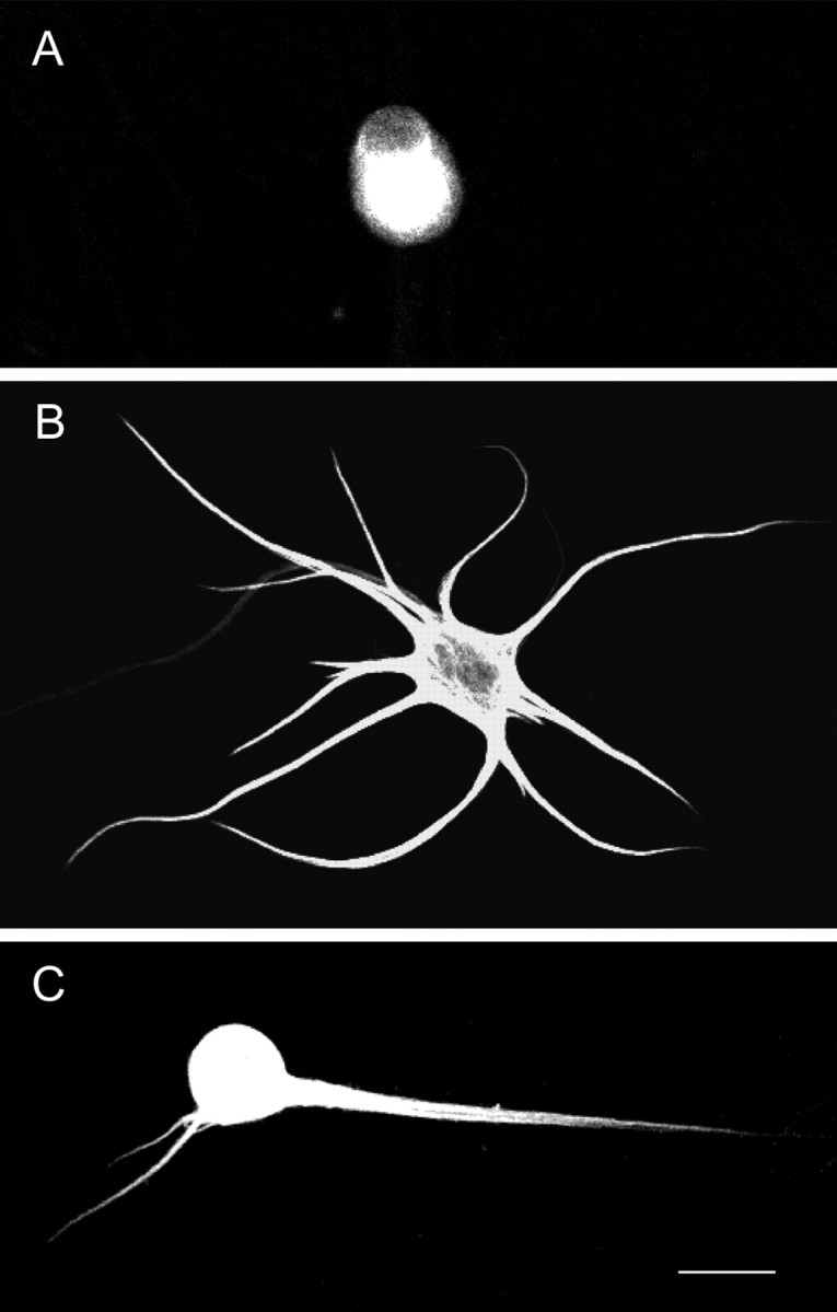
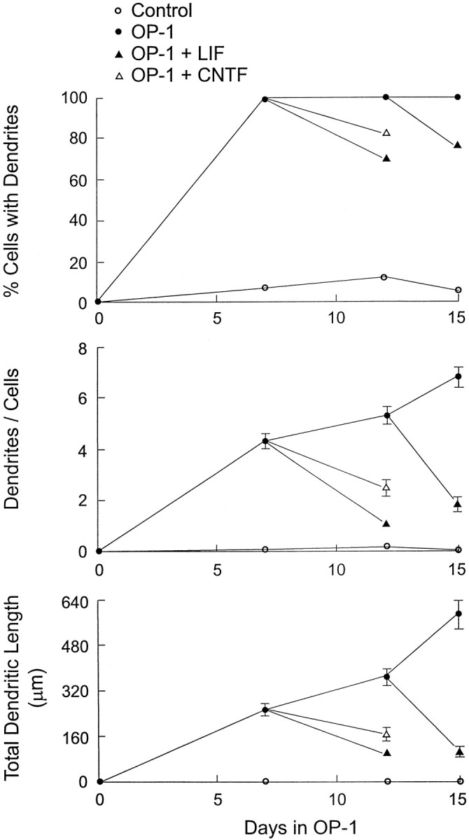
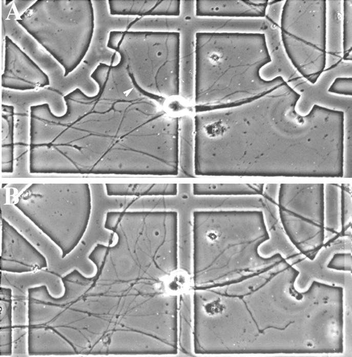
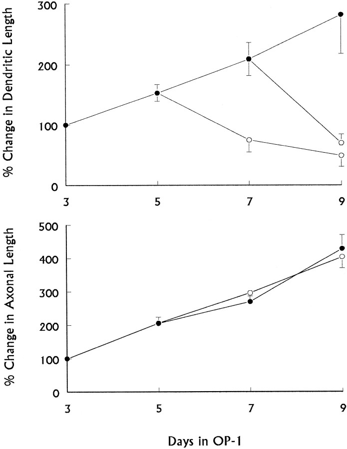
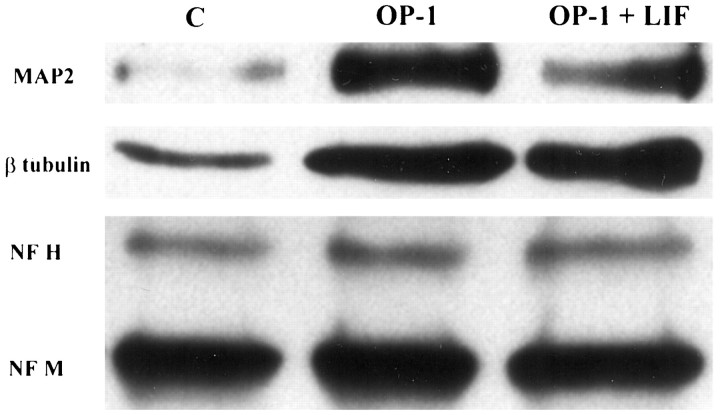
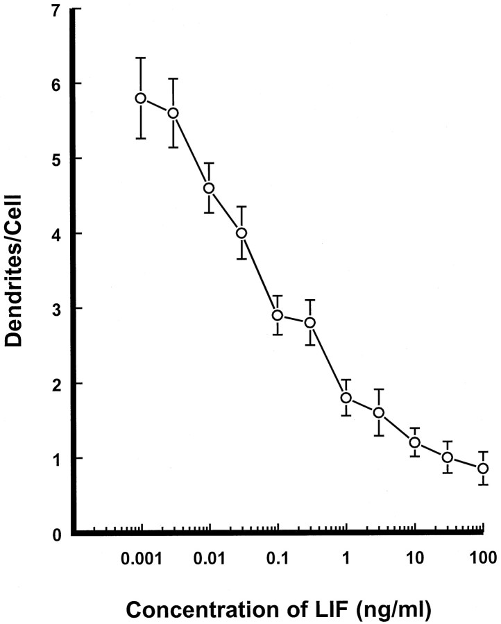
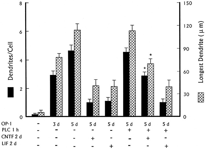
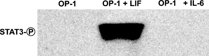
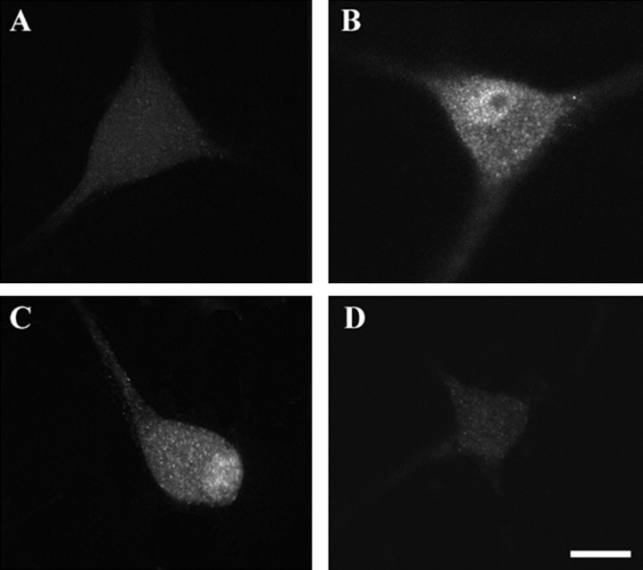
Dendritic retraction occurs in many regions of the developing brain and also after neural injury. However, the molecules that regulate this important regressive process remain largely unknown. Our data indicate that leukemia inhibitory factor (LIF) and ciliary neurotrophic factor (CNTF) cause sympathetic neurons to retract their dendrites in vitro, ultimately leading to an approximately 80% reduction in the size of the arbor. The dendritic retraction induced by LIF exhibited substantial specificity because it was not accompanied by changes in cell number, in the rate of axonal growth, or in the expression of axonal cytoskeletal elements. An antibody to gp130 blocked the effects of LIF and CNTF, and both cytokines induced phosphorylation and nuclear translocation of stat3. Moreover, addition of soluble interleukin-6 (IL-6) receptor to the medium endowed IL-6 with the ability to cause dendritic regression. These data indicate that ligands activating the gp130 pathway have the ability to profoundly alter neuronal cell shape and polarity by selectively causing the retraction of dendrites.


Figures

Fig. 1.
Fig. 1.
Effects of OP-1 and LIF on sympathetic neurons.A, Fluorescence micrograph of a sympathetic neuron that was grown under control conditions before being immunostained with a mAb (SMI52) to MAP2. Such cells typically lacked dendrites, whereas cells that had been treated with OP-1 (50 ng/ml) for 15 d (B) had complex arbors. C, The size of the dendritic arbor was reduced when cells that had initially been treated with OP-1 for 12 d were treated for an additional 3 d with the combination of LIF (10 ng/ml) and OP-1. Scale bar, 50 μm.
Fig. 2.
Fig. 2.
Time course of LIF- and CNTF-induced dendritic retraction in sympathetic neurons. Sympathetic neurons were plated onto polylysine-coated coverslips, and non-neuronal cells were eliminated by a 2 d exposure to an antimitotic agent. Cultures were then continuously treated with OP-1 (50 ng/ml). On day 7 or 12, some OP-1-treated cultures were also exposed to either LIF (30 ng/ml) or CNTF (30 ng/ml) for 3–5 d. Cellular morphology was analyzed after cultures had been immunostained with an mAb (SMI-52) to MAP2. Data are expressed as mean ± SEM (n = 30).
Fig. 3.
Fig. 3.
Serial observations of the effects of LIF on neuronal morphology. A, Phase-contrast image of a sympathetic neuron’s that had been treated with OP-1 (50 ng/ml) for 4 d. One of this neuron’s dendrites (arrowhead) could be examined in its entirety because it grew through an area where there were only a few axons. The cell was then treated with LIF (10 ng/ml) and OP-1 for 2 d. When the neuron was relocated (B), it was found that the dendrite had retracted. In contrast, the density of the axonal plexus increased and new processes appeared (bottom right and left). Note that the cell shown inA has a second, thick dendrite-like process that arises from the right side of the cell and that this process is associated with a bundle of axons. Such fasciculated processes were excluded from our analysis because we could not accurately determine where they ended.
Fig. 4.
Fig. 4.
Comparison of changes in dendritic and axonal length during LIF-induced dendritic retraction. Sympathetic neurons were plated onto gridded coverslips and exposed to OP-1 (●, 50 ng/ml) for a total of 9 d. On days 5 and 7, some cultures were also exposed to LIF (○, 30 ng/ml). Identified cells or regions on the grid were photographed daily to allow quantitation of dendritic or axonal growth, respectively. Mean ± SEM (n = 5–12).
Fig. 5.
Fig. 5.
LIF decreases OP-1-induced MAP2 expression. Cultures were exposed to OP-1 (50 ng/ml) for 3 d and then treated with either OP-1 alone or OP-1 + LIF (30 ng/ml) for an additional 4 d. Western blot analysis was performed to examine expression of MAP2 (280 kDa), β-tubulin (53 kDa), and the phosphorylated forms of the H (200 kDa) and M (160 kDa) neurofilament subunits. The chemiluminescent autographs are representative of three experiments that yielded similar results. In these experiments there was 2.5 ± 0.3-fold increase in MAP2 expression in cultures treated with OP-1 (p < 0.01 vs control). This was reduced to a 1.4 ± 0.2-fold increase in cultures treated with LIF and OP-1, a value that was not significantly different from the control condition. Similarly, there were no significant changes detected in the expression of phosphorylated forms of the M and H neurofilament subunits under any of the experimental conditions. Exposure to OP-1 produced a 1.9 ± 0.2-fold increase in tubulin expression (p < 0.01 vs control), and this change was unaffected by concomitant exposure to LIF (1.8 ± 0.2-fold increase over control).
Fig. 6.
Fig. 6.
Concentration–effect relationship for LIF-induced dendritic retraction. Cultures of sympathetic neurons were exposed to OP-1 (50 ng/ml) for 3 d and then treated with OP-1 plus various concentrations of LIF for an additional 4 d. The number of dendrites per cell was quantified after cultures had been immunostained with an mAb to the nonphosphorylated forms of the H and M neurofilament subunits. Mean ± SEM (n = 30).
Fig. 7.
Fig. 7.
Treatment with PI-PLC reverses CNTF- but not LIF-induced dendritic retraction. Cultures of sympathetic neurons were exposed to OP-1 (50 ng/ml) for 3 d. Subsequently, some cultures were exposed for an additional 2 d to OP-1 combined with CNTF (30 ng/ml) or LIF (30 ng/ml). Other cultures were treated with PI-PLC (1 U/ml) for 1 hr before receiving the aforementioned CNTF or LIF treatments. Cellular morphology was analyzed after immunostaining with an mAb to the nonphosphorylated forms of the H and M neurofilament subunits. Mean ± SEM (n = 30). *p < 0.01 versus OP-1 + CNTF.
Fig. 8.
Fig. 8.
The effects of soluble IL-6 receptor (sIL-6R) on dendritic retraction. Cultures of sympathetic neurons were exposed to OP-1 (50 ng/ml) for 3 d. Subsequently, cultures were treated with OP-1 + IL-6 (30 ng/ml), sIL-6R (100 ng/ml), or IL-6 + sIL-6R (10 or 100 ng/ml) for an additional 4 d. Cellular morphology was analyzed after immunostaining with a dendritic-specific antibody (SMI-32). Mean ± SEM (n = 30). *p < 0.01 versus OP-1 + IL-6.
Fig. 9.
Fig. 9.
LIF induces phosphorylation of stat3. Cultures of sympathetic neurons were exposed to OP-1 for 3 d and then treated with either LIF (30 ng/ml) or IL-6 (30 ng/ml) for 20 min. Cellular proteins were analyzed by Western blot using an antibody against the phosphorylated forms of stat3. Stat3 had an apparent molecular weight of 92 kDa. The chemiluminescent autographs are representative of three experiments that yielded similar results.
Fig. 10.
Fig. 10.
LIF and CNTF induce nuclear translocation of stat3. Cultures of sympathetic neurons were exposed to OP-1 (50 ng/ml) for 3 d (A) and then treated for 20 min with 30 ng/ml LIF (B), CNTF (C), or IL-6 (D). Cultures were then fixed and immunostained with an antibody to phosphorylated forms of stat3. The subcellular localization of stat3 was examined in a confocal microscope using 1 μm optical sections. Scale bar, 50 μm.

References

    1. Armengol JA, Sotelo C. Early dendritic development of Purkinje cells in the rat cerebellum. A light and electron microscopic study using axonal tracing in “in vitro” slices. Dev Brain Res. 1991;64:95–114. - PubMed
    1. Banner LK, Patterson PH. Major changes in the expression of the mRNAs for cholinergic differentiation factor/leukemia inhibitory factor and its receptor after injury to adult peripheral nerves and ganglia. Proc Natl Acad Sci USA. 1994;91:7109–7113. - PMC - PubMed
    1. Bauer J, Strauss S, Schreiter-Gasser U, Ganter U, Schlegel P, Witt I, Yolk B, Berger M. Interleukin-6 and alpha-2 macroglobulin indicate an acute phase state in Alzheimer’s disease cortices. FEBS Lett. 1991;285:111–114. - PubMed
    1. Carlson CD, Bai Y, Ding M, Jonakait GM, Hart RP. Interleukin 1 involvement in the induction of leukemia inhibitory factor mRNA expression following axotomy of sympathetic ganglia. J Neuroimmunol. 1996;70:181–190. - PubMed
    1. Carter BD, Lewin GR. Neurotrophins live or let die: does p75ntr decide? Neuron. 1997;18:187–190. - PubMed

Publication types

MeSH terms

LinkOut - more resources