Review
. 1999 Mar;63(1):21-53.
doi: 10.1128/MMBR.63.1.21-53.1999.

Metabolic engineering of poly(3-hydroxyalkanoates): from DNA to plastic

Affiliations
Review

Metabolic engineering of poly(3-hydroxyalkanoates): from DNA to plastic

L L Madison et al. Microbiol Mol Biol Rev. 1999 Mar.

Abstract

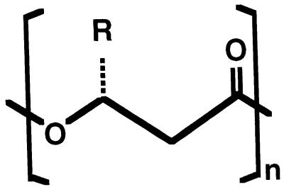
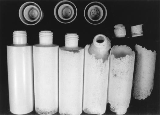
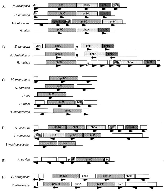
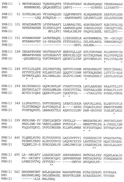
Poly(3-hydroxyalkanoates) (PHAs) are a class of microbially produced polyesters that have potential applications as conventional plastics, specifically thermoplastic elastomers. A wealth of biological diversity in PHA formation exists, with at least 100 different PHA constituents and at least five different dedicated PHA biosynthetic pathways. This diversity, in combination with classical microbial physiology and modern molecular biology, has now opened up this area for genetic and metabolic engineering to develop optimal PHA-producing organisms. Commercial processes for PHA production were initially developed by W. R. Grace in the 1960s and later developed by Imperial Chemical Industries, Ltd., in the United Kingdom in the 1970s and 1980s. Since the early 1990s, Metabolix Inc. and Monsanto have been the driving forces behind the commercial exploitation of PHA polymers in the United States. The gram-negative bacterium Ralstonia eutropha, formerly known as Alcaligenes eutrophus, has generally been used as the production organism of choice, and intracellular accumulation of PHA of over 90% of the cell dry weight have been reported. The advent of molecular biological techniques and a developing environmental awareness initiated a renewed scientific interest in PHAs, and the biosynthetic machinery for PHA metabolism has been studied in great detail over the last two decades. Because the structure and monomeric composition of PHAs determine the applications for each type of polymer, a variety of polymers have been synthesized by cofeeding of various substrates or by metabolic engineering of the production organism. Classical microbiology and modern molecular bacterial physiology have been brought together to decipher the intricacies of PHA metabolism both for production purposes and for the unraveling of the natural role of PHAs. This review provides an overview of the different PHA biosynthetic systems and their genetic background, followed by a detailed summation of how this natural diversity is being used to develop commercially attractive, recombinant processes for the large-scale production of PHAs.


Figures

FIG. 1
FIG. 1
Chemical structure of PHAs. PHAs are generally composed of (R)-β-hydroxy fatty acids, where the pendant group (R) varies from methyl (C1) to tridecyl (C13). Other fatty acids that have been incorporated have the hydroxy group at the γ, δ, or ɛ position, while the pendant group may be saturated or unsaturated or contain substituents. The best-known PHAs are P(3HB) (R = methyl), P(3HB-3HV) (R = methyl or ethyl), and P(3HO-3HH) (R = pentyl or propyl).
FIG. 2
FIG. 2
Degradation of P(3HB-3HV) in aerobic sewage sludge. Bottles made of P(3HB-3HV) were incubated during the summer (average temperature, 20°C) in aerobic sewage sludge. The progress of degradation is demonstrated with bottles that have been subjected to this treatment for 0, 2, 4, 6, 8, and 10 weeks (from left to right). Photograph courtesy of Dieter Jendrossek, Georg-August-Universität, Göttingen, Germany.
FIG. 3
FIG. 3
Biosynthetic pathway for P(3HB). P(3HB) is synthesized in a three-step pathway by the successive action of β-ketoacyl-CoA thiolase (PhbA), acetoacetyl-CoA reductase (PhbB), and P(3HB) polymerase (PhbC). The three enzymes are encoded by the genes of the phbCAB operon. A promoter upstream of phbC transcribes the complete operon.
FIG. 4
FIG. 4
pha and phb operons. The loci encoding the genes for PHA formation have been characterized from 18 different species. Genes specifying enzymes for ssc-PHA formation are designated phb, and those specifying enzymes for msc-PHA formation are designated pha. Not all pathways have completely been elucidated in these strains. The emerging picture is that pha and phb genes are not necessarily clustered and that the gene organization varies from species to species. Other genes possibly related to PHA metabolism may be linked to the essential pha and phb genes. (A) Complete phbCAB operons. (B) Interrupted phb loci. (C) Incomplete phb loci. (D) phb loci from organisms that encode two subunit P(3HB) polymerases. (E) The phbCJ locus of A. caviae involved in P(3HB-3HH) formation. (F) pha loci for msc-PHA formation in Pseudomonas.
FIG. 5
FIG. 5
Sequence similarity of representatives of three types of PHA polymerases. R. eutropha ssc-PHA polymerase (PHB1), P. oleovorans msc-PHA polymerase (PHO), and the PhbC subunit of the two-subunit polymerase from Synechocystis sp. (PHB2) were aligned by using the program of Higgins (MacDNASIS; IntelliGenetics, Mountain View, Calif.). Residues conserved in all PHA polymerases identified to date are marked by an asterisk.
FIG. 6
FIG. 6
(A) Similarities between PHA polymerase and lipase. PHA polymerase (C) acts at the surface of a PHA granule, where soluble precursors are polymerized and deposited in the hydrophobic environment of the granule. PHA depolymerase (Z) also acts at this surface and liberates the monomers from the polymer. Both enzymatic reactions are reminiscent of that of lipase (L), which cleaves ester bonds at triglyceride (TG)/water interfaces, yielding free acids and alkanols. (B) Proposed mechanism for the formation of PHA granules. Soluble enzyme converts monomer-CoA to oligomers, which remain enzyme bound (step 1). At a critical oligomer length and enzyme-oligomer concentration, the enzyme-oligomer complexes form micelles with the enzyme located at the interface, separating the PHA from the cytosol (step 2). Because of this compartmentalization, PHA polymerization is facilitated. Because the hydrophobic polymer can now be extruded into a hydrophobic environment instead of the aqueous phase, the reaction proceeds faster. The micelles are expanded and now appear as intracellular, granular structures visible with the phase-contrast microscope (step 3). As the number of granules increase, they may fuse and coalesce, giving rise to large aggregates of PHA (step 4).
FIG. 7
FIG. 7
Biosynthetic pathway for P(3HB-3HH). P(3HB-3HH) monomers are derived from fatty acid degradation by converting enoyl-CoA intermediates directly to (R)-3-hydroxyacyl-CoA precursors by an (R)-specific enoyl-CoA hydratase (PhaJ).
FIG. 8
FIG. 8
Biosynthetic pathway for P(3HB-3HV) from carbohydrates. Some microorganisms accumulate P(3HB-3HV) without supplementation of propionate, valerate, or other Codd fatty acids. Propionyl-CoA in these species is formed through the methylmalonyl-CoA pathway, which originates from succinyl-CoA in the TCA cycle. Propionyl-CoA and acetyl-CoA are converted to P(3HB-3HV) by the typical Phb enzymes.
FIG. 9
FIG. 9
Biosynthetic pathway for msc-PHA from hydrocarbons. Fluorescent pseudomonads of rRNA homology group I can derive monomers for PHA from fatty acid degradation. Intermediates from the β-oxidation cycle can be converted to (R)-3-hydroxyacyl-CoA by a hydratase (H), epimerase (E), or reductase (R) activity, whose nature is currently unknown. Any or all of these three enzymes and PHA polymerase determine the limits to the substrate specificity, which is from C6 to C16 3-hydroxy fatty acids.
FIG. 10
FIG. 10
Biosynthetic pathway for msc-PHA from carbohydrates. Monomers for PHA are derived from the fatty acid biosynthesis pathway as (R)-3-hydroxyacyl-ACP intermediates and are converted to (R)-3-hydroxyacyl-CoA through an acyl-ACP:CoA transacylase encoded by the phaG gene.
FIG. 11
FIG. 11
P(3HB) metabolism and N2 fixation in Rhizobium. (A) In the bacteroid of R. meliloti in symbiosis with alfalfa, the Tme malic enzyme is not expressed while Dme is inhibited by excess acetyl-CoA. Consequently, the levels of NAD(P)H are too low to pull acetyl-CoA into the P(3HB) pathway. In the free-living state, however, both Tme and Dme are active and P(3HB) formation is initiated under the desired conditions. (B) A direct link in central metabolism between the TCA cycle, P(3HB) formation, and amino acid metabolism is apparent from studies of the R. leguminosarum amino acid permease. Mutants that are less sensitive to high levels of aspartate have an increased secretion of glutamate. This increased production of glutamate is caused by inhibition of the TCA cycle either by a mutation in one of the genes encoding a TCA cycle enzyme or by a mutation in the PHA polymerase gene. In the absence of P(3HB) synthesis, the TCA cycle cannot function optimally, since increased reducing equivalents inhibit α-ketoglutarate dehydrogenase. Both types of mutations cause accumulation of α-ketoglutarate, which is directly converted to glutamate. (C) Recycling of reducing equivalents in Rhizobium. The TCA cycle is the most important pathway for supplying precursors of amino acids. To keep the TCA cycle active in the anaerobic bacteroid, P(3HB) biosynthesis and nitrogenase oxidize reducing equivalents. Different Rhizobium spp. have evolved different means to regulate the three NAD(P)H-oxidizing pathways in the free-living or bacteroid state.
FIG. 12
FIG. 12
Ancillary genes encoding enzymes and proteins that affect PHA accumulation. Three enzymes encoded by three genes are essential for P(3HB) formation. Several other gene products, however, affect P(3HB) formation, and mutations in the corresponding genes may decrease P(3HB) levels. Such enzymes and proteins can act on different aspects of P(3HB) formation: monomer supply, cofactor regeneration, granule assembly, or polymer degradation.
FIG. 13
FIG. 13
Endogenous formation of propionyl-CoA in R. eutropha R3, which has altered metabolism of the branched-chain amino acids. This mutant overproduces the acetolactate synthase approximately 15-fold to compensate for a defective threonine dehydratase. The endogenous accumulation of propionyl-CoA under nitrogen-limiting conditions allows this strain to produce P(3HB-3HV) without the supplementation of the growth medium with propionate or other cofeeds.
FIG. 14
FIG. 14
Propionate is an additional carbon source which is supplied as a cosubstrate for the synthesis of P(3HB-3HV) in recombinant E. coli. Several pathways have been shown to be involved in the uptake of propionate and are important in defining the optimal genotype for P(3HB-3HV) production strains. Both the acetoacetate degradation pathway (the Ato system) and the acetate secretion pathway (Ack/Pta) have been identified as contributing to propionate transport.
FIG. 15
FIG. 15
Biosynthesis of P(3HB-4HB) in recombinant E. coli by using heterologous genes from Clostridium kluyveri. The 4HB monomer in the synthesis of P(3HB-4HB) is derived from succinate. Succinate is converted to 4HB-CoA by enzymes that are encoded by genes from the gram-positive, strictly anaerobic C. kluyveri microbe.
FIG. 16
FIG. 16
Model of the P(3HB)-Ca2+-polyphosphate complex from E. coli. This P(3HB) complex forms a channel in the membrane to transport Ca2+ ions out of the cell. It is proposed that the channel is also involved in DNA uptake by competent E. coli cells. In this model, the Ca2+ ions (green) are localized between the inner polyphosphate molecule (yellow phosphorus atoms and red oxygen atoms) and a P(3HB) helix (red oxygen atoms, blue carbon atoms, and white hydrogen atoms). The methyl side groups of the P(3HB) helix face the outside of the channel and are in contact with the hydrophobic lipids of the membrane. The carbonyl oxygen atoms face the interior of the channel and ligand the Ca2+ ions. The phosphate groups play a similar role. Extrusion of Ca2+ ions may result from physical constraints on the structure or from enzymatic synthesis and degradation of the polyphosphate chain at the membrane/cytosol and membrane/periplasm interfaces.

References

    1. Abe C, Taima Y, Nakamura Y, Doi Y. New bacterial copolyesters of 3-hydroxyalkanoates and 3-hydroxy-ω-fluoroalkanoates produced by Pseudomonas oleovorans. Polym Commun. 1990;31:404–406.
    1. Abe H, Doi Y, Fukushima T, Eya H. Biosynthesis from gluconate of a random copolyester consisting of 3-hydroxybutyrate and medium-chain-length 3-hydroxyalkanoates by Pseudomonas sp. 61-3. Int J Biol Macromol. 1994;16:115–119. - PubMed
    1. Ackermann J-U, Babel W. Growth-associated synthesis of poly(hydroxybutyric acid) in Methylobacterium rhodesianum as an expression of an internal bottleneck. Appl Microbiol Biotechnol. 1997;47:144–149.
    1. Akiyama M, Doi Y. Production of poly(3-hydroxyalkanoates) from α,ω-alkanedioic acids and hydroxylated fatty acids by Alcaligenes sp. Biotechnol Lett. 1993;15:163–168.
    1. Akiyama M, Taima Y, Doi Y. Production of poly(3-hydroxyalkanoates) by a bacterium of the genus Alcaligenes utilizing long-chain fatty acids. Appl Microbiol Biotechnol. 1992;37:698–701.

MeSH terms

LinkOut - more resources