Review
. 1999 Mar;63(1):128-48.
doi: 10.1128/MMBR.63.1.128-148.1999.

Mechanisms of arthropod transmission of plant and animal viruses

Affiliations
Review

Mechanisms of arthropod transmission of plant and animal viruses

S M Gray et al. Microbiol Mol Biol Rev. 1999 Mar.

Abstract

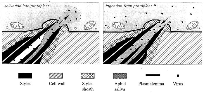
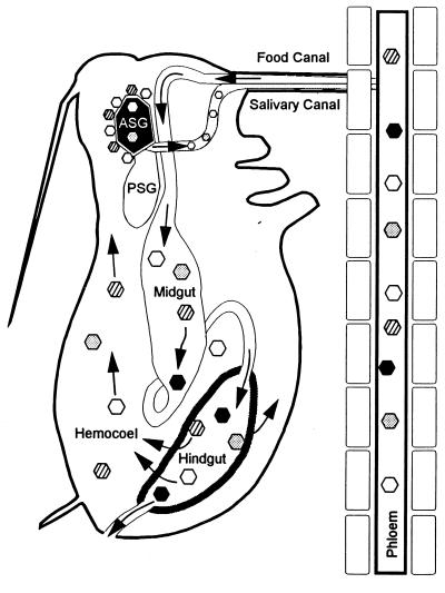
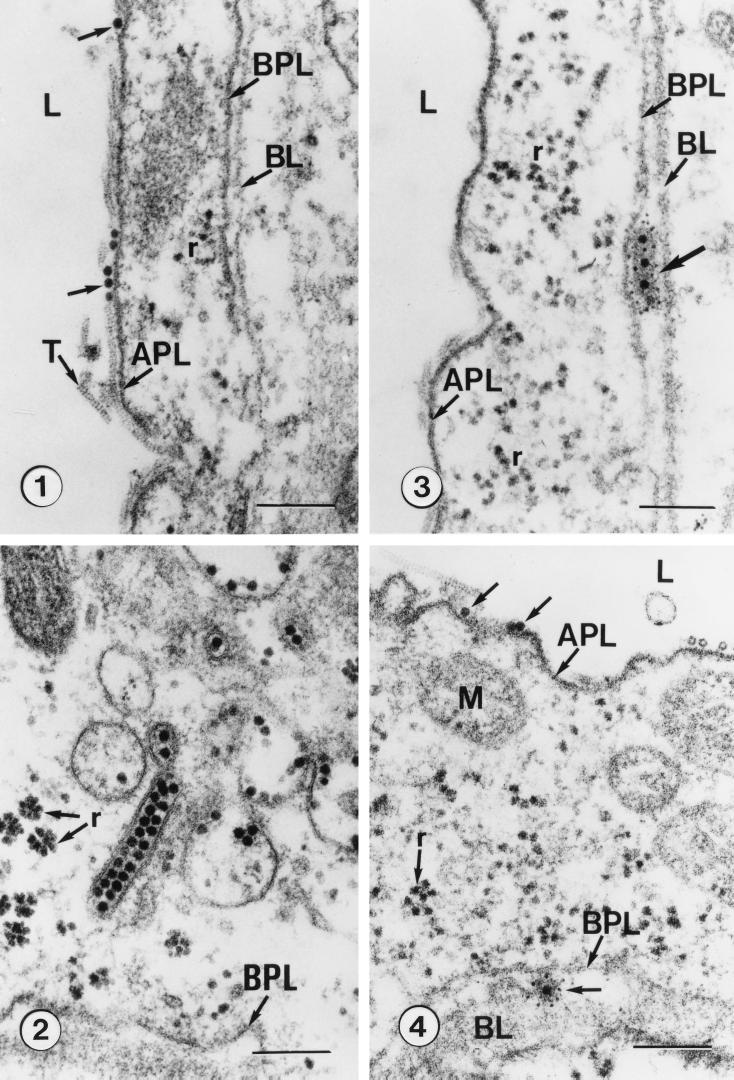
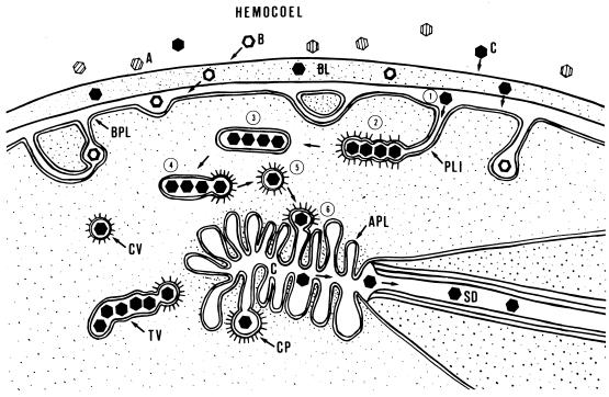
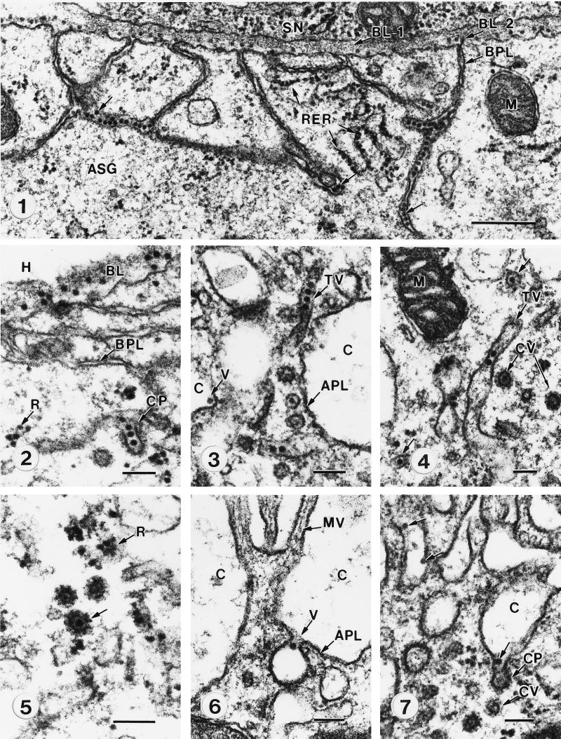
A majority of the plant-infecting viruses and many of the animal-infecting viruses are dependent upon arthropod vectors for transmission between hosts and/or as alternative hosts. The viruses have evolved specific associations with their vectors, and we are beginning to understand the underlying mechanisms that regulate the virus transmission process. A majority of plant viruses are carried on the cuticle lining of a vector's mouthparts or foregut. This initially appeared to be simple mechanical contamination, but it is now known to be a biologically complex interaction between specific virus proteins and as yet unidentified vector cuticle-associated compounds. Numerous other plant viruses and the majority of animal viruses are carried within the body of the vector. These viruses have evolved specific mechanisms to enable them to be transported through multiple tissues and to evade vector defenses. In response, vector species have evolved so that not all individuals within a species are susceptible to virus infection or can serve as a competent vector. Not only are the virus components of the transmission process being identified, but also the genetic and physiological components of the vectors which determine their ability to be used successfully by the virus are being elucidated. The mechanisms of arthropod-virus associations are many and complex, but common themes are beginning to emerge which may allow the development of novel strategies to ultimately control epidemics caused by arthropod-borne viruses.


Figures

FIG. 1
FIG. 1
Light micrograph of a longitudinal section through an aphid head and leaf as the aphid is feeding on the plant. The aphid stylet protrudes from the proboscis (A) and penetrates intracellularly through the mesophyll cells (B) and into the vascular bundle (C).
FIG. 2
FIG. 2
Autoradiographs of stylets of Myzus persicae given acquisition access to 125I-labeled tobacco etch potyvirus virions. (A) Stylets of an aphid that has not fed on an infected plant. (B) Stylets of an aphid that acquired labeled virus through a plastic membrane. (C) Distribution of label in stylets that have separated, showing label associated only with the food canal formed by the maxillary stylets. MA, mandibular stylets; MX, maxillary stylets; P, proboscis; S, stylet. Magnification, ×420. Reproduced from reference with permission of the publisher.
FIG. 3
FIG. 3
Model of the ingestion-salivation mechanism of noncirculative, nonpersistent transmission. Virus is ingested into the food canal (right), along with the cytoplasm. Virus adheres to the epicuticular lining of the food canal and the common duct at the very distal tip of the stylet, which is shared with the salivary canal. When the aphid first probes a cell after acquiring virus (left), saliva is injected into the cell. The watery salivary secretions will release virus from the cuticle lining the common duct, but virus farther inside the food canal would not be released by this mechanism. Reproduced from reference with permission of the publisher.
FIG. 4
FIG. 4
Circulative route of barley yellow dwarf luteoviruses (BYDVs) through aphids. All BYDV strains can be ingested from phloem into the aphid’s alimentary canal and arrive in the hindgut intact. The hindgut epithelium is the first transmission barrier; most BYDVs can bind to hindgut epithelial cells and be transported into the hemocoel, but some are excluded (solid hexagons). BYDVs acquired in the hemocoel must migrate to the ASG. The basal lamina of the ASG may selectively filter BYDVs or may concentrate virions, thereby increasing the efficiency of transport into the ASG. BYDVs (gray hexagons) not concentrated at the ASG may be transported into the ASG if they encounter it, but the efficiency of transmission is low. BYDVs may be concentrated at the ASG but be prevented from entering the ASG by an inability to bind to the ASG plasmalemma and initiate endocytosis (striped hexagons). Efficiently transmitted BYDVs are concentrated at the ASG and efficiently transported in the ASG and the salivary canal (open hexagons). Reprinted from reference with permission of the publisher.
FIG. 5
FIG. 5
Electron micrographs of the hindgut of Rhopalosiphum padi microinjected with anti-barley yellow dwarf luteovirus (BYDV) antibodies for immunolabeling following acquisition feedings on Parafilm membranes containing purified BYDV or on oats infected with BYDV. (Panel 1) Ingested virions (arrows) in the hindgut lumen (L) adsorbed to the apical plasmalemma (APL). Note the longitudinal views of extracellular tubules (T), ribosomes (r), basal plasmalemma (BPL), and basal lamina (BL). (Panel 2) Unlabeled virions concentrated in receptosome-like vesicles and in a tubular vesicle adjacent to the basal plasmalemma (BPL) and basal lamina. Ribosomes (r) are also shown. (Panel 3) Ferritin-labeled virions (arrow) captured between the basal plasmalemma (BPL) and basal lamina (BL) upon release from the hindgut cell into the hemocoel. Apical plasmalemma (APL), hindgut lumen (L), and ribosomes (r) are also shown. (Panel 4) Unlabeled virions (arrows) in the hindgut lumen (L) adjacent to the apical plasmalemma (APL) and an anti-BYDV-labeled virion adjacent to the basal plasmalemma (BPL) following transport to the hemocoel. The basal lamina (BL), mitochondria (M), and ribosomes (r) are also shown. Bars, 200 nm. Reproduced from reference with permission of the publisher.
FIG. 6
FIG. 6
Model of the interactions of luteoviruses with the ASG of aphid vector species. Three types of interactions were observed when virions of the MAV isolate of barley yellow dwarf virus were acquired by aphids that fed on infected plants or were injected with purified virions into the hemocole. In the first type of interaction, MAV virions had no affinity for the salivary basal lamina (BL) of specific aphid species and did not attach to or penetrate the basal lamina (A, nonpenetrating, nontransmitted virions). In other species, MAV virions did exhibit affinity for the salivary basal lamina and were able to attach to and, in some cases, penetrate the basal lamina. However, these virions were unable to initiate endocytosis at the basal plasmalemma (BPL) and were not transmitted (B, penetrating, nontransmitted virions). In the third type of interaction, virions consistently penetrated the basal lamina (step 1), were aggregated in plasmalemma invaginations (PLI), and were endocytosed into the cell by coated-pit formation (step 2). Virions acquired in the cytoplasm accumulated at the apical end of the cell in tubular vesicles (step 3). Individual virions budded from the tubular vesicles by coated-pit formation (step 4) and were transported to the salivary canal (Cn) in coated vesicles (step 5) that fused to the apical plasmalemma (APL), releasing the virion into the canal lumen (step 6). Transcytosed virions were then able to move into the salivary duct (SD) (C, penetrating-transmitted virions; TV, tubular vesicle; CP, coated pit; CV, coated vesicle). Reproduced from reference with permission of the publisher.
FIG. 7
FIG. 7
Ultrastructure of membranes associated with transcellular transport of microinjected virions of the MAV isolate of barley yellow dwarf virus through the ASG of Sitobion avenae. (Panel 1) Virions of MAV embedded in the ASG basal lamina (BL-2) and concentrated in basal plasmalemma (BPL) invaginations (arrows). Note the absence of virions in the basal lamina (BL-1) of the adjacent subesophageal nerve ganglion (SN). Particles in the cytoplasm are ribosomes associated with the rough endoplasmic reticulum (RER). M, mitochondria. Bar, 500 nm. (Panel 2) Virions penetrating the basal lamina (BL) from the hemocole (H) and in a coated pit (CP) during endocytosis through the basal plasmalemma (BPL). The small, irregularly shaped particles observed free in the cytoplasm are ribosomes (r), as determined by RNase digestion (3) and observation of ultrastructure under higher magnification. (Panel 3) Virions packaged in tubular vesicles (TV) adjacent to the apical plasmalemma (APL) lining a microvillus-lined canal (C). V, virion in canal. (Panel 4) Virions in tubular vesicles (TV) and associated coated vesicles (CV). M, mitochondria. (Panel 5) Comparison of a virion in a coated vesicle (arrow) to cytoplasmic ribosomes (R). (Panel 6) A virion (V) being released from the accessory salivary gland cell into the canal lumen (c) by exocytosis through the apical plasmalemma (APL). MV, microvilli. (Panel 7) Virions (arrows) in the canal lumen (C) released from coated pits (CP) following fusion of coated vesicles (CV) with the cell membrane. Bars, 100 nm. Reproduced from reference with permission of the publisher.

References

    1. Al K N, Covey S N. Variation in biological properties of cauliflower mosaic virus clones. J Gen Virol. 1994;75:3137–3145. - PubMed
    1. Ammar E D. Propagative transmission of plant and animal viruses by insects: factors affecting vector specificity and competence. Adv Dis Vector Res. 1994;10:289–331.
    1. Ammar E D, Jarlfors U, Pirone T P. Association of potyvirus helper component protein with virions and the cuticle lining the maxillary food canal and foregut of an aphid vector. Phytopathology. 1994;84:1054–1060.
    1. Atreya C D, Pirone T P. Mutational analysis of the helper component proteinase gene of a potyvirus: effects of amino acid substitutions, deletions, and gene replacement on virulence and aphid transmissibility. Proc Natl Acad Sci USA. 1993;90:11919–11923. - PMC - PubMed
    1. Backus E A. Anatomical and sensory mechanisms of planthopper and leafhopper feeding behavior. In: Nault L R, Rodriguez J G, editors. The leafhoppers and planthoppers. New York, N.Y: John Wiley & Sons, Inc.; 1985. pp. 163–194.

LinkOut - more resources