. 1999 Mar 15;515 ( Pt 3)(Pt 3):639-51.
doi: 10.1111/j.1469-7793.1999.639ab.x.

Kir2.1 encodes the inward rectifier potassium channel in rat arterial smooth muscle cells

Affiliations

Kir2.1 encodes the inward rectifier potassium channel in rat arterial smooth muscle cells

K K Bradley et al. J Physiol. .

Abstract

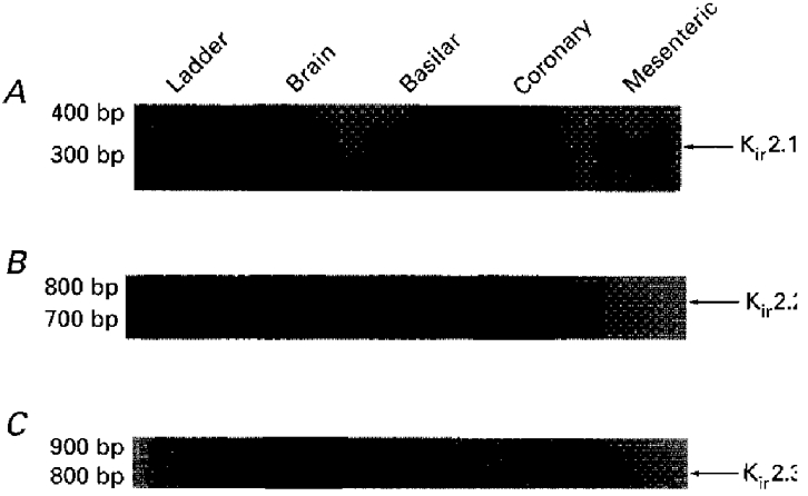
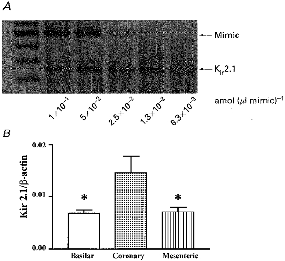
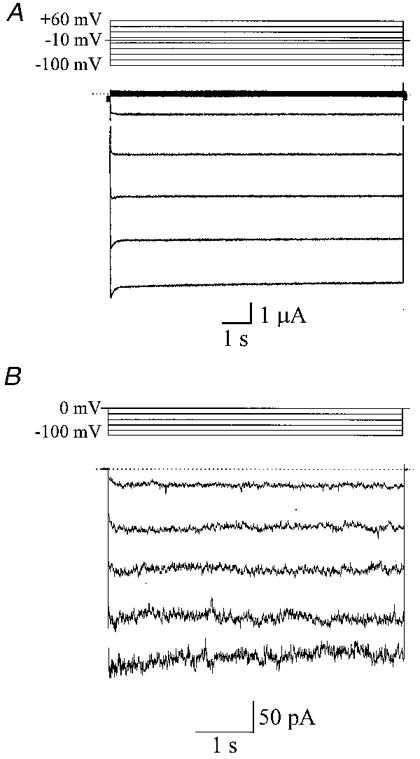
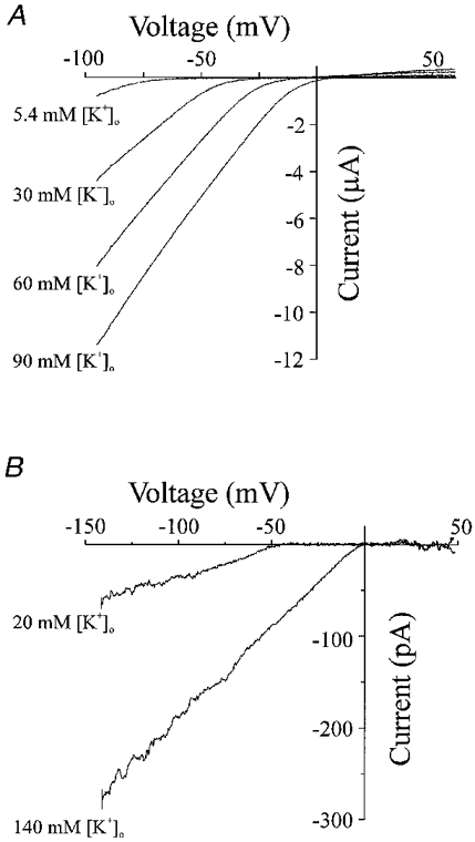
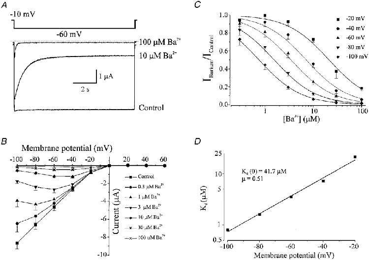
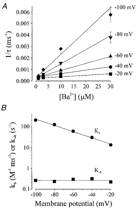
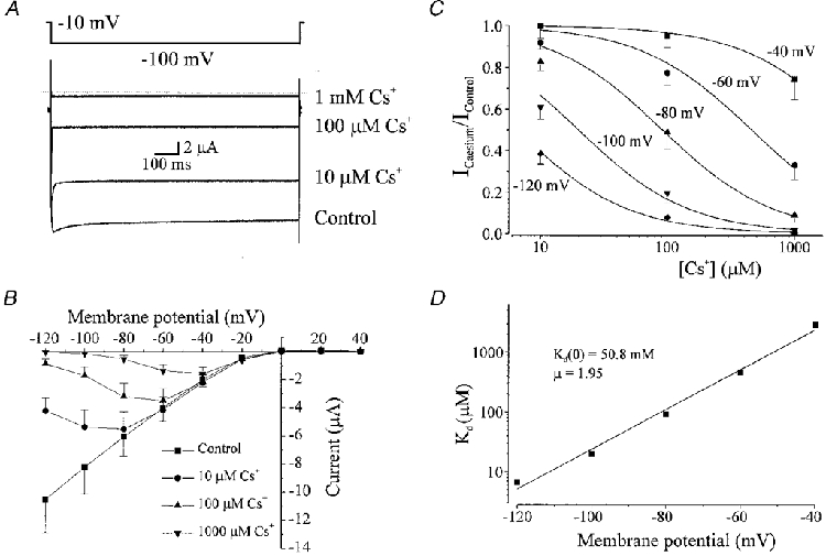
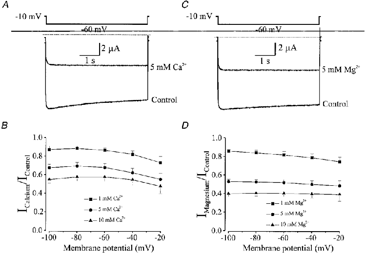
1. The molecular nature of the strong inward rectifier K+ channel in vascular smooth muscle was explored by using isolated cell RT-PCR, cDNA cloning and expression techniques. 2. RT-PCR of RNA from single smooth muscle cells of rat cerebral (basilar), coronary and mesenteric arteries revealed transcripts for Kir2.1. Transcripts for Kir2.2 and Kir2.3 were not found. 3. Quantitative PCR analysis revealed significant differences in transcript levels of Kir2.1 between the different vascular preparations (n = 3; P < 0.05). A two-fold difference was detected between Kir2.1 mRNA and beta-actin mRNA in coronary arteries when compared with relative levels measured in mesenteric and basilar preparations. 4. Kir2.1 was cloned from rat mesenteric vascular smooth muscle cells and expressed in Xenopus oocytes. Currents were strongly inwardly rectifying and selective for K+. 5. The effect of extracellular Ba2+, Ca2+, Mg2+ and Cs2+ ions on cloned Kir2.1 channels expressed in Xenopus oocytes was examined. Ba2+ and Cs+ block were steeply voltage dependent, whereas block by external Ca2+ and Mg2+ exhibited little voltage dependence. The apparent half-block constants and voltage dependences for Ba2+, Cs+, Ca2+ and Mg2+ were very similar for inward rectifier K+ currents from native cells and cloned Kir2.1 channels expressed in oocytes. 6. Molecular studies demonstrate that Kir2.1 is the only member of the Kir2 channel subfamily present in vascular arterial smooth muscle cells. Expression of cloned Kir2.1 in Xenopus oocytes resulted in inward rectifier K+ currents that strongly resemble those that are observed in native vascular arterial smooth muscle cells. We conclude that Kir2.1 encodes for inward rectifier K+ channels in arterial smooth muscle.


Figures

Figure 1
Figure 1. RT-PCR detection of Kir2.1 in anatomically different vascular smooth muscle cell preparations
Amplified PCR products generated using gene-specific primers for Kir2.1 (A), Kir2.2 (B), or Kir2.3 (C) were fractionated on 2 % agarose gels and size markers were used to indicate the size of the experimental fragments (lane 1). Transcriptional expression for Kir2.1 was detected in basilar, coronary and mesenteric arteries, whereas no expression for Kir2.2 and Kir2.3 was observed. Rat cerebellum was used as a positive control for the Kir2 primers.
Figure 2
Figure 2. Differential transcriptional expression of Kir2.1 in rat arteries
Competitive PCR products were resolved on 2 % ethidium bromide agarose gels. A representative gel of quantitative RT-PCR for Kir2.1 in rat basilar, coronary and mesenteric arteries is shown in A; two-fold serial dilutions of mimic DNA were included in the PCR reactions while the target cDNA (Kir2.1) concentration remained constant. The actual concentrations of target cDNA were calculated and expressed relative to β-actin cDNA concentration (B). Results are expressed as means ±s.e.m.; *significant difference when compared with transcript levels detected in coronary artery.
Figure 3
Figure 3. Arterial smooth muscle inward rectifier currents
A, inwardly rectifying potassium currents recorded from a Xenopus oocyte injected with RNA encoding a Kir2.1 channel protein cloned from vascular smooth muscle. The oocyte was held at a membrane potential of −10 mV and voltage pulses (of 10 s duration), from +60 to −100 mV were applied every 4 s in 20 mV increments. The bathing solution contained 90 mM K+. B, Ba2+-sensitive (500 μM) currents in an isolated coronary (septal) artery smooth muscle cell in a bathing solution containing 140 mM K+ (i.e. EK = 0 mV). The cell was held at a membrane potential of 0 mV with voltage pulses of between −20 and −100 mV applied every 10 s in 20 mV increments.
Figure 4
Figure 4. Inward rectification shifts with a change in external [K+]
A, current-voltage relationship from the same Xenopus oocyte in a bathing solution containing 90, 60, 30 or 5.4 mM K+. I-V relationships were obtained by performing voltage ramps initially by holding at a membrane potential of 0 mV, followed by a pulse to −100 mV for 15 ms before a voltage ramp of 480 ms duration from −100 to +60 mV (0.3 mV ms−1). B, Ba2+-sensitive currents (average of 5 voltage ramps from −140 to +50 mV) observed in an isolated coronary (septal) artery smooth muscle cell with 20 and 140 mM external (bathing) K+. The slope conductance and Erev for this cell were 0.77 and 1.99 nS, and −47.3 mV (EK =−49.5 mV) and −3.4 mV (EK = 0 mV) with 20 and 140 mM external K+, respectively.
Figure 5
Figure 5. Elevation of external [K+] increases the conductance of inward rectifier potassium currents and shifts the reversal potential
A, the slope conductance of the inward rectifier current increased with external [K+] (n = 8 oocytes). Slope conductance (C) was fitted with (C)([K+]o)n, giving C = 36.7μS and n = 0.28. B, changing the concentration of K+ in the bathing solution shifted the reversal potential (30 mM K+, n = 8 oocytes; 60 mM K+, n = 8 oocytes; 90 mM K+, n = 24 oocytes). The line represents the best fit to the relationship between external K+ and the reversal potential, with a slope corresponding to 53.1 mV shift in Erev for a 10-fold change in [K+].
Figure 6
Figure 6. Ba2+ inhibition of inward rectifier potassium currents in Xenopus oocytes injected with RNA encoding Kir2.1 cloned from vascular smooth muscle cells
Membrane current recorded from the same oocyte with voltage steps from a holding potential of −10 mV to −60 mV in the presence of 0 (control), 10 and 100 μM Ba2+. External K+ was 90 mM. Dotted line indicates zero current level. B, current-voltage relationship demonstrating the effect of applying 0.3, 1, 3, 10, 30 and 100 μM Ba2+ on membrane currents recorded at the end of 10 s voltage pulses (n = 5). Bathing K+ was 90 mM. C, relationship between external Ba2+ concentration and the fractional inhibition of inward current at −20, −40, −60, −80 and −100 mV. Data were fitted with eqn (1), to give Kd values of (μm): 21.4, 7.2, 3.5, 1.6 and 0.8 at −20, −40, −60, −80 and −100 mV, respectively. D, voltage dependence of the dissociation constants (Kd) from C. Data were fitted with eqn (2) with a Kd at 0 mV of 41.7 μM, and a slope (μ) of 0.51.
Figure 7
Figure 7. Concentration and voltage dependence of blocking kinetics of Ba2+ on inward rectifier K+ currents from vascular smooth muscle expressed in Xenopus oocytes
A, the onset of Ba2+ block was fitted with a single exponential after which the mean reciprocal values (±s.e.m.) of the time constant (τ) of current decay was plotted against Ba2+ concentration (n = 5 oocytes). These data were fitted by eqn (3) to determine the kb, the slope of the line, and k−b, the intercept (see Table 1). B, voltage dependence of the blocking rate, kb, and unblocking rate k−b, obtained from the slope and intercept of lines fitted in A. Data were fitted with an equation equivalent to eqn (2) with kb values of 7.4 m−1 s−1 at 0 mV and slope (μ) of −0.43, and k−b values of 0.26 s−1 at 0 mV and μ of −1.02.
Figure 8
Figure 8. External Cs+ block of inward rectifier potassium currents expressed in Xenopus oocytes by injection with RNA encoding Kir2.1 cloned from vascular smooth muscle
A, membrane current recorded from the same oocyte with voltage steps from a holding potential of −10 to −100 mV in the presence of 0 (control), 10, 100 and 1000 μM Cs+. Note that the current recorded at −100 mV in the presence of 1 mM Cs+ is smaller than that observed at −10 mV in control, demonstrating the voltage dependence of block. External K+ was 90 mM. B, current-voltage relationship demonstrating the voltage dependence of block of Cs+ at different membrane potentials (n = 8 oocytes). Currents were measured at the end of 1 s voltage pulses. C, relationship between Cs+ concentration and fractional inhibition of inward current at −40, −60, −80, −100 and −120 mV. Data were fitted using eqn (1) demonstrating Kd values of (μm): 2911.7, 458.3, 91.6, 19.9 and 6.5 at −40, −60, −80, −100 and −120 mV, respectively. D, voltage dependence of Kd. Data were fitted using eqn (2) with a Kd (0) of 50.8 mM and μ of 1.95.
Figure 9
Figure 9. External Ca2+ and Mg+ block of inward rectifier currents expressed in Xenopus oocytes by injection of RNA encoding Kir2.1 cloned from vascular smooth muscle
A, membrane current recorded from an oocyte in response to a 5 s voltage step from −10 to −60 mV in control (100 μM Ca2+) and after addition of 5 mM Ca2+. The bathing solution contained 90 mM K+ and nominal Mg2+. B, fractional inhibition of inward rectifier currents by addition of 1, 5 and 10 mM Ca2+ at different voltages (n = 8 oocytes). Inhibition of currents by Ca2+ is not voltage dependent. C, membrane current recorded from an oocyte in response to a 5 s voltage step from −10 to −60 mV in control (0 Mg2+) and in the presence of 5 mM Mg2+. The bathing solution contained 90 mM K+ and nominal Mg2+. D, fractional inhibition of inward rectifier currents by 1, 5 and 10 mM Mg2+ at different voltages (n = 8 oocytes). Inhibition of currents by Mg2+ is not voltage dependent.

References

    1. Abrams CJ, Davies NW, Shelton PA, Stanfield PR. The role of a single aspartate residue in ionic selectivity and block of a murine inward rectifier K+ channel Kir2.1. The Journal of Physiology. 1996;493:643–649. - PMC - PubMed
    1. Bonaccorsi A, Hermsmeyer K, Aprigliano O, Smith CB, Bohr DF. Mechanism of potassium-induced relaxation in arterial smooth muscle. Blood Vessels. 1977;14:261–276. - PubMed
    1. Edwards FR, Hirst GD. Inward rectification in submucosal arterioles of guinea-pig ileum. The Journal of Physiology. 1988;404:437–454. - PMC - PubMed
    1. Edwards FR, Hirst GD, Silverberg GD. Inward rectification in rat cerebral arterioles; involvement of potassium ions in autoregulation. The Journal of Physiology. 1988;404:455–466. - PMC - PubMed
    1. Hamill OP, Marty A, Neher B, Sigworth FJ. Improved patch-clamp techniques for high resolution current recording from cells and cell-free membrane patches. Pflügers Archiv. 1981;391:85–100. - PubMed

Publication types

MeSH terms

LinkOut - more resources