. 1999 Mar;10(3):525-36.
doi: 10.1091/mbc.10.3.525.

Phosphorylation of tyrosine 992, 1068, and 1086 is required for conformational change of the human epidermal growth factor receptor c-terminal tail

Affiliations

Phosphorylation of tyrosine 992, 1068, and 1086 is required for conformational change of the human epidermal growth factor receptor c-terminal tail

A Bishayee et al. Mol Biol Cell. 1999 Mar.

Abstract

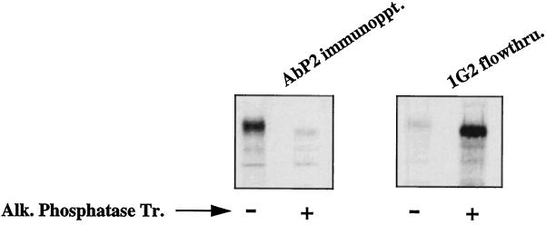
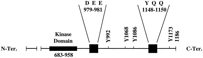
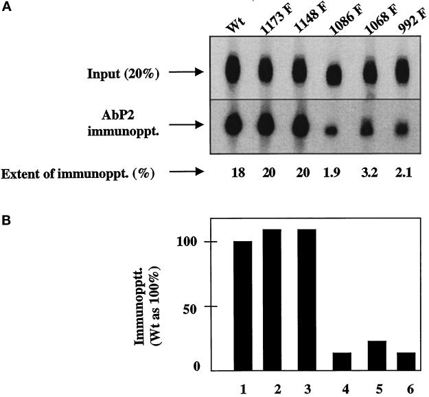
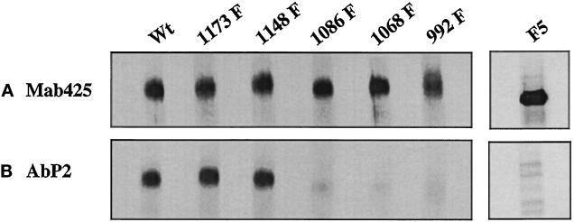
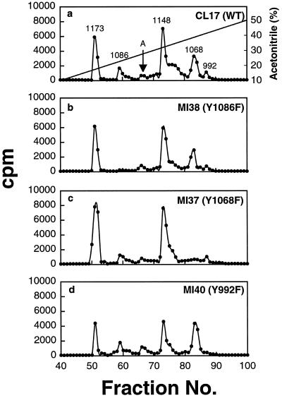
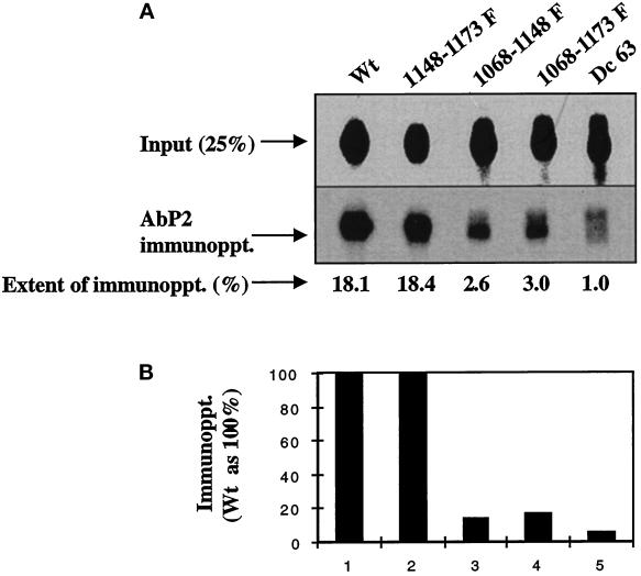
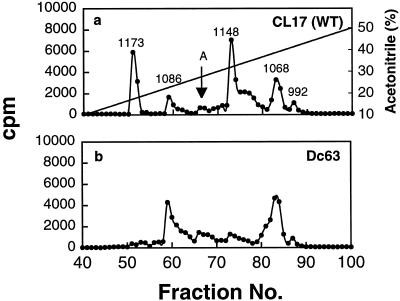
We reported previously that a conformation-specific antibody, Ab P2, to a 16-amino acid peptide (Glu-Gly-Tyr-Lys-Lys-Lys-Tyr-Gln-Gln-Val-Asp-Glu-Glu-Phe-Leu-Arg) of the cytoplasmic domain of the beta-type platelet-derived growth factor receptor also recognizes the epidermal growth factor (EGF) receptor. Although the antibody is not directed to phosphotyrosine, it recognizes in immunoprecipitation the activated and hence phosphorylated form of both receptors. In P2 peptide, there are two tripeptide sequences, Asp-Glu-Glu and Tyr-Gln-Gln, that are also present in the EGF receptor. Our present studies using either EGF receptor C-terminal deletion mutants or point mutations (Tyr-->Phe) and our previous studies on antibody inhibition by P2-derived peptides suggest that Gln-Gln in combination with Asp-Glu-Glu forms a high-affinity complex with Ab P2 and that such complex formation is dependent on tyrosine phosphorylation. Of the five phosphate acceptor sites in the EGF receptor, clustered in the extreme C-terminal tail, phosphorylation of three tyrosine residues (992, 1068, and 1086) located between Asp-Glu-Glu and Gln-Gln is necessary for Ab P2 binding. In contrast, the acceptor sites Tyr 1173 and 1148 play no role in the conformation change. Asp-Glu-Glu and Gln-Gln are located 169 amino acids apart, and it is highly likely that the interactions among three negatively charged phosphotyrosine residues in the receptor C terminus may result in the bending of the peptide chain in such a way that these two peptides come close to each other to form an antibody-binding site. Such a possibility is also supported by our finding that receptor dephosphorylation results in complete loss of Ab P2-binding activity. In conclusion, we have identified a domain within the cytoplasmic part of the EGF receptor whose conformation is altered by receptor phosphorylation; furthermore, we have identified the tyrosine residues that positively regulate this conformation.


Figures

Figure 1
Figure 1
The phosphorylation-induced conformational change is reversible. A431 cells were labeled with Tran 35S-label for 10 h. The EGF receptors in the detergent-solubilized cell lysates were phosphorylated with unlabeled ATP in the presence of 1 μM EGF as described in MATERIALS AND METHODS. After purification of the EGF receptors by anti-phosphotyrosine monoclonal antibody (1G2), the receptors were allowed to bind with wheat germ agglutinin–agarose. The bound receptor was eluted from the beads by 0.4 M N-acetylglucosamine in 20 mM HEPES, pH 7.4, 0.15 M NaCl, 0.2% NP-40, and protease inhibitors. An aliquot of the purified receptor was left untreated (−) or treated with alkaline phosphatase (50 units/ml) (+) coupled to agarose in the presence of 1 mg/ml bovine serum albumin. After incubation at 4°C for 1 h, both samples were centrifuged, and the supernatants were subjected to immunoprecipitation in the presence of 1 mM vanadate with Ab P2 (AbP2 immunoppt.; left) or anti-phosphotyrosine antibody 1G2 (right). 1G2 flowthru. denotes the labeled receptor that did not bind to 1G2. Alk. Phosphatase Tr., Alkaline phosphatase treatment.
Figure 2
Figure 2
Locations of the phosphate acceptor sites and the tripeptides (Asp-Glu-Glu and Tyr-Gln-Gln) with respect to the kinase domain of the human EGF receptor. The amino acids are identified by their single-letter codes: D for Asp, E for Glu, Q for Gln, and Y for Tyr. C-Ter., C-terminal; N-Ter., N-terminal.
Figure 3
Figure 3
Single Tyr→Phe substitution at 992, 1068, or 1086 drastically reduces the binding of Ab P2 to the 32P-labeled EGF receptor. Detergent-solubilized membranes from the wild type (Wt) or the single Y→F EGF receptor mutants were phosphorylated with labeled ATP (specific radioactivity of 350 cpm/fmol) in the presence of 1 μM EGF. After purification of the labeled receptor by 1G2–Sepharose, the 32P-labeled receptor was quantified as described in MATERIALS AND METHODS. For immunoprecipitation, 1.25 fmol of the EGF receptor was incubated with 5 μg of protein A–purified Ab P2 in a total volume of 15 μl under conditions described in MATERIALS AND METHODS. After isolation of the immune complexes with formaldehyde-fixed S. aureus, the labeled proteins were analyzed by SDS-PAGE and autoradiography, and the region containing the 170-kDa EGF receptor band was densitometrically scanned. (A) Top, 20% of labeled samples that have not been subjected to immunoprecipitation [Input (20%)]. Middle, the EGF receptor bands after immunoprecipitation with Ab P2 (AbP2 immunoppt.). Bottom, the extent of immunoprecipitation. (B) The results of the immunoprecipitation relative to that of the wild-type receptor.
Figure 4
Figure 4
Interaction of Ab P2 with 35S-labeled EGF receptor is dependent on the phosphorylation of Tyr 992, 1068, and 1086. 35S-labeled EGF receptors from the wild type and the single Y→F substitution mutants were phosphorylated with unlabeled ATP in the presence of EGF, and the labeled receptors were purified with 1G2–Sepharose. (A) The relative concentrations of the EGF receptor in different cell types were quantified by precipitation with mAb 425 (Mab425), a monoclonal antibody to an external peptide epitope of the receptor. (B) Equal amounts of the EGF receptor from different mutants as shown in A were subjected to immunoprecipitation with Ab P2 (AbP2) followed by SDS-PAGE (3.5–10%) and fluorography. In a separate experiment, 35S-labeled cell lysates from the EGF receptor F5 mutant in which all five known phosphate acceptor sites were mutated to Phe were incubated with unlabeled ATP in the presence of EGF, and the labeled receptors were purified with wheat germ agglutinin. The purified receptor preparation was immunoprecipitated with mAb 425, Ab P2, or nonimmune serum and was analyzed by SDS-PAGE as described above.
Figure 5
Figure 5
Phosphopeptide maps of the EGF receptors after trypsin digestion. (a–d) Receptors bound to EGF–Affi-Gel 15 were phosphorylated with [γ-32P]ATP under autophosphorylation conditions. After dissociation from the gel beads by heating with SDS-sample buffer, the 32P-labeled receptors were subjected to SDS-PAGE and digested twice with 20 μg of trypsin, and then 50,000 cpm were analyzed by a reverse phase HPLC C18 column as described in MATERIALS AND METHODS. Fractions (0.5 ml) were collected, and Cerenkov counts were determined. The arrow labeled A indicates the unidentified peak eluted between Tyr 1068 and 1148. WT, wild type.
Figure 6
Figure 6
A deletion mutant with a truncation of the C-terminal 63 amino acids (Dc 63) does not bind to Ab P2. Anti-phosphotyrosine antibody (1.25 fmol; 1G2)-purified 32P-labeled EGF receptors from the wild-type or the mutated receptors were immunoprecipitated with 5 μg of Ab P2 under the conditions described in Figure 3. (A) Top, 25% of labeled samples that have not been subjected to immunoprecipitation [Input (25%)]. Middle, the EGF receptor bands after immunoprecipitation with Ab P2 (AbP2 immunoppt.). Bottom, the extent of immunoprecipitation. (B) The results of the immunoprecipitation relative to that of the wild-type receptor.
Figure 7
Figure 7
The phosphopeptide maps of the tryptic digest from the EGF receptor deletion mutant Dc63. (a–d) The conditions of the experiment and the methods used are the same as those described in Figure 5.

Similar articles

Cited by

References

    1. Alvarez CV, Shon K-J, Miloso M, Beguinot L. Structural requirements of the epidermal growth factor receptor for tyrosine phosphorylation of esp8 and esp15, substrates lacking Src SH2 homology domains. J Biol Chem. 1995;270:16271–16276. - PubMed
    1. Bangalore L, Tanner AJ, Laudano AP, Stern D. Antiserum raised against a synthetic phosphotyrosine-containing peptide selectively recognizes p185neu/erbB-2 and the epidermal growth factor receptor. Proc Natl Acad Sci USA. 1992;89:11637–11641. - PMC - PubMed
    1. Bishayee S, Majumdar S, Scher CD, Khan S. Characterization of a novel anti-peptide antibody that recognizes a specific conformation of the platelet-derived growth factor receptor. Mol Cell Biol. 1988;8:3696–3702. - PMC - PubMed
    1. Bishayee S, Ross AH, Womer R, Scher CD. Purified human platelet-derived growth factor receptor has ligand stimulated tyrosine kinase activity. Proc Natl Acad Sci USA. 1986;83:6756–6760. - PMC - PubMed
    1. Biswas R, Basu M, Sen-Majumdar A, Das M. Intrapeptide autophosphorylation of the epidermal growth factor receptor: regulation of kinase catalytic function by receptor dimerization. Biochemistry. 1985;24:3795–3802. - PubMed

Publication types

MeSH terms

LinkOut - more resources