. 1999 Mar;10(3):537-49.
doi: 10.1091/mbc.10.3.537.

High-affinity binding of the AP-1 adaptor complex to trans-golgi network membranes devoid of mannose 6-phosphate receptors

Affiliations

High-affinity binding of the AP-1 adaptor complex to trans-golgi network membranes devoid of mannose 6-phosphate receptors

Y Zhu et al. Mol Biol Cell. 1999 Mar.

Abstract

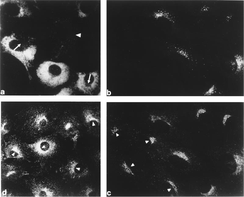
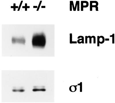
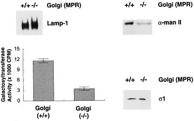
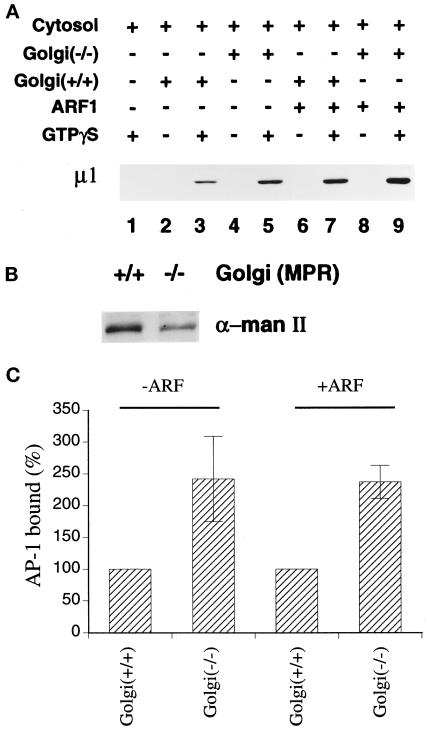
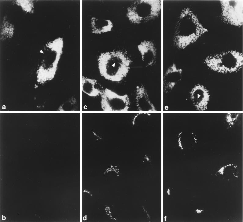
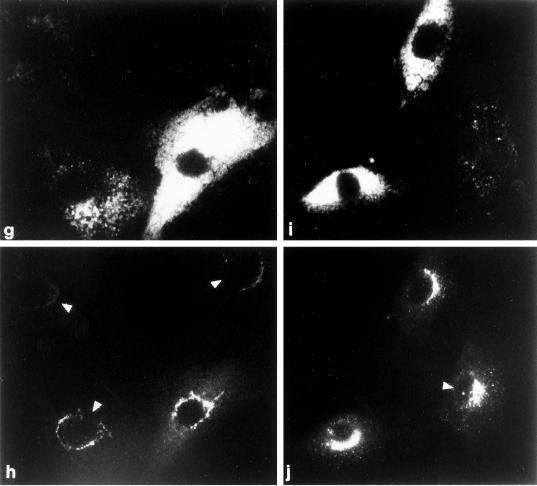
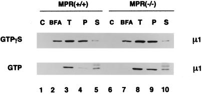
The GTP-binding protein ADP-ribosylation factor (ARF) initiates clathrin-coat assembly at the trans-Goli network (TGN) by generating high-affinity membrane-binding sites for the AP-1 adaptor complex. Both transmembrane proteins, which are sorted into the assembling coated bud, and novel docking proteins have been suggested to be partners with GTP-bound ARF in generating the AP-1-docking sites. The best characterized, and probably the major transmembrane molecules sorted into the clathrin-coated vesicles that form on the TGN, are the mannose 6-phosphate receptors (MPRs). Here, we have examined the role of the MPRs in the AP-1 recruitment process by comparing fibroblasts derived from embryos of either normal or MPR-negative animals. Despite major alterations to the lysosome compartment in the MPR-deficient cells, the steady-state distribution of AP-1 at the TGN is comparable to that of normal cells. Golgi-enriched membranes prepared from the receptor-negative cells also display an apparently normal capacity to recruit AP-1 in vitro in the presence of ARF and either GTP or GTPgammaS. The AP-1 adaptor is recruited specifically onto the TGN and not onto the numerous abnormal membrane elements that accumulate within the MPR-negative fibroblasts. AP-1 bound to TGN membranes from either normal or MPR-negative fibroblasts is fully resistant to chemical extraction with 1 M Tris-HCl, pH 7, indicating that the adaptor binds to both membrane types with high affinity. The only difference we do note between the Golgi prepared from the MPR-deficient cells and the normal cells is that AP-1 recruited onto the receptor-lacking membranes in the presence of ARF1.GTP is consistently more resistant to extraction with Tris. Because sensitivity to Tris extraction correlates well with nucleotide hydrolysis, this finding might suggest a possible link between MPR sorting and ARF GAP regulation. We conclude that the MPRs are not essential determinants in the initial steps of AP-1 binding to the TGN but, instead, they may play a regulatory role in clathrin-coated vesicle formation by affecting ARF.GTP hydrolysis.


Figures

Figure 1
Figure 1
Steady-state distribution of the AP-1 adaptor complex and clathrin in normal and MPR-negative cells in vivo. Normal and MPR-negative mouse embryo fibroblasts were cocultured on glass coverslips, fixed with formaldehyde, and then probed either with mAb 1D4B directed against mouse LAMP-1 (panel a), a mixture of affinity-purified rabbit antibodies against the CI-MPR (panel b), and a mAb (clone 41) recognizing the γ subunit of AP-1 (panel c). The MPR-negative cells are easily distinguished from the MPR-positive cells either by the presence of numerous swollen LAMP-1–positive structures (panel a) or by the lack of CI-MPR labeling (panel b). A control, MPR-containing cell is indicated (panel a, arrowhead) as is the region in some of the MPR-negative cells that is devoid of LAMP-1–positive structures and contains the bulk of the other organelles in these cells (panel a, arrows). Similar levels of AP-1 staining are observed in both the MPR-lacking (panel c, arrowheads) and the normal cells. A culture of the MPR-negative cells alone was processed for analysis with mAb X22, directed against the clathrin heavy chain (panel d). The concentration of clathrin over the juxtanuclear region of each cell is clearly visible.
Figure 2
Figure 2
Increased LAMP-1 content in MPR-negative cells. Aliquots of 10 μg protein of normal or MPR-negative fibroblast total cell lysates were boiled in 1× SDS-sample buffer, resolved on polyacrylamide gels, and transferred to nitrocellulose. The blot was probed with the anti-mouse LAMP-1 mAb 1D4B and the anti–AP-1 ς1 subunit antibody, DE/1. The nomenclature used to indicate the presence of the MPRs is +/+ for the normal cells, and −/− for the CD- and CI-MPR double-negative cells. Only the relevant portions of the blot are shown. The faint band seen above the ς1 subunit might represent ς1B (Takatsu et al., 1998).
Figure 3
Figure 3
Characterization of Golgi-enriched membrane fractions. Aliquots of 10 μg protein from the Golgi-enriched membrane fractions derived from either normal (+/+) or the MPR-negative (−/−) cells were boiled in 1× SDS-sample buffer, resolved on polyacrylamide gels, and then transferred to nitrocellulose filters. The blots were probed with anti-mouse LAMP-1 antibody, mAb 1D4B, polyclonal anti–α-mannosidase II antiserum, or the anti–AP-1 ς1 subunit antibody, DE/1. Only the relevant portion of each blot is shown. Galactosyltransferase activity in 10-μg protein aliquots from the normal (+/+) or MPR-negative (−/−) Golgi-enriched fractions was determined. At this protein concentration, the enzyme activity is within the linear range of this assay.
Figure 4
Figure 4
Recruitment of AP-1 onto MPR-positive and MPR-negative Golgi-enriched membranes. (A) Recruitment assays containing 50 μg/ml normal (+/+) or MPR-negative (−/−) Golgi-enriched membranes, 5 mg/ml gel-filtered rat liver cytosol, 4 μM recombinant myristoylated ARF1, and 100 μM GTPγS were prepared on ice as indicated. After incubation at 37°C for 15 min, the Golgi-enriched membrane pellets were recovered, resolved on polyacrylamide gels, and transferred to nitrocellulose. The blot was probed with the anti–μ1-subunit antibody, RY/1. Only the relevant portion of the blot is shown. (B) Aliquots of 10 μg protein of the Golgi-enriched membrane fractions derived from either normal (+/+) or the MPR-negative (−/−) cells used in panel A were analyzed by immunoblotting with the polyclonal anti–α-mannosidase II antiserum. (C) The μ1-subunit signal from six (−ARF) or four (+ARF) independent experiments was quantitated by densitometry and the average values (± SEM) are expressed relative to the Golgi marker content of the membranes. The values for the MPR-positive membranes were arbitrarily set to 100%.
Figure 5
Figure 5
Recruitment of AP-1 in permeabilized MPR-negative fibroblasts. (a–f) Digitonin-permeabilized receptor-negative cells were incubated at 37°C for 20 min with ∼5 mg/ml gel-filtered rat liver cytosol and either 1 mM GDP (panels a and b) or 100 μM GTPγS (panels c–f). After washing, the cells were fixed and then prepared for indirect immunofluorescence using a mixture of the anti–LAMP-1 mAb 1D4B (panels a and c) and affinity purified anti–γ-subunit antibody AE/1 (panels b and d) or mAb 1D4B (panel e) and affinity purified anti–β-subunit antibody GD/1 (panel f). The conditions for photography and printing of panels b, d, and f were identical. In some cells, the region that is devoid of LAMP-1–positive structures containing the bulk of the other perinuclear organelles is indicated (panels a, c, and e, arrowheads). (g–j) Cocultures of MPR-positive and MPR-negative fibroblasts were permeabilized, mixed with 5 mg/ml gel-filtered cytosol and 100 μM GTPγS, and incubated at 37°C for 20 min. After washing the cells were fixed and prepared for immunofluorescence analysis using a mixture of the anti–LAMP-1 mAb 1D4B (panels g and i) and affinity-purified anti–γ-subunit antibody AE/1 (panels h and j). Two representative images are shown, and the normal, MPR-positive cells are indicated by the arrowheads.
Figure 5
Figure 5
Recruitment of AP-1 in permeabilized MPR-negative fibroblasts. (a–f) Digitonin-permeabilized receptor-negative cells were incubated at 37°C for 20 min with ∼5 mg/ml gel-filtered rat liver cytosol and either 1 mM GDP (panels a and b) or 100 μM GTPγS (panels c–f). After washing, the cells were fixed and then prepared for indirect immunofluorescence using a mixture of the anti–LAMP-1 mAb 1D4B (panels a and c) and affinity purified anti–γ-subunit antibody AE/1 (panels b and d) or mAb 1D4B (panel e) and affinity purified anti–β-subunit antibody GD/1 (panel f). The conditions for photography and printing of panels b, d, and f were identical. In some cells, the region that is devoid of LAMP-1–positive structures containing the bulk of the other perinuclear organelles is indicated (panels a, c, and e, arrowheads). (g–j) Cocultures of MPR-positive and MPR-negative fibroblasts were permeabilized, mixed with 5 mg/ml gel-filtered cytosol and 100 μM GTPγS, and incubated at 37°C for 20 min. After washing the cells were fixed and prepared for immunofluorescence analysis using a mixture of the anti–LAMP-1 mAb 1D4B (panels g and i) and affinity-purified anti–γ-subunit antibody AE/1 (panels h and j). Two representative images are shown, and the normal, MPR-positive cells are indicated by the arrowheads.
Figure 6
Figure 6
Sensitivity of bound AP-1 to extraction with Tris-HCl. AP-1 recruitment assays containing cytosol, normal (+/+) or MPR-negative (−/−) Golgi-enriched membranes, and exogenous ARF1 were performed in the absence of nucleotides, or in the presence of either 100 μM GTPγS or 1 mM GTP, with or without 100 μg/ml BFA. After incubation, pellets from assays without added nucleotides (C), or with guanine nucleotide and BFA together (BFA), or with guanine nucleotide alone (T) were recovered and resuspended in SDS-sample buffer. A fourth pellet, identical to that in sample T, was resuspended in 100 μl of 1.0 M Tris-HCl, pH 7.0, on ice. After incubation on ice for 10 min, the membranes were collected again by centrifugation. The resulting supernatant was aspirated and protein was precipitated with methanol/chloroform after addition of 5 μg BSA as a carrier. The pellet (P) and supernatant (S) samples from the Tris extraction were resuspended in identical volumes of SDS-sample buffer and, together with the other pellets, analyzed by immunoblotting with the anti–AP-1 μ1-subunit antibody, RY/1. The diffuse band above the μ1 subunit seen in the lower panel (lanes 5 and 10) corresponds to nonspecific reactivity with the carrier BSA.

Similar articles

Cited by

References

    1. Ahle S, Mann A, Eichelsbacher U, Ungewickell U. Structural relationships between clathrin assembly proteins from the Golgi and the plasma membrane. EMBO J. 1988;7:919–929. - PMC - PubMed
    1. Alconada A, Bauer U, Hoflack B. A tyrosine-based motif and a casein kinase II phosphorylation site regulate the intracellular trafficking of the varicella-zoster virus glycoprotein I, a protein localized in the trans-Golgi network. EMBO J. 1996;15:6096–6110. - PMC - PubMed
    1. Aoe T, Cukierman E, Lee A, Cassel D, Peters PJ, Hsu VW. The KDEL receptor, ERD2, regulates intracellular traffick by recruiting a GTPase-activating protein for ARF1. EMBO J. 1997;16:7305–7316. - PMC - PubMed
    1. Aridor M, Bannykh SI, Rowe T, Balch WE. Sequential coupling between COPII and COPI vesicle coats in endoplasmic reticulum to Golgi transport. J Cell Biol. 1995;131:875–893. - PMC - PubMed
    1. Balch WE, Dunphy WG, Braell WA, Rothman JE. Reconstitution of the transport of protein between successive compartments of the Golgi measured by the coupled incorporation of N-acetylglucosamine. Cell. 1984;39:405–416. - PubMed

Publication types

MeSH terms

Substances

LinkOut - more resources