. 1999 Mar;10(3):581-96.
doi: 10.1091/mbc.10.3.581.

GCS1, an Arf guanosine triphosphatase-activating protein in Saccharomyces cerevisiae, is required for normal actin cytoskeletal organization in vivo and stimulates actin polymerization in vitro

Affiliations

GCS1, an Arf guanosine triphosphatase-activating protein in Saccharomyces cerevisiae, is required for normal actin cytoskeletal organization in vivo and stimulates actin polymerization in vitro

I J Blader et al. Mol Biol Cell. 1999 Mar.

Abstract

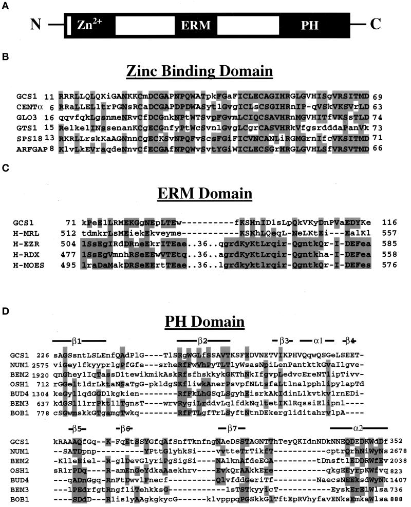
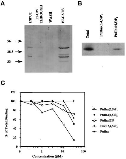
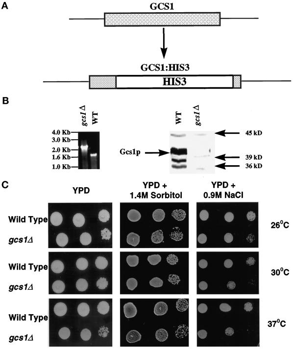
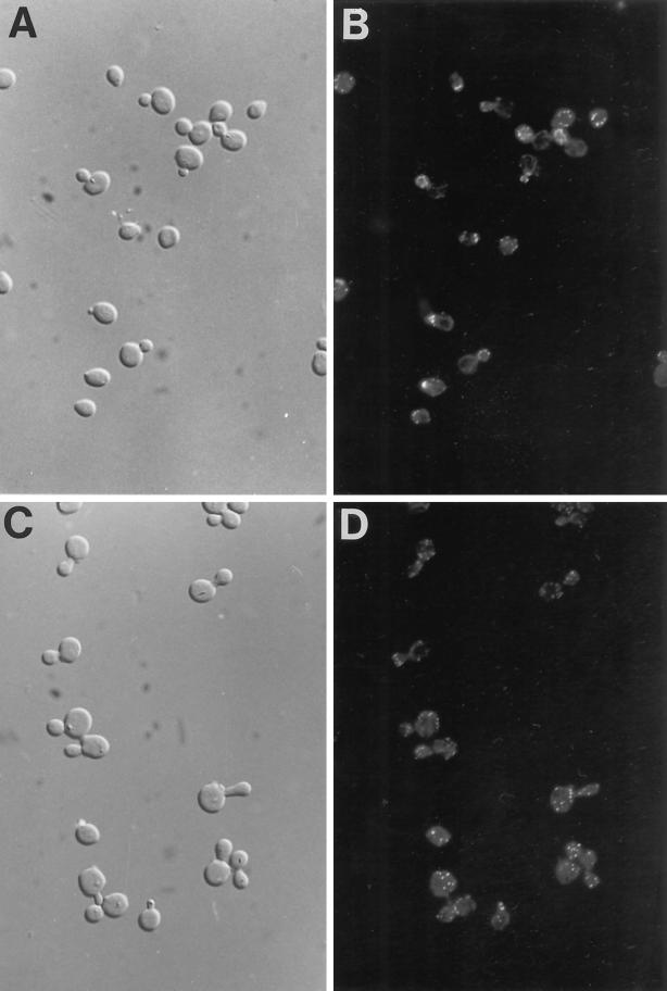
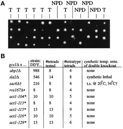
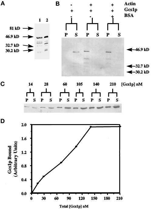
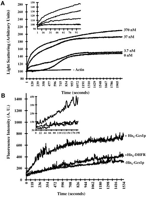
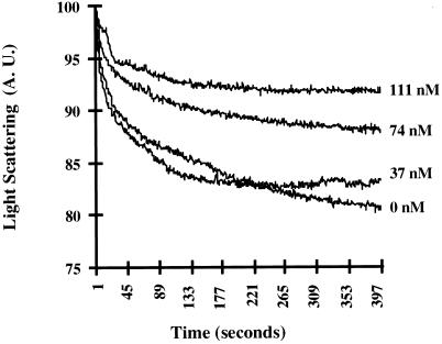
Recent cloning of a rat brain phosphatidylinositol 3,4, 5-trisphosphate binding protein, centaurin alpha, identified a novel gene family based on homology to an amino-terminal zinc-binding domain. In Saccharomyces cerevisiae, the protein with the highest homology to centaurin alpha is Gcs1p, the product of the GCS1 gene. GCS1 was originally identified as a gene conditionally required for the reentry of cells into the cell cycle after stationary phase growth. Gcs1p was previously characterized as a guanosine triphosphatase-activating protein for the small guanosine triphosphatase Arf1, and gcs1 mutants displayed vesicle-trafficking defects. Here, we have shown that similar to centaurin alpha, recombinant Gcs1p bound phosphoinositide-based affinity resins with high affinity and specificity. A novel GCS1 disruption strain (gcs1Delta) exhibited morphological defects, as well as mislocalization of cortical actin patches. gcs1Delta was hypersensitive to the actin monomer-sequestering drug, latrunculin-B. Synthetic lethality was observed between null alleles of GCS1 and SLA2, the gene encoding a protein involved in stabilization of the actin cytoskeleton. In addition, synthetic growth defects were observed between null alleles of GCS1 and SAC6, the gene encoding the yeast fimbrin homologue. Recombinant Gcs1p bound to actin filaments, stimulated actin polymerization, and inhibited actin depolymerization in vitro. These data provide in vivo and in vitro evidence that Gcs1p interacts directly with the actin cytoskeleton in S. cerevisiae.


Figures

Figure 1
Figure 1
Sequence comparison of GCS1. (A) Domain structure of Gcs1p. (B) Comparison of the CxxCx16CxxC amino-terminal zinc-binding domain of GCS1 to rat brain centaurin α, the yeast proteins GLO3, GTS1, and SPS18, and the rat liver Arf GAP. (C) Comparison of Gcs1p with merlin and the ERM family of proteins: ezrin, radixin, and moesin. (D) Comparison of the PH domain from GCS1 to six yeast proteins that contain PH domains according to the Stanford Saccharomyces Genome Database (SGD): NUM1, BEM2, OSH1, BUD4, BEM3, and BOB1. Shaded amino acids represent conserved amino acids in a majority of the family members; capital letters are amino acids conserved between GCS1 and other proteins. Conserved amino acids belong to the same Dayhoff group (GPAST, MILV, KRH, NQED, FWY, C).
Figure 2
Figure 2
His6-Gcs1p binding to PtdIns(3,4,5)P3 analogs. (A) Ni2+-NTA agarose-purified His6-Gcs1p (total) was incubated with the Affigel-aminopropyl-InsP4 resin. The flow through was collected (flow through), and the resin was washed (wash) and eluted with 2× SDS-sample buffer (eluate). Fractions were separated by SDS-PAGE and subjected to immunoblot analysis using an anti-RGS-His4 antibody. (B) His6-Gcs1p, eluted from the Affigel-aminopropyl-InsP4 resin with 1.5 M NaCl buffer, was dialyzed, and then incubated with 110 nCi [3H]BZDC-Ins(1,3,4,5)P4 photolabel, in the absence (total) or presence of 10 μM unlabeled phosphoinositide shown. Proteins were separated by 10% SDS-PAGE, and the gels were fixed, dried, and fluorographed. (C) Competition of His6-Gcs1p binding to the Affigel-aminopropyl-InsP4 resin by including in the binding assay increasing concentrations of unlabeled phosphoinositides. The resin was washed, and bound protein was eluted with sample buffer and separated by SDS-PAGE. Gels were transferred to nitrocellulose, and the His6-Gcs1p was detected with the anti-RGS-His4 antibody. Blots were densitized, and the results are presented as a percentage of total binding in the absence of unlabeled phosphoinositide; each value represents at least three independent determinations.
Figure 3
Figure 3
Disruption of GCS1. (A) GCS1 was disrupted by replacing nucleotides 224-1021 with the HIS3 gene by homologous recombination. (B) Genomic DNA isolated from wild-type and gcs1Δ was screened by PCR (left). Whole-cell lysates were prepared from midlogarithmic wild-type and gcs1Δ cells grown at 30°C were separated by SDS-PAGE and transferred to nitrocellulose. Gcs1p was detected by immunoblot analysis using antisera generated against full-length His6-Gcs1p fusion protein (right). (C) Serially diluted cell suspensions (10 μl) were spotted onto YPD and YPD plates supplemented with either 1.4 M sorbitol or 0.9 M NaCl. The plates were incubated for 48–72 h at the indicated temperatures.
Figure 4
Figure 4
Actin cytoskeleton distribution of log-phase wild-type andgcs1Δ cells. Wild-type (A and B) and gcs1Δ (C and D) cells grown at 30°C were fixed, and stained with rhodamine-conjugated phalloidin as described in MATERIALS AND METHODS. Cells were viewed with Nomarski optics (left) and fluorescence rhodamine filter (right) with a 100× oil immersion objective. Representative fields are shown; note the increase in the number of actin patches in gcs1Δ. Focal planes were chosen to maximize the number of actin patches observed.
Figure 5
Figure 5
Sensitivity of gcs1 mutants to Lat-B. (A) Halo assays were used to determine the sensitivity of wild-type (left) and gcs1Δ (right) cells to the indicated concentration of Lat-B. (B) Sensitivity of wild-type (left) and gcs1–6 (right) cells expressing the gcs1ts plasmid at either 30 or 37°C.
Figure 6
Figure 6
Genetic interactions between GCS1, actin-associated proteins, and actin mutants. gcs1Δ yeast were crossed with strains containing mutations in the single actin gene, ACT1, and with strains containing null mutants of a number of actin-associated proteins. (A) GCS1 and SLA2 are synthetic lethal. Shown are tetrads that were tetratype (T) or nonparental ditype (NPD), based on auxotrophies. In all cases, the haploid progeny predicted to be gcs1Δ, sla2Δ failed to grow at 25°C. (B) A table indicating the crosses made and the resulting temperature sensitivity of the double-mutant progeny. Note that certain of the mutants to which the gcs1Δ strain was crossed were already temperature sensitive. The only synthetic effect observed, other than with sla2Δ, was with sac6Δ. , gcs1Δ, sac6Δ strains showed some variability in phenotype; some predicted double mutants failed to grow. The least severe temperature sensitivity is indicated. *, These crosses were made using GWK9A (gcs1Δ::URA3) provided by Dr. G.C. Johnston (see also Table 1).
Figure 7
Figure 7
Gcs1p interacts with actin in vitro. (A) His6-Gcs1p was purified using Ni2+-NTA resin. The purified protein was analyzed by Coomassie stain (lane 1) and by immunoblot analysis using an anti-RGS-His4 antibody (lane 2). (B) Purified His6-Gcs1p was incubated with 3 μM polymerized yeast F-actin in the presence or absence of BSA and centrifuged at 270,000 × g. The pellets (P) and supernatants (S) were separated by SDS-PAGE and subjected to immunoblot analysis using an anti-RGS-His4 antibody. (C) Increasing concentrations (14–210 nM) of purified His6-Gcs1p were incubated with 3 μM polymerized yeast F-actin and centrifuged at 270,000 × g. The pellets (P) and supernatants (S) were separated by SDS-PAGE and subjected to immunoblot analysis using an anti-RGS-His4 antibody. (D) Quantification of immunoblot in panel C using a densitometer.
Figure 8
Figure 8
Time course for actin polymerization in the presence of His6-Gcs1p. (A) Monomeric yeast G-actin (2 μM) was polymerized in the absence or presence of increasing concentrations of His6-Gcs1p. Actin polymerization was monitored as an increase in light scattering as described in MATERIALS AND METHODS. Inset trace is a magnification of the first 105 s of the polymerization assay. In addition, 370 nM His6-Gcs1p was incubated in the absence of actin to determine the intrinsic light scattering of His6-Gcs1p. (B) Monomeric 1:10 pyrene-labeled rabbit muscle actin (5 μM): muscle actin was polymerized in the absence or presence of 0.2 μM His6-Gcs1p or His6-DHFR. Polymerization was monitored as an increase in fluorescence intensity as described in MATERIALS AND METHODS. Inset trace is a magnification of the first 300 s of the polymerization assay.
Figure 9
Figure 9
Time course for actin depolymerization in the presence of His6-Gcs1p. F-actin (2 μM) was diluted 30-fold into G-buffer containing increasing concentrations of His6-Gcs1p. Actin depolymerization was monitored as an decrease in light scattering as described in MATERIALS AND METHODS.

Similar articles

Cited by

References

    1. Adams AE, Botstein D, Drubin DG. Requirement of yeast fimbrin for actin organization and morphogenesis in vivo. Nature. 1991;354:404–408. - PubMed
    1. Amatruda JF, Gattermeir DJ, Karpova TS, Cooper JA. Effects of null mutations and overexpression of capping protein on morphogeneis, actin distribution and polarized secretion in yeast. J Cell Biol. 1992;119:1151–1162. - PMC - PubMed
    1. Antonny B, Huber I, Paris S, Chabre M, Cassel D. Activation of ADP-ribosylation factor 1 GTPase-activating protein by phosphatidylcholine-derived diacylglycerols. J Biol Chem. 1997;272:30848–30851. - PubMed
    1. Ayscough KR, Drubin DG. ACTIN: general principles from studies in yeast. Annu Rev Cell Dev Biol. 1996;12:129–160. - PubMed
    1. Ayscough KR, Stryker J, Pokala N, Sanders M, Crews P, Drubin DG. High rates of actin filament turnover in budding yeast and roles for actin in establishment and maintenance of cell polarity using the actin inhibitor latrunculin-A. J Cell Biol. 1997;137:399–416. - PMC - PubMed

Publication types

MeSH terms