. 1999 Mar;10(3):677-91.
doi: 10.1091/mbc.10.3.677.

Cluster of differentiation antigen 4 (CD4) endocytosis and adaptor complex binding require activation of the CD4 endocytosis signal by serine phosphorylation

Affiliations

Cluster of differentiation antigen 4 (CD4) endocytosis and adaptor complex binding require activation of the CD4 endocytosis signal by serine phosphorylation

C Pitcher et al. Mol Biol Cell. 1999 Mar.

Abstract

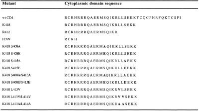
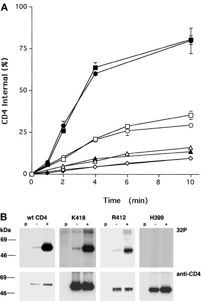
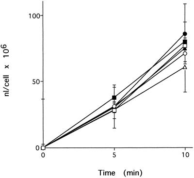
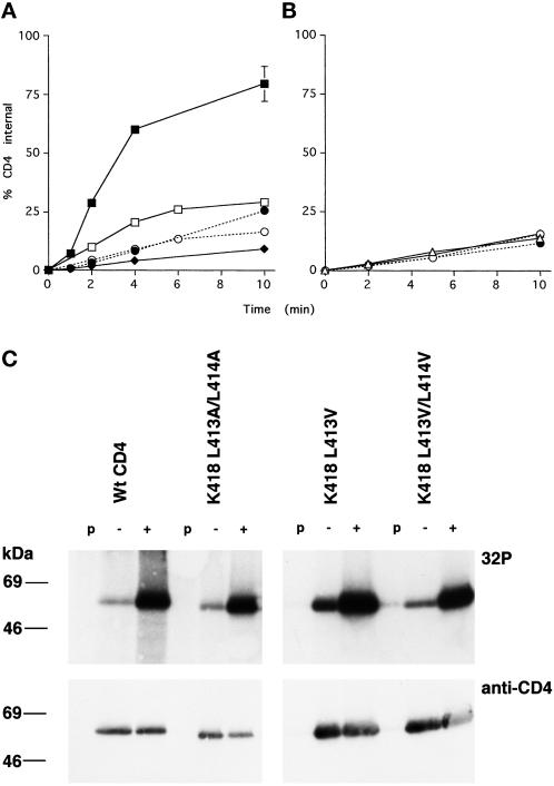
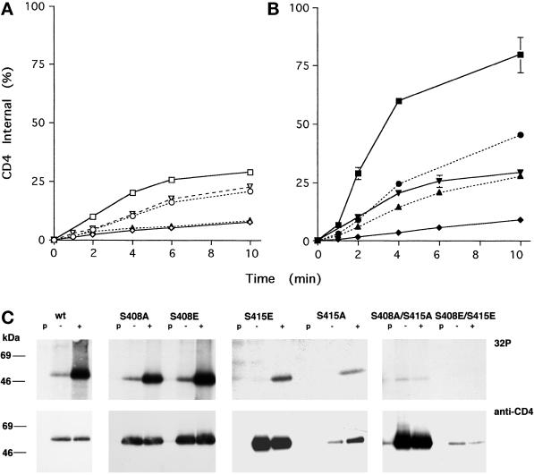
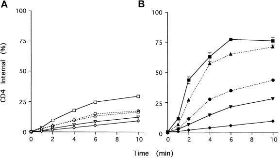
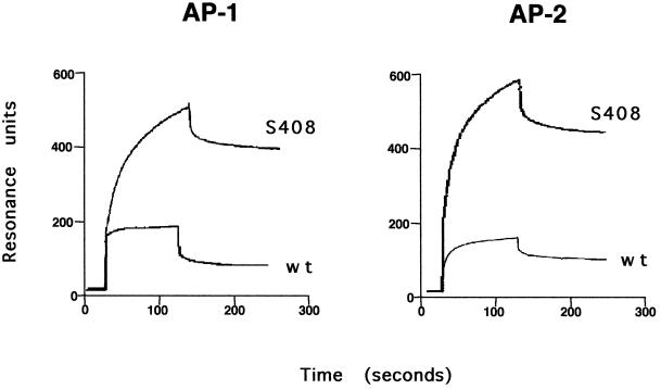
Cluster of differentiation antigen 4 (CD4), the T lymphocyte antigen receptor component and human immunodeficiency virus coreceptor, is down-modulated when cells are activated by antigen or phorbol esters. During down-modulation CD4 dissociates from p56(lck), undergoes endocytosis through clathrin-coated pits, and is then sorted in early endosomes to late endocytic organelles where it is degraded. Previous studies have suggested that phosphorylation and a dileucine sequence are required for down-modulation. Using transfected HeLa cells, in which CD4 endocytosis can be studied in the absence of p56(lck), we show that the dileucine sequence in the cytoplasmic domain is essential for clathrin-mediated CD4 endocytosis. However, this sequence is only functional as an endocytosis signal when neighboring serine residues are phosphorylated. Phosphoserine is required for rapid endocytosis because CD4 molecules in which the cytoplasmic domain serine residues are substituted with glutamic acid residues are not internalized efficiently. Using surface plasmon resonance, we show that CD4 peptides containing the dileucine sequence bind weakly to clathrin adaptor protein complexes 2 and 1. The affinity of this interaction is increased 350- to 700-fold when the peptides also contain phosphoserine residues.


Figures

Figure 1
Figure 1
CD4 and CD4 cytoplasmic domain mutants used in this study. The full-length cytoplasmic domain is illustrated for wt CD4 commencing at residue R396. Single amino acid substitutions at S408, L413, L414, and S415 are marked in bold.
Figure 2
Figure 2
Endocytosis and phosphorylation of CD4 truncations mutants. (A) HeLa cells stably expressing CD4 or CD4 truncation mutants (Figure 1) were labeled with 0.3 nM 125I-Q4120 at 4°C and then warmed to 37°C in binding medium (open symbols) or medium containing 100 ng/ml PMA (solid symbols) for the indicated times. The total and acid-resistant intracellular radioactivities were determined for each time point as described in MATERIALS AND METHODS. Data from one representative experiment are illustrated, and each point represents the mean ± SD for duplicate samples. □ and ▪, wt CD4; ○ and ●, K418; ▵ and ▴, R412; ⋄ and ♦, H399. (B) Cells were labeled with [32P]orthophosphate (as described in MATERIALS AND METHODS) and incubated in medium with (+) or without (−) 100 ng/ml PMA for 3 min. The cells were rapidly cooled to 4°C and lysed, and the CD4 molecules were immunoprecipitated, separated by SDS-PAGE, and transferred to nitrocellulose paper. CD4 protein was visualized and quantitated by Western blot, and the 32P-activity was determined on the same blots using a phosphorimager. The positions of the molecular weight standards are shown; p, preclear lane (see MATERIALS AND METHODS). Note that the K418 32P lanes illustrated here have been exposed for shorter times than the other panels.
Figure 3
Figure 3
Effect of PMA on fluid phase endocytosis. Cells expressing CD4, K418, or H399 were incubated in BM containing HRP in the presence (filled symbols) or absence (open symbols) of 100 ng/ml PMA. At the end of the incubation period the cells were rapidly cooled to 4°C and washed extensively, and the cell-associated HRP was determined. □ and ▪, wt CD4; ○ and ●, K418; ▵ and ▴, H399.
Figure 4
Figure 4
Effects of leucine substitutions on K418 CD4 endocytosis and phosphorylation. (A and B) Endocytosis of K418 CD4 L substitutions (Figure 1) was measured as described in Figure 2A. Open symbols indicate the constitutive endocytosis of constructs in the absence of PMA, and solid symbols indicate endocytosis in the presence of 100 ng/ml PMA. (A) □ and ▪, K418; ○ and ●, K418 L413A/L414A; ⋄ and ♦, H399. (B) ▵ and ▴, K418 L413V/L414V; ○ and ●, K418 L413V. (C) Cells expressing K418 CD4 L413/L414 substitutions were labeled with [32P]orthophosphate, incubated in medium with (+) or without (−) 100 ng/ml PMA for 3 min, and CD4 immunoprecipitated as described in Figure 2B. The left six lanes of the top panel were exposed to film for twice as long as the right six lanes.
Figure 5
Figure 5
Effects of serine substitutions on K418 CD4 endocytosis and phosphorylation. (A and B) Endocytosis of K418 CD4 S to A substitutions (Figure 1) was measured as described in Figure 2A. (A) Constitutive endocytosis of the constructs in the absence of PMA. (B) Endocytosis in the presence of 100 ng/ml PMA. □ and ▪, K418; ○ and ●, K418 S408A; ▿ and ▾, K418 S415A; ▵ and ▴, K418 S408A/S415A; ⋄ and ♦, H399. (C) Cells expressing K418 CD4 S to A or E (see Figure 5) substitutions were labeled with [32P]orthophosphate, incubated in medium with (+) or without (−) PMA, and CD4 immunoprecipitated as described in Figure 2B.
Figure 6
Figure 6
Effects of serine to glutamic acid substitutions on K418 CD4 endocytosis. Endocytosis of K418 CD4 S to E substitutions (Figure 1) was measured as described in Figure 2A. (A) Constitutive endocytosis of the constructs in the absence of PMA. (B) Endocytosis in the presence of 100 ng/ml PMA. □ and ▪, wt CD4; ▵ and ▴, K418 S408E; ○ and ●, K418 S415E; ▿ and ▾, K418 S408E/S415E; ⋄ and ♦, = H399.
Figure 7
Figure 7
SPR analysis of CD4 peptide–adaptor complex binding. Biotinylated CD4 tail peptides were immobilized on an SA5 sensor surface and analyzed for binding of purified AP-2 and AP-1 adaptor complexes. The wt peptide bound both adaptor complexes with very low affinity (see Table 3); however, high-affinity adaptor binding was observed when peptides containing phosphorylated S408 were used.

References

    1. Bar-Zvi D, Mosley ST, Branton D. In vivo phosphorylation of clathrin-coated vesicle proteins from rat reticulocytes. J Biol Chem. 1998;263:4408–4415. - PubMed
    1. Bremnes T, Lauvrak V, Lindqvist B, Bakke O. A region from the medium chain adaptor subunit (μ) recognizes leucine- and tyrosine-based sorting signals. J Biol Chem. 1998;273:8638–8645. - PubMed
    1. Bremnes B, Madsen T, Geddedahl M, Bakke O. An LI and ML motif in the cytoplasmic tail of the MHC- associated invariant chain mediate rapid internalization. J Cell Sci. 1994;107:2021–2032. - PubMed
    1. Corvera S, Chawla A, Chakrabarti R, Joly M, Buxton J, Czech MP. A double leucine within the GLUT4 glucose transporter COOH-terminal domain functions as an endocytosis signal. J Cell Biol. 1994;126:979–989. - PMC - PubMed
    1. Darsow T, Burd CG, Emr SD. Acidic di-leucine motif essential for AP-3-dependent sorting and restriction of the functional specificity of the Vam3p vacuolar t-SNARE. J Cell Biol. 1998;142:913–922. - PMC - PubMed

Publication types

MeSH terms