. 1999 Mar;10(3):741-56.
doi: 10.1091/mbc.10.3.741.

Interaction of the Doa4 deubiquitinating enzyme with the yeast 26S proteasome

Affiliations

Interaction of the Doa4 deubiquitinating enzyme with the yeast 26S proteasome

F R Papa et al. Mol Biol Cell. 1999 Mar.

Abstract

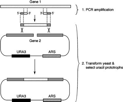
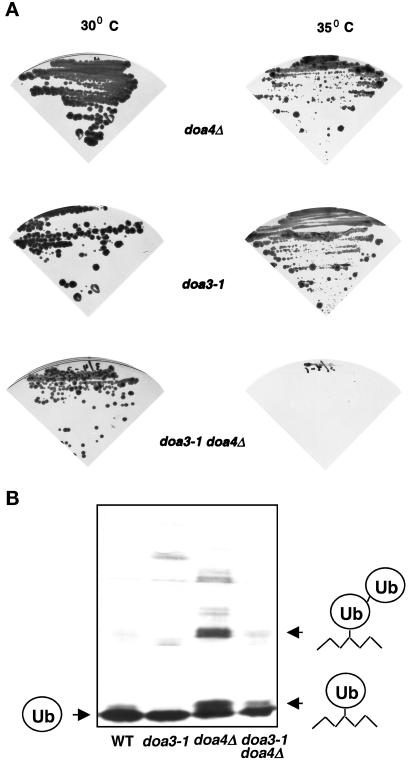
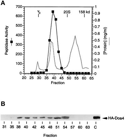
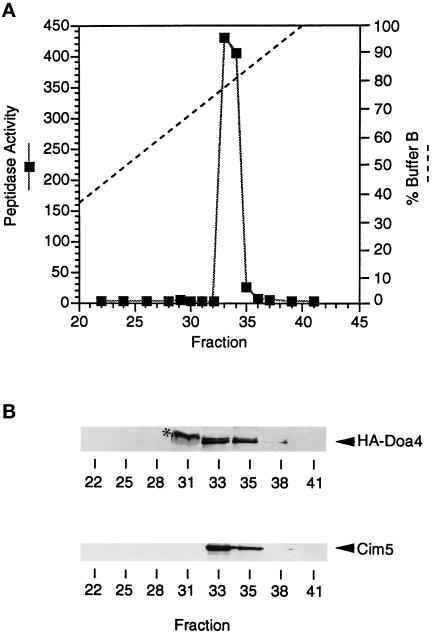
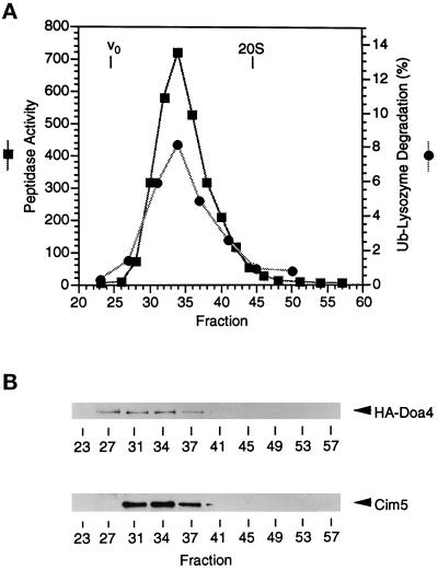
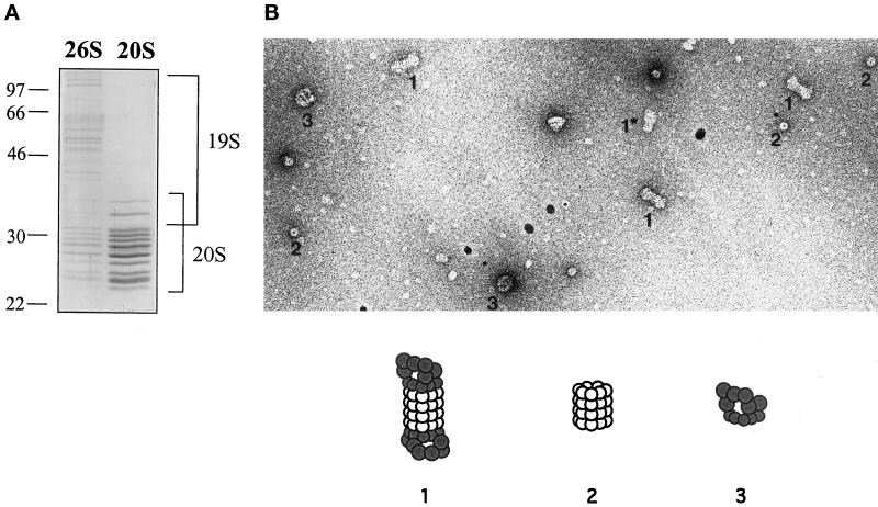
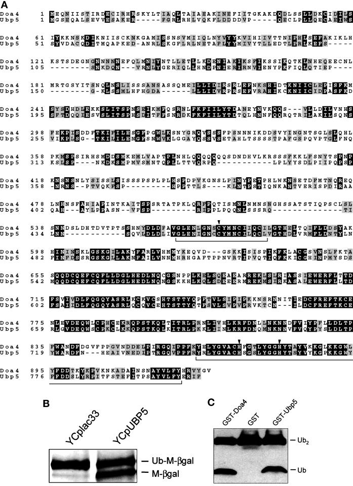
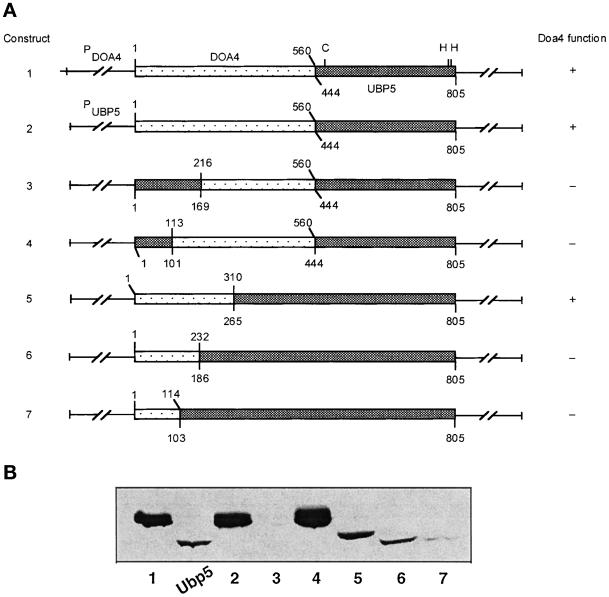
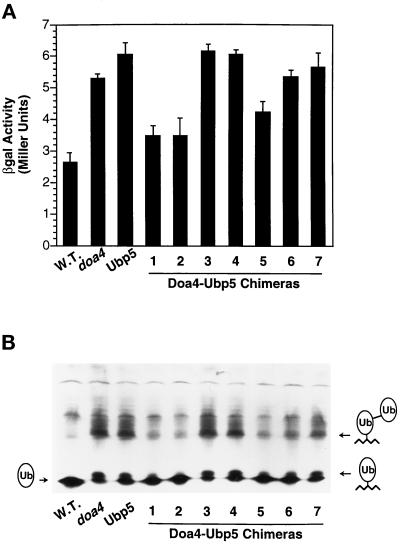
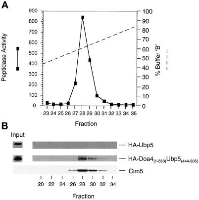
e Saccharomyces cerevisiae Doa4 deubiquitinating enzyme is required for the rapid degradation of protein substrates of the ubiquitin-proteasome pathway. Previous work suggested that Doa4 functions late in the pathway, possibly by deubiquitinating (poly)-ubiquitin-substrate intermediates associated with the 26S proteasome. We now provide evidence for physical and functional interaction between Doa4 and the proteasome. Genetic interaction is indicated by the mutual enhancement of defects associated with a deletion of DOA4 or a proteasome mutation when the two mutations are combined. Physical association of Doa4 and the proteasome was investigated with a new yeast 26S proteasome purification procedure, by which we find that a sizeable fraction of Doa4 copurifies with the protease. Another yeast deubiquitinating enzyme, Ubp5, which is related in sequence to Doa4 but cannot substitute for it even when overproduced, does not associate with the proteasome. DOA4-UBP5 chimeras were made by a novel PCR/yeast recombination method and used to identify an N-terminal 310-residue domain of Doa4 that, when appended to the catalytic domain of Ubp5, conferred Doa4 function, consistent with Ubp enzymes having a modular architecture. Unlike Ubp5, a functional Doa4-Ubp5 chimera associates with the proteasome, suggesting that proteasome binding is important for Doa4 function. Together, these data support a model in which Doa4 promotes proteolysis through removal of ubiquitin from proteolytic intermediates on the proteasome before or after initiation of substrate breakdown.


Figures

Figure 1
Figure 1
Outline of the PCR-based in vivo gap repair procedure (PCR–GR).
Figure 2
Figure 2
Genetic interaction between doa4Δ and doa3. (A) Enhanced growth defect of a doa3-1 doa4Δ double mutant. Doubly mutant doa3-1 doa4Δ cells, as well as the corresponding single mutants, were grown at 30°C and 35°C for one week. The wild-type parents grow equally well at both temperatures. (B) Suppression of the low molecular mass ubiquitin conjugates found in doa4Δ cells by impairment of the proteasome. Extracts from exponentially growing wild-type, doa3-1, doa4Δ, and doa3-1 doa4Δ cells were separated on an 18% polyacrylamide gel, blotted to a polyvinylidene difluoride membrane, and probed with an affinity-purified antiubiquitin antibody (a gift of Cecile Pickart, Department of Biochemistry, School of Public Health, Johns Hopkins University, Baltimore, MD). The positions of ubiquitin and the mono-ubiquitinated and di-ubiquitinated conjugates specific to doa4 cells are indicated.
Figure 3
Figure 3
Peptidase activity profile and anti–HA-Doa4 immunoblot analysis of yeast extracts fractionated by Sephacryl S-400 gel filtration. (A) Cleavage of suc-LLVY-AMC in fractions collected from an S-400 column fractionation of extracts from doa4Δ cells carrying pDOA4–8(HA). Protein concentration is also plotted. Dextran blue was used to determine the exclusion volume, Vo, and purified 20S proteasomes and aldolase (molecular mass 158 kDa) were used to calibrate the column. (B) Anti-HA immunoblot analysis of HA-Doa4 protein. C, crude lysate.
Figure 4
Figure 4
Fractionation of pooled 26S proteasome-containing fractions from a Sephacryl S-400 column by Mono Q anion exchange chromatography. (A) Peptidase activities in fractions collected from a Mono Q HR (5/5) anion exchange column. Elution of proteins was with a linear gradient of 26S buffer containing 0.8 M NaCl (buffer B). (B) Immunoblot analysis of HA-Doa4 and Cim5 proteins in the Mono Q fractions. Asterisk, weak anti-HA cross-reactivity with an abundant yeast protein (not always observed).
Figure 5
Figure 5
Proteasome fractionation by Superose 6 gel filtration. (A) Peptidase activity and polyubiquitin(Ub)–[125I]-lysozyme degradation in fractions from a Superose 6 gel filtration column. The column was loaded with the pooled and concentrated 26S proteasome-containing Mono Q fractions (Figure 4). Ub–lysozyme degradation is reported as the percentage of total 125I radioactivity that was acid-soluble after a 45 min reaction with 50 μl of each fraction. The elution peak of 20S proteasomes (700 kDa) and the void volume, Vo (5 × 10⁶ D, calibrated with blue dextran) are indicated. (B) Immunoblot analysis of HA-Doa4 and Cim5 proteins in the Superose 6 fractions.
Figure 6
Figure 6
Characterization of purified yeast 26S proteasomes. (A) Analysis of purified 20S and 26S proteasomes by SDS-PAGE followed by Coomassie Blue staining. Protein size standards (in kilodaltons) are indicated. (B) Electron micrograph of purified 26S proteasome complexes negatively stained with uranyl acetate. Several different species are visible: 26S proteasomes with two PA700 complexes attached at either end (1); 20S proteasome with a single PA700 complex (1*); core 20S proteasomes (2); and complexes that are likely to correspond to free PA700 (3).
Figure 7
Figure 7
An enzyme closely related to Doa4 encoded by the yeast UBP5 (YER144c) gene. (A) Sequence alignment of Doa4 and Ubp5. The two proteins were aligned with the ClustalW algorithm followed by manual adjustment. Identical residues are boxed in black, and structurally related residues are boxed in gray. The Cys and His boxes are indicated by brackets, and the conserved Cys and His residues in these two motifs are marked by arrowheads. (B) Cleavage of a ubiquitin–protein fusion by Ubp5. Shown is an anti-βgal Western immunoblot analysis of extracts from MC1061 cells harboring a plasmid expressing Ub-M-βgal and either YCplac33 vector or YCplac33-UBP5. (C) Ubiquitin isopeptidase activity of Ubp5 using a Lys48-linked diubiquitin substrate. Substrate was incubated at 30°C in E. coli extracts expressing either GST or GST-Ubp5. Partially purified GST-Doa4 was used as a control.
Figure 8
Figure 8
Expression of chimeric Doa4-Ubp5 proteins in doa4Δ cells. (A) Schematics of chimeric DOA4-UBP5 genes specifying the sites of fusion. Lightly stippled boxes correspond to DOA4 coding sequences, and darkly stippled boxes correspond to those of UBP5. Doa4 function was assayed by growth of doa4Δ haploid cells on 0.8 μg/ml canavanine and by sporulation of doa4Δ/doa4Δ diploid cells. (B) Anti-HA immunoblot analysis of extracts from doa4Δ cells expressing the chimeras diagrammed in A and from cells expressing Ubp5-HA (lane marked Ubp5).
Figure 9
Figure 9
Close correlation between Deg1-βgal accumulation and levels of low molecular mass (poly)ubiquitin conjugates in doa4Δ cells expressing Doa4-Ubp5 chimeras. (A) βgal activity assays of extracts made from yeast cells with an integrated Deg1-lacZ reporter gene. W.T., wild-type strain (MHY501). The remaining strains harbor a chromosomal deletion of DOA4 and carry the empty YEplac195 vector (doa4) or plasmids expressing Ubp5 or the Doa4-Ubp5 chimeras. Numbering of chimeras as in Figure 8. (B) Antiubiquitin immunoblot analysis of same strains as in A.
Figure 10
Figure 10
Cofractionation of a Doa4-Ubp5 chimera with 26S proteasomes. Top, suc-LLVY-AMC–cleaving activity of Mono Q column fractions. Pooled 26S proteasome-containing fractions from an S-400 gel filtration provided the input. Lysate was made from doa4Δ cells expressing the HA-tagged Doa41–560–Ubp5444–805 chimera from a YEplac195-based plasmid. Bottom, Immunoblot detection of HA-Doa4-Ubp5, HA-Ubp5, and Cim5 proteins. At left are shown anti-HA immunoblots of HA-tagged proteins in the pooled S-400 fractions that provided the inputs to the Mono Q column.

Similar articles

Cited by

References

    1. Akopian TN, Kisselev AF, Goldberg AL. Processive degradation of proteins and other catalytic properties of the proteasome from Thermoplasma acidophilum. J Biol Chem. 1997;272:1791–1798. - PubMed
    1. Amerik AY, Swaminathan S, Krantz BA, Wilkinson KD, Hochstrasser M. In vivo disassembly of free polyubiquitin chains by yeast Ubp14 modulates rates of protein degradation by the proteasome. EMBO J. 1997;16:4826–4838. - PMC - PubMed
    1. Ausubel FM, Brent R, Kingston RE, Moore DD, Seidman JG, Smith JA, Struhl K. Current Protocols in Molecular Biology. New York: John Wiley & Sons; 1989.
    1. Baker RT, Tobias JW, Varshavsky A. Ubiquitin-specific proteases of Saccharomyces cerevisiae. Cloning of UBP2 and UBP3, and functional analysis of the UBP gene family. J Biol Chem. 1992;267:23364–23375. - PubMed
    1. Baudin A, Ozier-Kalogeropoulos O, Denouel A, Lacroute F, Cullin C. A simple and efficient method for direct gene deletion in Saccharomyces cerevisiae. Nucleic Acids Res. 1993;21:3329–3330. - PMC - PubMed

Publication types

MeSH terms

Substances