. 1999 Mar;10(3):771-84.
doi: 10.1091/mbc.10.3.771.

Gene knockouts reveal separate functions for two cytoplasmic dyneins in Tetrahymena thermophila

Affiliations

Gene knockouts reveal separate functions for two cytoplasmic dyneins in Tetrahymena thermophila

S Lee et al. Mol Biol Cell. 1999 Mar.

Abstract

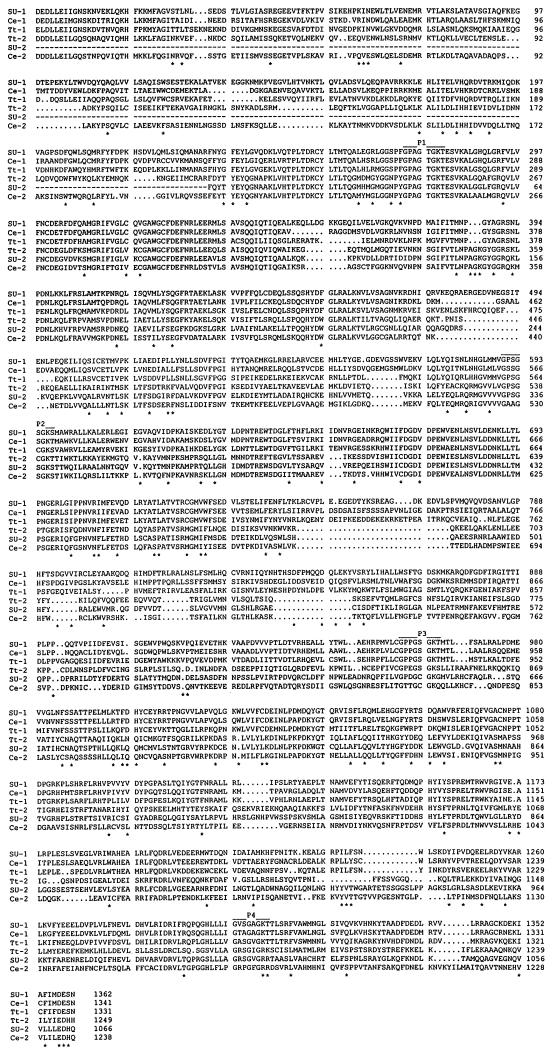
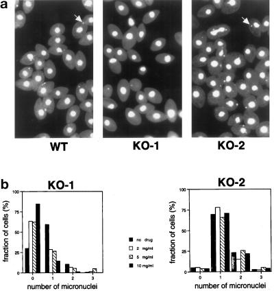
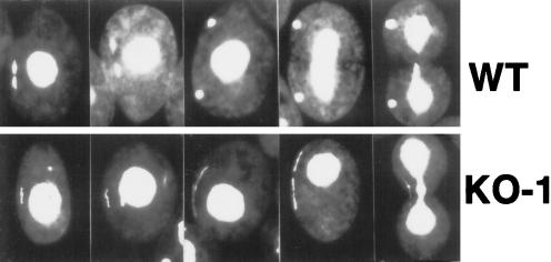
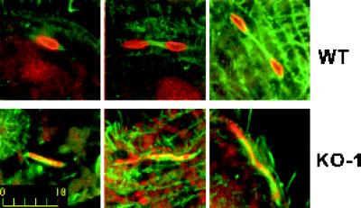
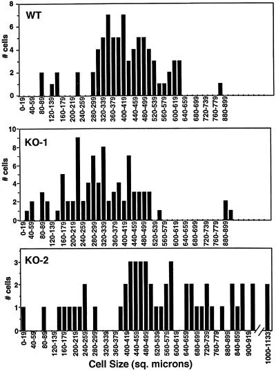
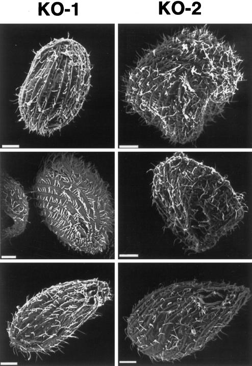
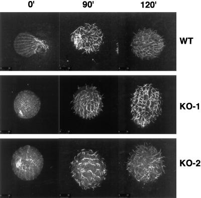
In many organisms, there are multiple isoforms of cytoplasmic dynein heavy chains, and division of labor among the isoforms would provide a mechanism to regulate dynein function. The targeted disruption of somatic genes in Tetrahymena thermophila presents the opportunity to determine the contributions of individual dynein isoforms in a single cell that expresses multiple dynein heavy chain genes. Substantial portions of two Tetrahymena cytoplasmic dynein heavy chain genes were cloned, and their motor domains were sequenced. Tetrahymena DYH1 encodes the ubiquitous cytoplasmic dynein Dyh1, and DYH2 encodes a second cytoplasmic dynein isoform, Dyh2. The disruption of DYH1, but not DYH2, resulted in cells with two detectable defects: 1) phagocytic activity was inhibited, and 2) the cells failed to distribute their chromosomes correctly during micronuclear mitosis. In contrast, the disruption of DYH2 resulted in a loss of regulation of cell size and cell shape and in the apparent inability of the cells to repair their cortical cytoskeletons. We conclude that the two dyneins perform separate tasks in Tetrahymena.


Figures

Figure 1
Figure 1
Alignment of catalytic domain sequences of Dyh1 and Dyh2. The deduced amino acid sequences of Dyh1 and Dyh2 from Tripneustes gratilla (SU-1 and SU-2; accession numbers Z21941 and U03969), C. elegans (Ce-1 and Ce-2; accession numbers L33260 and Z75536), and Tetrahymena thermophila (Tt-1 and Tt-2; accession numbers AF025312 and AF025313) were aligned by the PILEUP program (GCG, University of Wisconsin). The four P-loops are identified. Each position (*) at which there is a conserved residue in the three Dyh1 sequences that is different from a conserved residue in the three Dyh2 sequences is identified.
Figure 2
Figure 2
Stimulation of DYH2 in response to deciliation. Total RNA was isolated from mock- and twice-deciliated wild-type cells. (a) The Northern blots were repeatedly probed, exposed to x-ray film, stripped, and then reprobed to obtain the data shown. The autoradiography signals were measured by densitometry. (b) The relative densities are plotted. The steady-state concentration of DYH2 RNA, but not that of DYH1, increased during reciliation. The positive control in this experiment was ciliary dynein β heavy chain DYH4 (accession number AF072878) that also increased in expression in response to deciliation.
Figure 3
Figure 3
Targeted disruptions of DYH1 and DYH2. (a and b) Diagrams of portions of the macronuclear wild-type (WT) genes (top) and the disruption constructs (bottom) for DYH1 (a) and DYH2 (b). The DYH1 disruption construct was made by inserting the neomycin-resistance gene at the chromosomal KpnI site. The DYH2 disruption construct was made by deleting the EcoRV-EcoRV fragment and replacing it with the neomycin-resistance gene. In each diagram, the positions of the genes encoding the P-loops (P1–P4) are indicated. The locations of the gene-specific probes used in the Southern and Northern blots are underlined. (c and d) Southern blots demonstrating the targeted disruptions of the DYH1 (c) and DYH2 (d) genes. In each panel, the blot on the left was probed with the gene-specific probe and showed the loss of the appropriately sized hybridizing fragment in the KO cell lines. The blots on the right of each panel were probed with the coding region of the neomycin-resistance gene and showed that the neo gene was inserted in the appropriate locations. In each case, the neo probe hybridized with a single band. (e) Northern blots of total RNAs obtained from wild-type (B2086), KO-2, and KO-1 cells. The ∼14.5-kb dynein heavy chain bands and the 1.4-kb neo bands were identified with gene-specific probes. The disruption of each dynein gene affected the expression of only the targeted gene. (f) Southern blots of DNAs from wild-type (B2086) and KO-1 cells probed with neo and DYH1. Genomic DNA digested with BglII was probed in this experiment. The paromomycin-selected KO-1 cells (lane labeled KO-1) possessed a large amount of the neo gene and very little of the wild-type version of DYH1. After growth for 3 d in the absence of paromomycin, the cells (lane labeled KO-1 rescued) possessed a high copy number of wild-type DYH1 and only a small amount of the neomycin-resistance gene. This experiment demonstrates that the KO-1 cells were incomplete knockouts of the DYH1 gene and that the copy number of the DYH1 gene could be manipulated by changing the selection pressure.
Figure 4
Figure 4
Dyh1 is required for phagocytosis. Cells were incubated for 1 h with fluorescent latex beads, washed, fixed with formaldehyde, and counterstained with DiOC6. In these confocal fluorescence micrographs, the beads are red, and the membranes stained with DiOC6 are green. The KO-1 cells were unable to phagocytose the beads. However, KO-1 cells grown for 3 d in the absence of paromomycin (KO-1 rescued) possessed phagocytic activity. All photographs are at the same magnification (bar, in micrometers, in lower left corner of KO–2). Note the larger size of the KO-2 cells.
Figure 5
Figure 5
Many KO-1 cells lack a distinct micronucleus. (a) Fields of cells stained with DAPI and viewed by fluorescence microscopy. Most of the wild-type (B2086) and KO-2 cells contained a micronucleus (examples indicated with arrows), but most of the KO-1 cells did not. The KO-1 and KO-2 cells were grown for several weeks in paromomycin at 10 mg/ml before this experiment. (b) Effects of paromomycin concentration on the number of visible micronuclei per cell in KO-1 and KO-2. Cells were grown in paromomycin at 10 mg/ml and then transferred to medium containing different concentrations of the drug (0–10 mg/ml). After 3 d of vegetative growth, the cells were fixed and stained with DAPI. Approximately 200 cells at each drug concentration were viewed by fluorescence microscopy in a blind protocol. The number of distinct micronuclei in the KO-1 cells was affected by the paromomycin. At high concentrations of the drug, most of the KO-1 cells lacked a visible micronucleus, whereas in the absence of drug, most of the KO-1 cells had one micronucleus. The different paromomycin concentrations did not affect the micronuclear phenotype of the KO-2 cells, demonstrating that the effect in the KO-1 cells was not simply attributable to the drug. The KO-2 cells with two micronuclei included dividing cells in these nonsynchronized cultures.
Figure 6
Figure 6
Dyh1 is required for normal micronuclear chromosome distribution during mitosis. Wild-type and KO-1 cells were fixed with formaldehyde, stained with DAPI, and viewed by fluorescence microscopy. To capture KO-1 cells with micronuclei, we grew the cells for 3 d in the absence of paromomycin, returned the cells to drug at 10 mg/ml for 1 d, and then fixed and stained the cells. A gallery of cells is shown in this figure; in each photograph, the dividing micronucleus is at the viewer’s left. Top, wild-type micronuclear mitosis. Bottom, KO-1 micronuclear mitosis. In the wild-type cells, the two sets of chromosomes separated very early in mitosis. In the KO-1 cells, the chromosomes never segregated to opposite poles. Instead, chromosomal DNA was spread through the micronucleus as mitosis proceeded. This resulted in one or more thread-like DAPI-stained structures.
Figure 7
Figure 7
Double immunofluorescence microscopy of micronuclear mitotic apparatuses. Cells were permeabilized, fixed, and stained with anti-tubulin and anti-phosphorylated histone antibodies. The cells were viewed by confocal scanning laser fluorescence microscopy. The microtubules are green, the chromatin is red, and yellow is the superposition of the two stains. Top, wild-type micronuclear mitosis. Bottom, KO-1 micronuclear mitosis. Bar, 10 μm.
Figure 8
Figure 8
Dyh2 contributes to cell size. Surface areas of wild-type (B2086), KO-1, and KO-2 cells were measured from cells photographed using differential interference contrast microscopy. The distributions by surface area for each cell type are shown in the graphs. The KO-2 cells were found to have a wide range of sizes, with many cells being significantly larger than wild-type and KO-1 cells. The slightly smaller size of the KO-1 cells relative to wild-type cells may be related to the involvement of Dyh1 in phagocytosis.
Figure 9
Figure 9
Dyh2 contributes to cell shape. A gallery of KO-1 and KO-2 cells viewed by scanning electron microscopy are shown. Many KO-2 cells were misshapen. Bars, 6 μm.
Figure 10
Figure 10
Disruption of surface ciliary pattern in reciliated KO-2 cells. Wild-type (B2086), KO-1, and KO-2 cells were deciliated with calcium and then returned to Neff medium. At the indicated times after deciliation, the cells were fixed and stained with anti-tubulin antibodies and then viewed by confocal laser scanning fluorescence microscopy. The wild-type and KO-1 cells regrew their cilia in rows corresponding to the longitudinal cortical microtubules. In contrast, the KO-2 cells regrew their cilia in a disorganized manner.

Similar articles

Cited by

References

    1. Allen SL, Altschuler MA, Bruns PJ, Cohen J, Doerder FP, Gaertig J, Gorovsky M, Orias E, Turkewitz A. Proposed genetic nomenclature rules for Tetrahymena thermophila, Paramecium primaurelia and Paramecium tetraurelia. Genetics. 1998;149:459–462. - PMC - PubMed
    1. Aniento F, Emans N, Griffiths G, Gruenberg J. Cytoplasmic dynein-dependent vesicular transport from early to late endosomes. J Cell Biol. 1993;123:1373–1387. - PMC - PubMed
    1. Asai DJ. Functional and molecular diversity of dynein heavy chains. Semin Cell Dev Biol. 1996;7:311–320.
    1. Asai DJ, Brokaw CJ. Dynein heavy chain isoforms and axonemal motility. Trends Cell Biol. 1993;3:398–402. - PubMed
    1. Asai DJ, Brokaw CJ, Harmon RC, Wilson L. Monoclonal antibodies to tubulin and their effects on the movement of reactivated sea urchin spermatozoa. Cell Motil. 1982;1:175–180. - PubMed

Publication types

Associated data

LinkOut - more resources