. 1999 Mar;10(3):799-817.
doi: 10.1091/mbc.10.3.799.

Visualization of receptor-mediated endocytosis in yeast

Affiliations

Visualization of receptor-mediated endocytosis in yeast

J Mulholland et al. Mol Biol Cell. 1999 Mar.

Abstract

We studied the ligand-induced endocytosis of the yeast alpha-factor receptor Ste2p by immuno-electron microscopy. We observed and quantitated time-dependent loss of Ste2p from the plasma membrane of cells exposed to alpha-factor. This ligand-induced internalization of Ste2p was blocked in the well-characterized endocytosis-deficient mutant sac6Delta. We provide evidence that implicates furrow-like invaginations of the plasma membrane as the site of receptor internalization. These invaginations are distinct from the finger-like plasma membrane invaginations within actin cortical patches. Consistent with this, we show that Ste2p is not located within the cortical actin patch before and during receptor-mediated endocytosis. In wild-type cells exposed to alpha-factor we also observed and quantitated a time-dependent accumulation of Ste2p in intracellular, membrane-bound compartments. These compartments have a characteristic electron density but variable shape and size and are often located adjacent to the vacuole. In immuno-electron microscopy experiments these compartments labeled with antibodies directed against the rab5 homologue Ypt51p (Vps21p), the resident vacuolar protease carboxypeptidase Y, and the vacuolar H+-ATPase Vph1p. Using a new double-labeling technique we have colocalized antibodies against Ste2p and carboxypeptidase Y to this compartment, thereby identifying these compartments as prevacuolar late endosomes.


Figures

Figure 1
Figure 1
Anti-Ste2p antibodies localize to the cell surface and plasma membrane invaginations of wild-type cells (RH448) grown at 25°C. Note the clustered localization of Ste2p to the invaginations (arrows) in C–E. N, nucleus; V, vacuole. Bar, 0.5 μm.
Figure 2
Figure 2
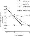
Ste2p localization is quantitatively lost, in a time- and α-factor-dependent manner from the cell surface of wild-type cells at 25 and 37°C. In the endocytosis mutant, sac6Δ Ste2p remains localized to the cell surface after exposure to α-factor at 25°C. To indicate nonspecific background, the average number of anti-Ste2p antibody localization to the cell surface of ste2Δ cells grown at 25°C is shown.
Figure 3
Figure 3
After exposure to α-factor, Ste2p is located in membrane-bound compartments. Wild-type cells (RH448) were taken after 5 min (A–D), 10 min (E), and 15 min (F–H) of continuous exposure to α-factor. Note small peripheral vesicles (long arrows) and tubular–vesicular compartments to which anti-Ste2p antibodies localize soon after exposure of α-factor (A–D). Note also the perivacuole compartments that localize antibodies directed against Ste2p (F–H). Many of these compartments appear to contain internal membranes and in sequential sections appear as interconnected tubular–vesicular structures (E and F). After 15 min of exposure to α-factor at 37°C, perivacuolar compartments accumulate (G and H). Note in F and G the localization of anti-Ste2p antibodies to tangentially cut invaginations of plasma membrane (F and G, short arrows) as well as to the membrane-bound compartments located proximal to these invaginations. V, vacuole; N, nucleus. Bars, 0.5 μm.
Figure 4
Figure 4
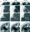
Ste2p is not located within the cortical actin patch before (A and B) or after (t = 5 min; C and D) exposure to α-factor. (A–C) Examples of double labeling with antibodies against cofilin (15-nm gold particles) and Ste2p (10-nm gold particles) on wild-type cells. Note that Ste2p is not observed in the cortical actin patch (long arrows) but is located in furrow-like invaginations of the plasma membrane (short arrows). (D) Example of adjacent-face double localization using anti-actin (red in merged image) and anti-Ste2p (yellow in merged image) antibodies. The asterisk in D indicates a furrow-like invagination that localizes both anti-Ste2p and anti-actin antibodies; long arrows indicate cortical actin patches (see text). Note that the section in D with anti-Ste2p localization does not appear to be associated with an invagination until the adjacent section is examined. Note also that some furrow-like invaginations are located next to actin patches (A, B, and D). Bars, 0.1 μm.
Figure 5
Figure 5
Ste2p is nonrandomly distributed between invaginated and noninvaginated areas of the cell surface. Percentage of cell surface–associated Ste2p that is in furrow-like plasma membrane invaginations (PMIs) in wild-type cells exposed to α-factor at 25 or 37°C and sac6Δ cells at 25°C is plotted. A total of 50 individual cell sections at each time point were counted for each sample. Background localization to invaginations was 33 ± 2.4% (averaged over all points) of the total cell surface background localization; this was subtracted from all data points shown.
Figure 6
Figure 6
Ste2p (yellow in merged image) is localized to vesicles that are proximal and perhaps continuous with furrow-like invaginations of the plasma membrane (arrows) and colocalizes with actin (red in merged image). Adjacent-face double localization of Ste2p and actin in wild-type cells before (A and B) and after (t = 5 min; C and D) exposure to α-factor is shown. Note that the anti-Ste2p section in A does not appear to be associated with a plasma membrane invagination until the adjacent section is examined. Note also in D that the “tip” of the furrow-like invagination (asterisk) has anti-actin antibodies localized around it. Bar, 0.1 μm.
Figure 7
Figure 7
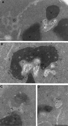
Antibodies directed against the vacuolar protease CPY (A) and the 90-kDa subunit of the vacuolar H+ ATPase Vph1p (B) localize to the vacuole and vacuole-associated compartments in wild-type cells. Note that the anti-CPY antibodies localize within the vacuole and the vacuole-associated compartment and that anti-Vph1p antibodies localize to the membrane of the vacuole and the perivacuole compartment. These perivacuole compartments, but not the vacuole, localize antibodies directed against Ypt51p (C and D). Note that the vacuole-associated compartments appear as tubular–vesicular structures having internal membranes. V, vacuole; g, Golgi. Bar, 0.5 μm.
Figure 8
Figure 8
CPY and internalized Ste2p colocalize in prevacuole compartments. (A and B) Adjacent-face double localization of anti-Ste2p (yellow in merged images) and anti-CPY (red in merged images) antibodies after 15 min of continuous exposure to α-factor at 25°C. Note that the prevacuole compartments appear to be interconnected. (C) Example of adjacent-face double labeling with Ste2p antibodies only. Note that the first section of C shows a grazing cut through a prevacuole compartment that contains vesicles to which Ste2p is localized (arrows). This prevacuole compartment appears to be fusing with the vacuole. V, vacuole. Bar, 0.1 μm.
Figure 9
Figure 9
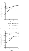
Prevacuole compartments are late endosomes. Quantitation of Ste2p and CPY in prevacuole compartments after exposure to α-factor at 25 or 37°C. For each time point 300 cell sections were observed, and the number of CPY- or Ste2p-containing endosomes was counted. (A) Percentage of cell sections that were observed with CPY or with Ste2p antibody localization. (B) Average number of marker-positive (Ste2p or CPY) endosomes that were observed within those cell sections.

Similar articles

Cited by

References

    1. Adams AE, Botstein D, Drubin D. Requirement of yeast fimbrin for actin organization and morphogenesis in vivo. Nature. 1991;354:404–408. - PubMed
    1. Anderson R, Vasile E, Mello R, Brown M, Goldstein J. Immunocytochemical visualization of coated pits and vesicles in human fibroblasts: relation to low density lipoprotein receptor distribution. Cell. 1978;15:919–933. - PubMed
    1. Benedetti H, Raths S, Crausaz F, Riezman H. The END3 gene encodes a protein that is required for the internalization step of endocytosis and for actin cytoskeleton organization in yeast. Mol Biol Cell. 1994;5:1023–1037. - PMC - PubMed
    1. Botstein D, Amberg D, Huffaker T, Mulholland J, Adams A, Drubin D, Stearns T. The yeast cytoskeleton. In: Broach JR, Pringle JR, Jones EW, editors. The Molecular and Cellular Biology of the Yeast Saccharomyces. Cold Spring Harbor, NY: Cold Spring Harbor Laboratory; 1997. pp. 1–90.
    1. Bryant N, Stevens T. Vacuole biogenesis in Saccharomyces cerevisiae: protein transport pathways to the yeast vacuole. Microbiol Mol Biol Rev. 1998;62:230–247. - PMC - PubMed

Publication types

MeSH terms

LinkOut - more resources