. 1999 Mar;119(3):817-28.
doi: 10.1104/pp.119.3.817.

NADH-glutamate synthase in alfalfa root nodules. Genetic regulation and cellular expression

Affiliations

NADH-glutamate synthase in alfalfa root nodules. Genetic regulation and cellular expression

G B Trepp et al. Plant Physiol. 1999 Mar.

Abstract

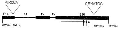
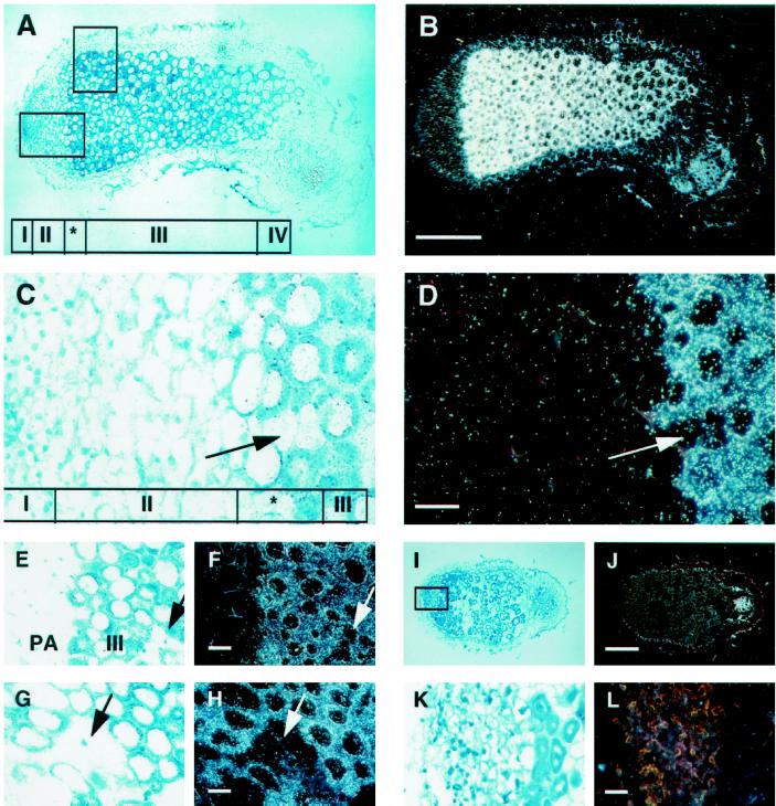
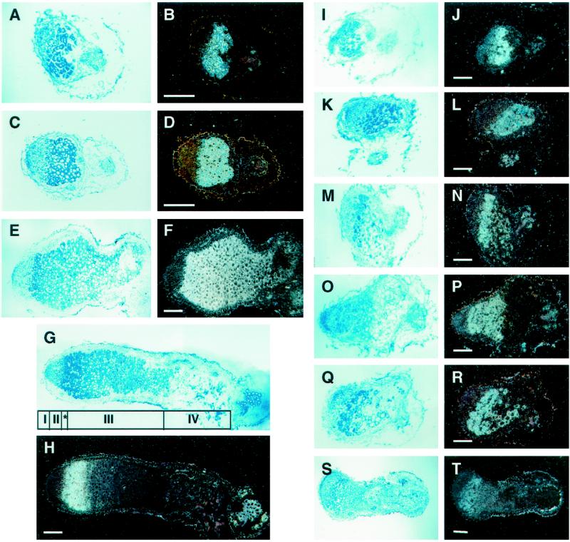
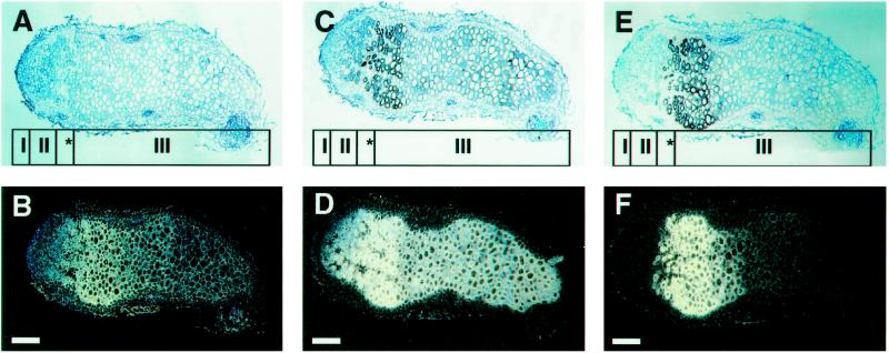
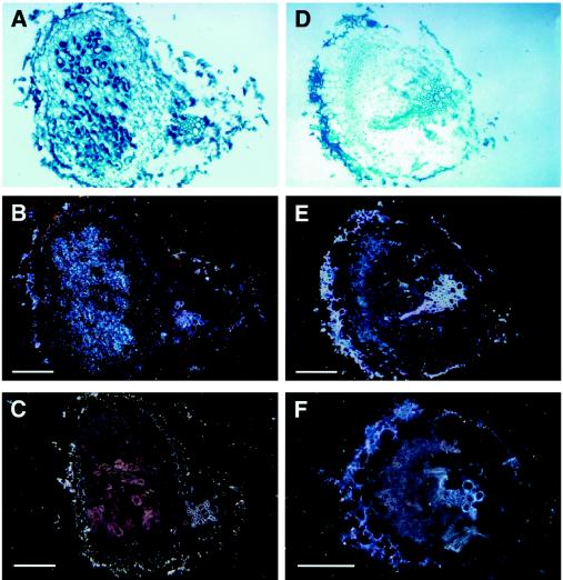
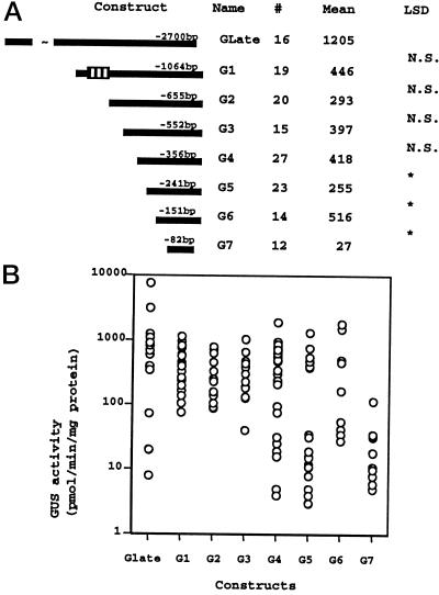
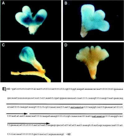
NADH-dependent glutamate synthase (NADH-GOGAT; EC 1.4.1.14) is a key enzyme in primary nitrogen assimilation in alfalfa (Medicago sativa L.) root nodules. Here we report that in alfalfa, a single gene, probably with multiple alleles, encodes for NADH-GOGAT. In situ hybridizations were performed to assess the location of NADH-GOGAT transcript in alfalfa root nodules. In wild-type cv Saranac nodules the NADH-GOGAT gene is predominantly expressed in infected cells. Nodules devoid of bacteroids (empty) induced by Sinorhizobium meliloti 7154 had no NADH-GOGAT transcript detectable by in situ hybridization, suggesting that the presence of the bacteroid may be important for NADH-GOGAT expression. The pattern of expression of NADH-GOGAT shifted during root nodule development. Until d 9 after planting, all infected cells appeared to express NADH-GOGAT. By d 19, a gradient of expression from high in the early symbiotic zone to low in the late symbiotic zone was observed. In 33-d-old nodules expression was seen in only a few cell layers in the early symbiotic zone. This pattern of expression was also observed for the nifH transcript but not for leghemoglobin. The promoter of NADH-GOGAT was evaluated in transgenic alfalfa plants carrying chimeric beta-glucuronidase promoter fusions. The results suggest that there are at least four regulatory elements. The region responsible for expression in the infected cell zone contains an 88-bp direct repeat.


Figures

Figure 1
Figure 1
Diagrammatic representation of the portion of the alfalfa NADH-GOGAT gene used for PCR. Exons are indicated by boxed regions, and introns are represented by lines. PCR was performed over three exons (exon 14 [E14], exon 15 [E15], exon 16 [E16]) and two introns (intron 14 [I14] and intron 15 [I15]) of the reported alfalfa gene using primer 1 (AIKQVA) and primer 2 (CEYMTGG). The putative binding for the (Fe-S) cluster is indicated by arrows, and the putative flavin mononucleotide binding site is underlined.
Figure 2
Figure 2
Localization of NADH-GOGAT mRNA in alfalfa root nodules by in situ hybridization. Bright-field (A, C, E, G, I, and K) and dark-field (B, D, F, H, J, and L) photographs of a longitudinal section through 19-d-old effective alfalfa nodules. Nodule ultrastructure was classified based on the nomenclature of Vasse et al. (1990): meristem (zone I), invasion zone (zone II), interzone (*), N2-fixing zone (zone III), and senescent zone (zone IV). The nodule in A to H was hybridized with NADH-GOGAT 35S-labeled antisense RNA probe. Enlargement of the lower boxed region in A shown in bright-field (C) and dark-field (D) photographs includes a portion of the meristem (zone I), invasion zone (zone II), and interzone (*). Enlargement of the upper box in A shown in bright-field (E) and dark-field (F) photographs includes the central zone (III), including parenchymal tissue (PA). In bright-field (G) and dark-field (H) photographs of infected and uninfected cells, arrows in C through H point to uninfected cells. Bright-field (I and K) and dark-field (J and L) photographs of a longitudinal section through a 19-d-old effective alfalfa nodule hybridized with NADH-GOGAT 35S-labeled sense RNA probe. Bars in B and J = 800 μm; bars in D, F, H, and L = 80 μm.
Figure 3
Figure 3
Localization of NADH-GOGAT transcript in effective and plant-gene-controlled ineffective alfalfa nodules during development. Longitudinal sections through effective (A–H) and plant-controlled ineffective (I–T) alfalfa root nodules. The bright-field and dark-field image pairs are of 7- (A, B, I, and J), 9- (C, D, K, L, M, and N), 19- (E, F, O, P, Q, and R), and 33- (G, H, S, and T) d-old root nodules. Bars = 400 μm.
Figure 4
Figure 4
In situ localization of the NADH-GOGAT, nifH, and leghemoglobin transcripts in 33-d-old alfalfa root nodules using serial sections. The bright-field and dark-field image pairs are of longitudinal sections through 33-d-old root nodules that were hybridized with 35S-labeled NADH-GOGAT (A and B), leghemoglobin (C and D), or nifH (E and F) antisense RNA probe. Bars = 400 μm.
Figure 5
Figure 5
In situ localization of NADH-GOGAT transcripts in nodules induced by S. meliloti F642 and 7154. The bright-field (A and D) and dark-field (B, C, E, and F) images are of longitudinal sections through 19-d-old root nodules induced by S. meliloti F642 (A–C) and S. meliloti 7154 (D–F). The sections in A, B, D, and E were hybridized with 35S-labeled NADH-GOGAT antisense RNA probe, whereas those in C and F were hybridized with sense riboprobes and photographed under dark-field conditions. Bars = 400 μm.
Figure 6
Figure 6
Diagram of the NADH-GOGAT promoter-GUS gene constructs. A, Eight sequences containing the putative NADH-GOGAT promoter were translational fusions to the GUS gene; the length of these truncations is measured with respect to the transcriptional start. The boxed region in construct G1 marks the location of the 88-bp direct repeat. The number of individual transformants tested for each construct is indicated (#). Mean GUS activity (pmol min−1 mg−1 protein) is indicated with the heading “Mean.” lsd values between adjacent deletions were calculated, and the level of significance was set at P < 0.01. N.S., Not significant; *, significant at P < 0.01. B, Distribution of GUS activity per construct measured from individual independently transformed plants.
Figure 7
Figure 7
GUS staining of the NADH-GOGAT promoter deletions. For deletions GLate (A) and G1 (B), individual transformants with GUS activity close to the mean had a GUS staining pattern similar to the results obtained for in situ hybridizations. A loss in tissue specificity was observed in deletion G2 (C), whereas no GUS staining was observed in deletion G7 (D). E, The 409-bp region between deletion G1 and G2. The 88-bp direct repeat is overlined, and the location of the conserved octanucleotide motif is underlined.

Similar articles

Cited by

References

    1. Anderson MP, Vance CP, Heichel GH, Miller SS. Purification and characterization of NADH-glutamate synthase from alfalfa root nodules. Plant Physiol. 1989;90:351–358. - PMC - PubMed
    1. Austin S, Bingham ET, Mathews DE, Shahan MN, Will J, Burgess RR. Production and field performance of transgenic alfalfa (Medicago sativa L.) expressing alpha-amylase and manganese-dependent lignin peroxidase. Euphytica. 1995;85:381–383.
    1. Avila C, Marquez AJ, Pajuelo P, Cannell ME, Wallsgrove RM, Forde BG. Cloning and sequence analysis of a cDNA for barley ferredoxin-dependent glutamate synthase and molecular analysis of photorespiratory mutants deficient in the enzyme. Planta. 1993;189:475–483. - PubMed
    1. Baron AC, Tobin TH, Wallsgrove RM, Tobin AK. A metabolic control analysis of the glutamine synthetase/glutamate synthase cycle in isolated barley (Hordeum vulgare L.) chloroplasts. Plant Physiol. 1994;105:415–424. - PMC - PubMed
    1. Becker TW, Perrot-Rechenmann C, Suzuki A, Hirel B. Subcellular and immunocytochemical localization of the enzymes involved in ammonia assimilation in mesophyll and bundle-sheath cells of maize leaves. Planta. 1993;191:129–136.

Publication types

MeSH terms

LinkOut - more resources