. 1999 Mar;119(3):1073-82.
doi: 10.1104/pp.119.3.1073.

Impacts of aluminum on the cytoskeleton of the maize root apex. short-term effects on the distal part of the transition zone

Affiliations

Impacts of aluminum on the cytoskeleton of the maize root apex. short-term effects on the distal part of the transition zone

M Sivaguru et al. Plant Physiol. 1999 Mar.

Abstract

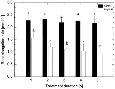
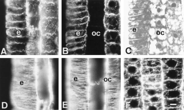
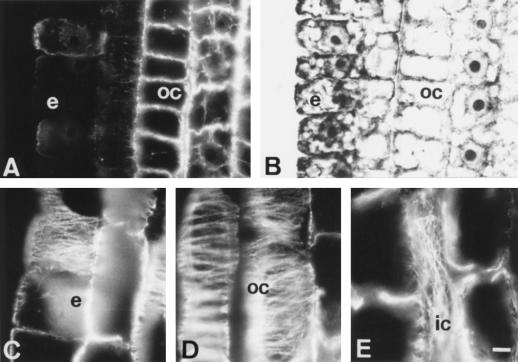
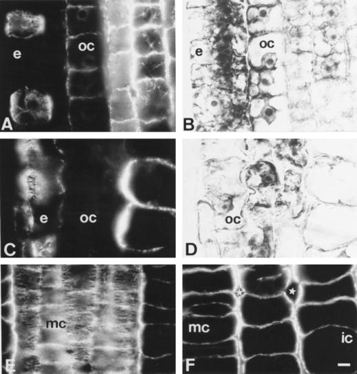
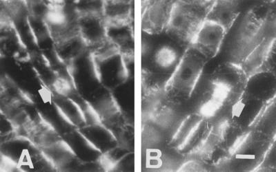
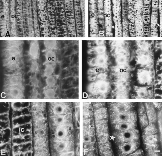
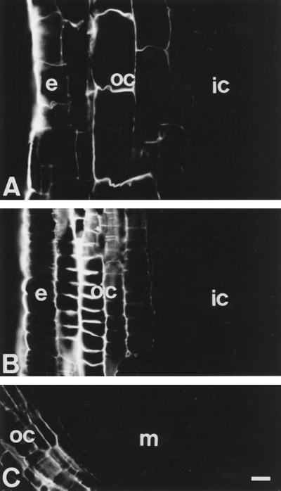
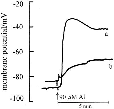
Using monoclonal tubulin and actin antibodies, Al-mediated alterations to microtubules (MTs) and actin microfilaments (MFs) were shown to be most prominent in cells of the distal part of the transition zone (DTZ) of an Al-sensitive maize (Zea mays L.) cultivar. An early response to Al (1 h, 90 μM) was the depletion of MTs in cells of the DTZ, specifically in the outermost cortical cell file. However, no prominent changes to the MT cytoskeleton were found in elongating cells treated with Al for 1 h in spite of severe inhibition of root elongation. Al-induced early alterations to actin MFs were less dramatic and consisted of increased actin fluorescence of partially disintegrated MF arrays in cells of the DTZ. These tissue- and development-specific alterations to the cytoskeleton were preceded by and/or coincided with Al-induced depolarization of the plasma membrane and with callose formation, particularly in the outer cortex cells of the DTZ. Longer Al supplies (>6 h) led to progressive enhancements of lesions to the MT cytoskeleton in the epidermis and two to three outer cortex cell files. Our data show that the cytoskeleton in the cells of the DTZ is especially sensitive to Al, consistent with the recently proposed specific Al sensitivity of this unique, apical maize root zone.


Figures

Figure 1
Figure 1
Effect of 90 μm monomeric Al on root-elongation rate of the Al-sensitive maize cv Lixis treated for 1 to 5 h. Values are means of five independent replicates. Means followed by the same letter do not differ significantly at P < 0.05 (Tukey's test).
Figure 2
Figure 2
Effects of short-term Al treatment (90 μm, 1 h) on MTs in cells of the DTZ (A, B, and C) and of the elongation region (D and E). A and D, Control. C is the DIC image of B. The most sensitive cells with respect to Al effects on the MT cytoskeleton proved to be the outermost cortical cells of the DTZ, as these lost all of their MTs after 1 h of Al treatment (B). In contrast, no effects on MTs were found in the elongation zone (D and E). Extremely dense endoplasmic MTs were induced by 1 h of Al in the apical meristem (F). e, Epidermis; oc, outer cortex. Bar = 8 μm for A to E and 6 μm for F.
Figure 3
Figure 3
Effects of medium-term Al treatment (90 μm, 6 h) on MTs of cells in the DTZ (A and B) and in the elongation zone (C, D, and E). In the DTZ the outermost cortical and epidermal cells are devoid of MTs (A) whereas cells of the elongation region show dense arrays of transverse CMTs (C and D). In some inner cortex cells, longitudinal arrangement of CMTs was found (E). e, Epidermis; oc, outer cortex; ic, inner cortex. Bar = 10 μm.
Figure 4
Figure 4
Effects of long-term Al treatment (90 μm, 12 h) on MTs and cell shapes in DTZ (A and B) and proximal (C and D) part of the TZ. B and D are DIC images of A and C. Note that epidermal and outer cortex cells are already distorted in the proximal part of the TZ (C and D), whereas middle cortex cells are abnormally expanded (F). Note the presence of unusually large intercellular spaces (stars in F). Although most cells of the middle cortex have disoriented CMTs, some cells still show dense transversal arrays (E). e, Epidermis; oc, outer cortex; ic, inner cortex; mc, middle cortex. Bar = 8 μm.
Figure 5
Figure 5
Induction of periclinal divisions in meristematic outer cortex by 10 μm NPA (A) and 90 μm Al (B), both supplied for 6 h. Bar = 8 μm.
Figure 6
Figure 6
Effects of Al (90 μm) treatment on the actin MFs in apical root cells of an Al-sensitive maize cv Lixis. All images are from the DTZ region of the root apex. Comparable regions of control (A, C, and E) and 6-h (B) or 1-h (D and F) Al-treated root apices. Note the more prominent actin fluorescence and absence of filamentous actin in epidermal and outer cortex cells after Al treatments (B and D) when compared with their respective controls (A and C). Al-induced fragmentation or altered polymerization of actin MFs transforming almost all filamentous actin of inner cortex cells into actin-positive dots or F-actin fragments (F, compare the comparable control position, E). Note increased intercellular spaces in Al-treated root apices (stars in B and F). All root apices are facing the bottom of this figure. Bar = 16 μm (A and B) or 7 μm (all others). e, Epidermis; oc, outer cortex; ic, inner cortex.
Figure 7
Figure 7
Al- (90 μm, 6 h) induced callose deposits in cells within the elongation zone (A). Cells of the DTZ accumulated the highest amounts of callose (B). Low amounts of callose were found in the outer cortex cells behind the quiescent center (C). The callose formation was restricted to the epidermis (e) and outer cortex (oc) cells throughout the root apex. All root apices are facing the bottom of this figure. ic, Inner cortex; m, meristem. Bar = 23 μm.
Figure 8
Figure 8
Effect of 90 μm Al supply on the PM potentials in the outer cortex cells of an Al-sensitive maize cv Lixis root apex. Impalement was at 1.8 mm (a) and at 2.8 mm (b) DFT (n = 11). Data are representative of single recordings.

References

    1. Alfano F, Russell A, Gambardella R, Duckett JG. The actin cytoskeleton of the liverwort Riccia fluitans: effects of cytochalasin B and aluminium ions on rhizoid tip growth. J Plant Physiol. 1993;142:569–574.
    1. Baluška F, Barlow PW, Hauskrecht M, Kubica Š, Parker JS, Volkmann D. Microtubule arrays in maize root cells: interplay between the cytoskeleton, nuclear organization and postmitotic cellular growth patterns. New Phytol. 1995;130:177–192.
    1. Baluška F, Barlow PW, Volkmann D. Complete disintegration of the microtubular cytoskeleton precedes auxin-mediated reconstruction in postmitotic maize root cells. Plant Cell Physiol. 1996a;37:1013–1021. - PubMed
    1. Baluška F, Brailsford RW, Hauskrecht M, Jackson MB, Barlow PW. Cellular dimorphism in the maize root cortex: involvement of microtubules, ethylene and gibberelin in the differentiation of cellular behaviour in postmitotic growth zones. Bot Acta. 1993a;106:394–403.
    1. Baluška F, Hasenstein KH. Root cytoskeleton: its role in perception of and response to gravity. Planta. 1997;203:S-69–78. - PubMed

LinkOut - more resources