. 1999 Mar 1;13(5):532-44.
doi: 10.1101/gad.13.5.532.

The conserved protein kinase Ipl1 regulates microtubule binding to kinetochores in budding yeast

Affiliations

The conserved protein kinase Ipl1 regulates microtubule binding to kinetochores in budding yeast

S Biggins et al. Genes Dev. .

Abstract

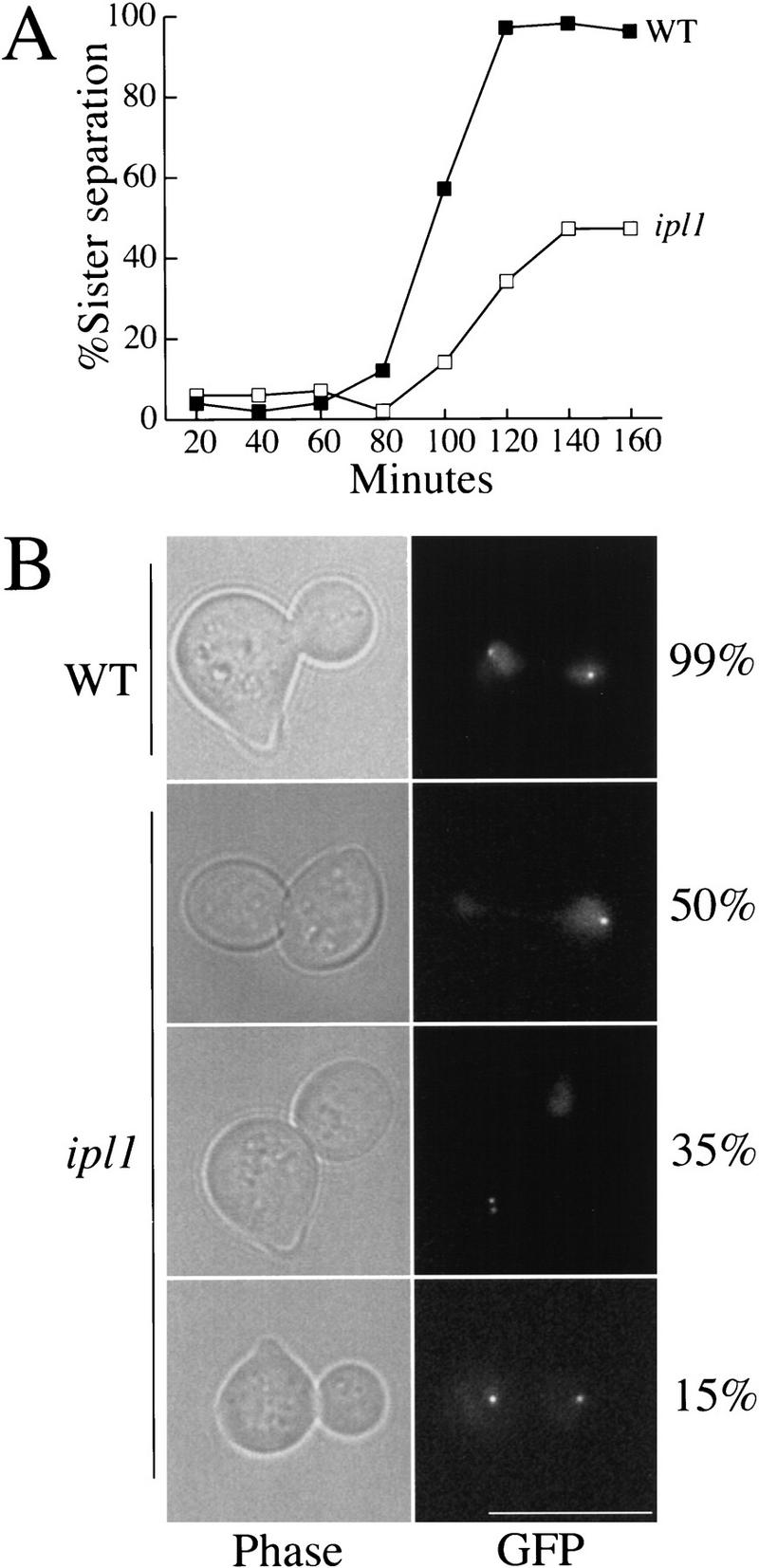
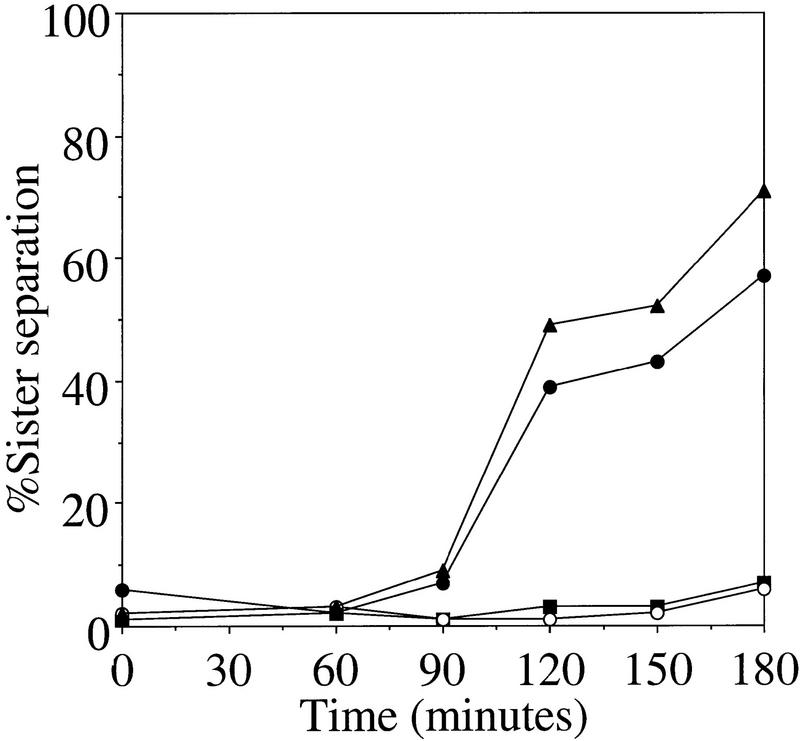
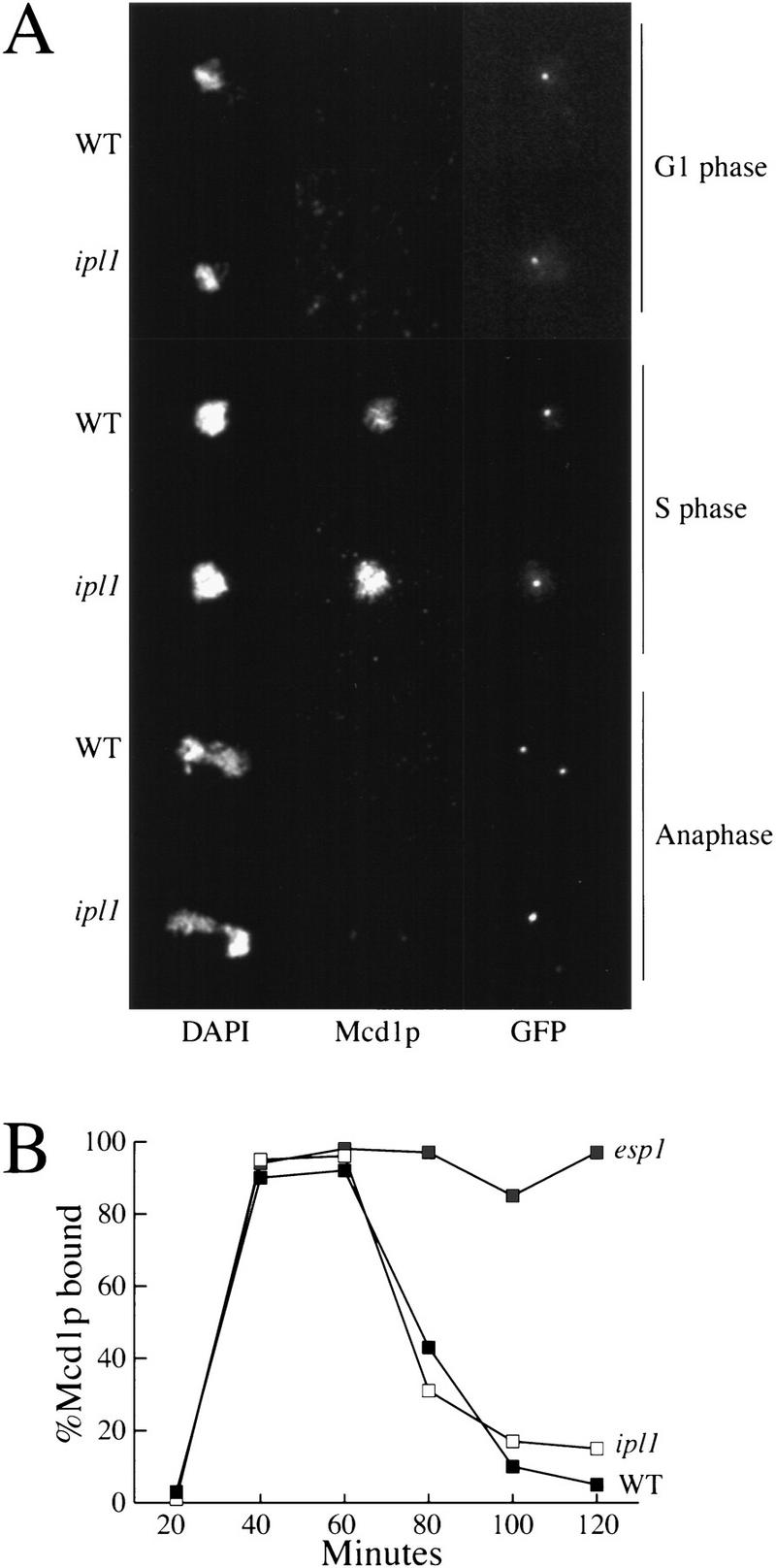
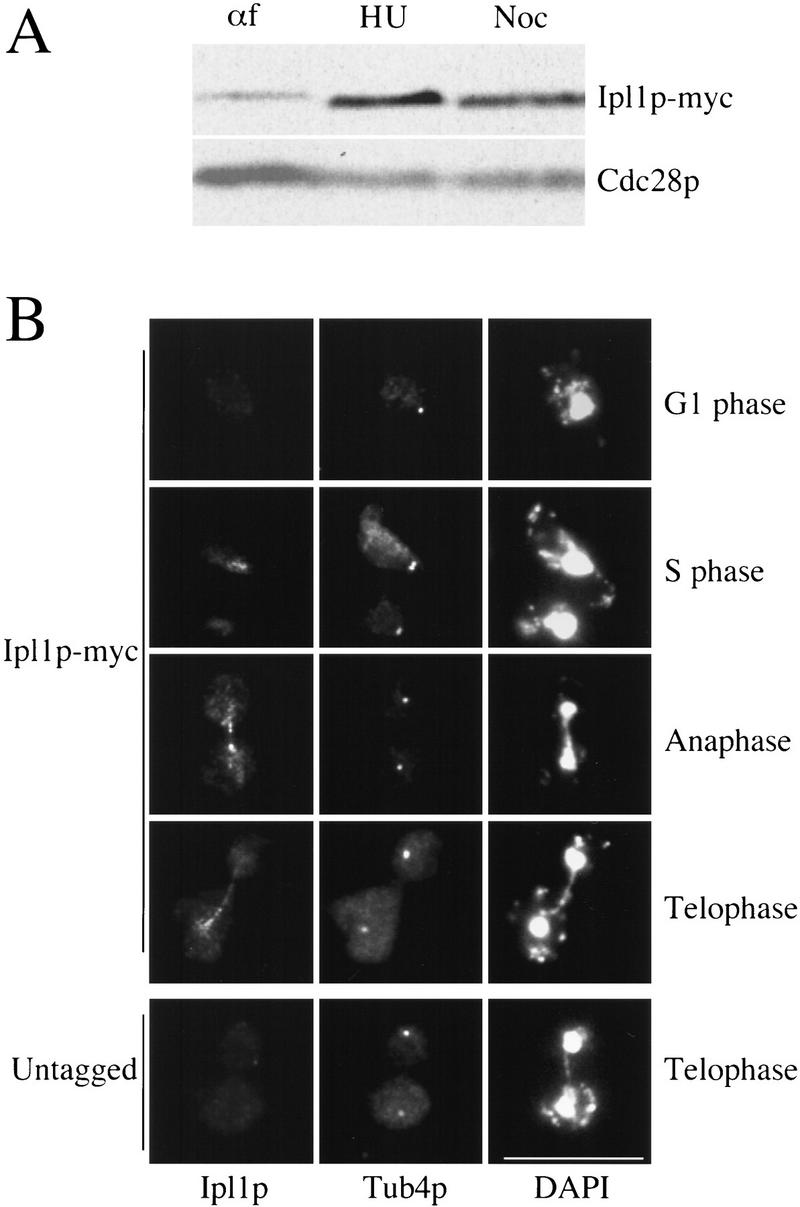
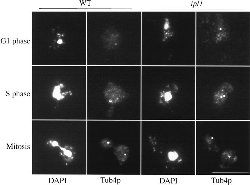
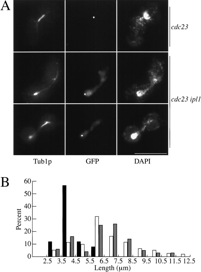
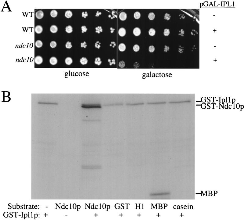
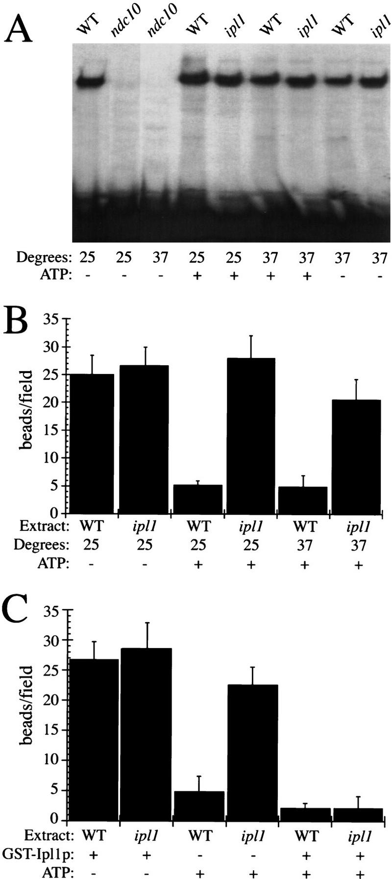
Chromosome segregation depends on kinetochores, the structures that mediate chromosome attachment to the mitotic spindle. We isolated mutants in IPL1, which encodes a protein kinase, in a screen for budding yeast mutants that have defects in sister chromatid separation and segregation. Cytological tests show that ipl1 mutants can separate sister chromatids but are defective in chromosome segregation. Kinetochores assembled in extracts from ipl1 mutants show altered binding to microtubules. Ipl1p phosphorylates the kinetochore component Ndc10p in vitro and we propose that Ipl1p regulates kinetochore function via Ndc10p phosphorylation. Ipl1p localizes to the mitotic spindle and its levels are regulated during the cell cycle. This pattern of localization and regulation is similar to that of Ipl1p homologs in higher eukaryotes, such as the human aurora2 protein. Because aurora2 has been implicated in oncogenesis, defects in kinetochore function may contribute to genetic instability in human tumors.


Figures

Figure 1
Figure 1
(A) Sister chromatids in ipl1 mutants. Wild-type and ipl1-321 cells released from α-factor arrest (T = 0) into the nonpermissive temperature (37°C) were scored for sister chromatid separation by microscopy. These strains contained integrated lactose operators at the centromere of chromosome IV. Although sister chromatids completely separate in wild-type cells (SBY214; █), they appear to separate only 50% of the time in ipl1-321 (SBY322; □ ). (B) The phenotypes of cells at 140 min after α-factor release are shown. Sister chromatids were visualized by GFP–LacI fluorescence and the nuclear DNA was visualized by the background GFP fluorescence. Three ipl1 mutant phenotypes are observed: Sisters appear to be held together 50% of the time, both sisters separate at one pole 35% of the time, and sisters separate to opposite poles 15% of the time. Phase contrast is shown in the left panels; GFP fluorescence is shown in the right panels. Bar, 10 μm.
Figure 2
Figure 2
Sister chromatids separate in ipl1 mutants. Wild-type (SBY214; █), ipl1-321 (SBY322; ○), mad2Δ (SBY99; ▴), and mad2Δ ipl1-321 (SBY100; ●) strains were arrested in G1 with α-factor (T = 0), released to the nonpermissive temperature (37°C) in nocodazole and benomyl, and scored for the percentage sister chromatid separation over time. In the absence of a spindle, ipl1 mutants are competent to separate sister chromatids.
Figure 3
Figure 3
Mcd1p localization is normal in ipl1 mutants. (A) Mcd1p was localized to chromosomes by indirect immunofluorescence on chromosome spreads in wild-type (SBY376) and ipl1-321 (SBY378) strains containing HA3-epitope-tagged Mcd1p throughout the cell cycle. DAPI staining is shown in the left panels, anti-HA antibody staining is shown in the middle panels, and anti-GFP antibody staining is shown in the right panels. G1-phase, S-phase, and anaphase cells are indicated. Mcd1p is localized to chromosomes during S phase but not during G1 or anaphase in both wild-type and ipl1 cells. Bar, 10 μm. (B) The results of Mcd1p localization to chromosomes were quantified and the percentage of chromosome spreads with Mcd1p localized to chromosomes is graphed vs. time during the cell cycle for wild-type (SBY214; WT), ipl1-321 (SBY322; ipl1), and esp1-1 strains (SBY102; esp1).
Figure 4
Figure 4
Ipl1p is a cell-cycle-regulated protein associated with the spindle. (A) The levels of Ipl1p–Myc12 (SBY388) in α-factor-arrested cells (αf, left lane), hydroxyurea-arrested cells (HU, middle lane), and nocodazole-arrested cells (Noc, right lane) were compared by Western blotting protein extracts with anti-Myc antibodies. The levels of Cdc28p are shown as a control. (B) Indirect immunofluorescence microscopy was performed on Ipl1p–Myc12 (SBY146) and an untagged control (SBY3) strain. Anti-Myc antibodies that recognize Ipl1p are shown in the left panels, anti-Tub4 antibodies are in the middle panels, and DAPI staining is in the right panels. The experiment was performed on cells throughout the cell cycle as indicated. Ipl1p localizes to the mitotic spindle. Bar, 10 μm.
Figure 5
Figure 5
Spindle pole body duplication is normal in ipl mutants. Indirect immunofluorescence microscopy was performed to detect spindle pole bodies in wild-type (SBY214) and ipl1-321 (SBY322) strains throughout the cell cycle at the nonpermissive temperature. Strains were harvested to analyze spindle pole bodies in G1, S phase, and mitosis as indicated in wild-type (left panels) and ipl1-321 cells (right panels). DAPI staining and anti-Tub4p staining are indicated. Bar, 10 μm.
Figure 6
Figure 6
The spindle elongates in ipl1 mutants during a metaphase arrest. (A) Indirect immunofluorescence was performed on cdc23-1 (SBY134) and cdc23-1 ipl1-321 (SBY133) mutants shifted to the nonpermissive temperature (37°C) for 4 hr as indicated. Anti-tubulin staining is shown in the left panels, anti-GFP staining (to recognize the marked sister chromatid) is in the middle panels, and DAPI staining is shown in the right panels. Bar, 10 μm. (B) The spindle length (μm) in cdc23-1 (solid bars), cdc23-1 ipl1-321 (open bars), and cdc23-1 pds1-296 (SBY340; shaded bars) strains was measured in 100 cells, and the percentage of cells containing each spindle length graphed.
Figure 7
Figure 7
Ipl1p interacts with Ndc10p. (A) Serial dilutions of wild-type (WT; SBY214) and ndc10-1 (SBY164) cells in the presence or absence of pGAL–IPL1 (pSB169) at 23°C on glucose and galactose media are shown. Although pGAL–IPL1 does not affect wild-type cells (SBY394), it severely inhibits the growth of ndc10-1 cells (SBY196), suggesting a specific interaction between NDC10 and IPL1. (B) Ipl1p phosphorylates Ndc10p. Kinase assays were performed using the GST–Ipl1p kinase (lanes 1, 3–7) or GST as a control (lane 2). The substrates tested were GST–Ipl1, GST–Ndc10p, GST, histone HI, myelin basic protein (MBP), and casein, as indicated. The GST–Ndc10p is a carboxy-terminal fragment of Ndc10p that contains codons 679–894 of the 956-amino-acid protein. Two micrograms of substrate was used in each reaction except GST–Ipl1p autophosphorylation. The autophosphorylation of GST–Ipl1p and the phosphorylation of GST–Ndc10p and MBP by GST–Ipl1p is indicated at right. There are GST–Ndc10p breakdown products in addition to the GST–Ndc10 fusion protein in lane 3.
Figure 8
Figure 8
ipl1 mutants affect kinetochore function. (A) Nondenaturing gel analysis of kinetochore assembly was performed in the presence or absence of ATP using radiolabeled centromere DNA and extracts prepared from wild-type (SBY214), ndc10-1 (SBY164), or ipl1-321 (SBY322) cells grown at either 23°C or 37°C. The ndc10-1 kinetochore mutant is defective in assembling kinetochores but ip11 mutants and wild-type cells assemble normal kinetochores. (B) The microtubule-binding activity (no. of beads bound per field) in the presence or absence of ATP of wild-type amd ipl1-321 extracts made from cells grown at 25°C or 37°C is graphed. The ipl1-321 mutant is not sensitive to the addition of ATP at either temperature. (C) The microtubule-binding activity (no. of beads bound per field) in the presence or absence of ATP, and recombinant GST–Ipl1p from cells grown at 25°C is graphed. The addition of GST–Ipl1p to ipl1-321 extracts restores the microtubule inhibition by ATP.

References

    1. Axton JM, Dombradi V, Cohen PT, Glover DM. One of the protein phosphatase 1 isoenzymes in Drosophila is for mitosis. Cell. 1990;63:33–46. - PubMed
    1. Biggins S, Murray A. Sister chromatid cohesion in mitosis. Curr Opin Cell Biol. 1998;10:769–775. - PubMed
    1. Bischoff JR, Anderson L, Zhu Y, Mossie K, Ng L, Souza B, Schryver B, Flanagan P, Clairvoyant F, Ginther C, Chan CS, Novotny M, Slamon DJ, Plowman GD. A homologue of Drosophila aurora kinase is oncogenic and amplified in human colorectal cancers. EMBO J. 1998;17:3052–3065. - PMC - PubMed
    1. Cahill DP, Lengauer C, Yu J, Riggins GJ, Willson JK, Markowitz SD, Kinzler KW, Vogelstein B. Mutations of mitotic checkpoint genes in human cancers. Nature. 1998;392:300–303. - PubMed
    1. Chan CS, Botstein D. Isolation and characterization of chromosome-gain and ploidy mutants in yeast. Genetics. 1993;135:677–691. - PMC - PubMed

Publication types