. 1999 Mar 1;13(5):556-68.
doi: 10.1101/gad.13.5.556.

Nuclear import of the Drosophila Rel protein Dorsal is regulated by phosphorylation

Affiliations

Nuclear import of the Drosophila Rel protein Dorsal is regulated by phosphorylation

E A Drier et al. Genes Dev. .

Abstract

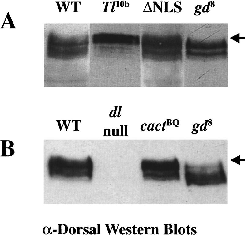
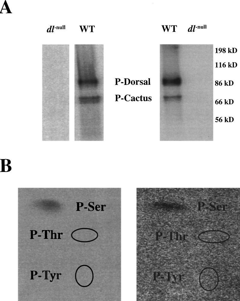
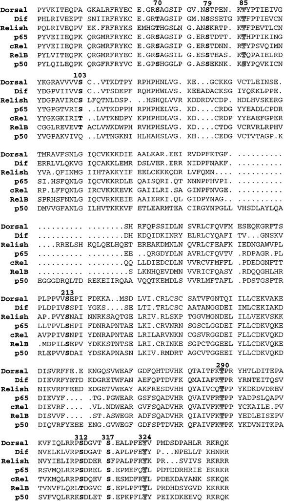
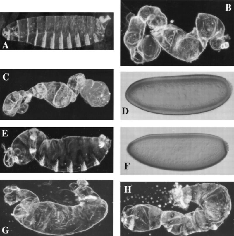
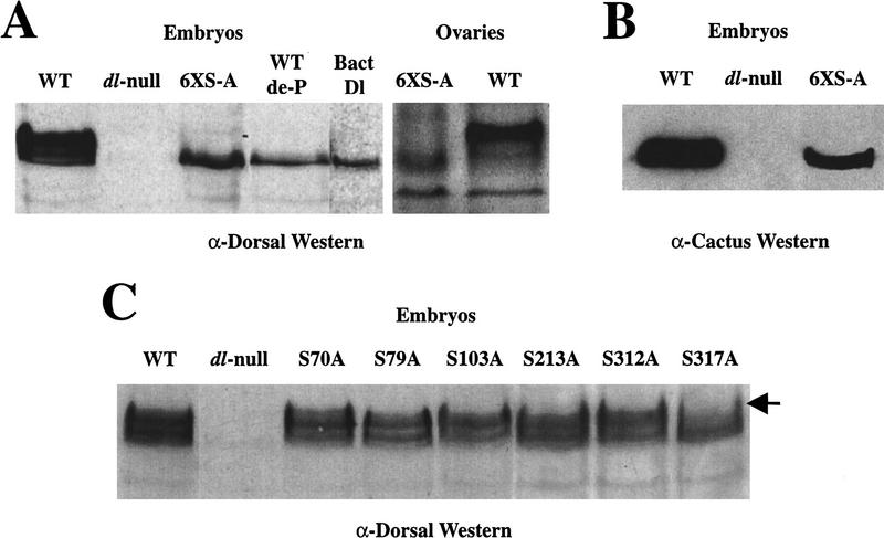
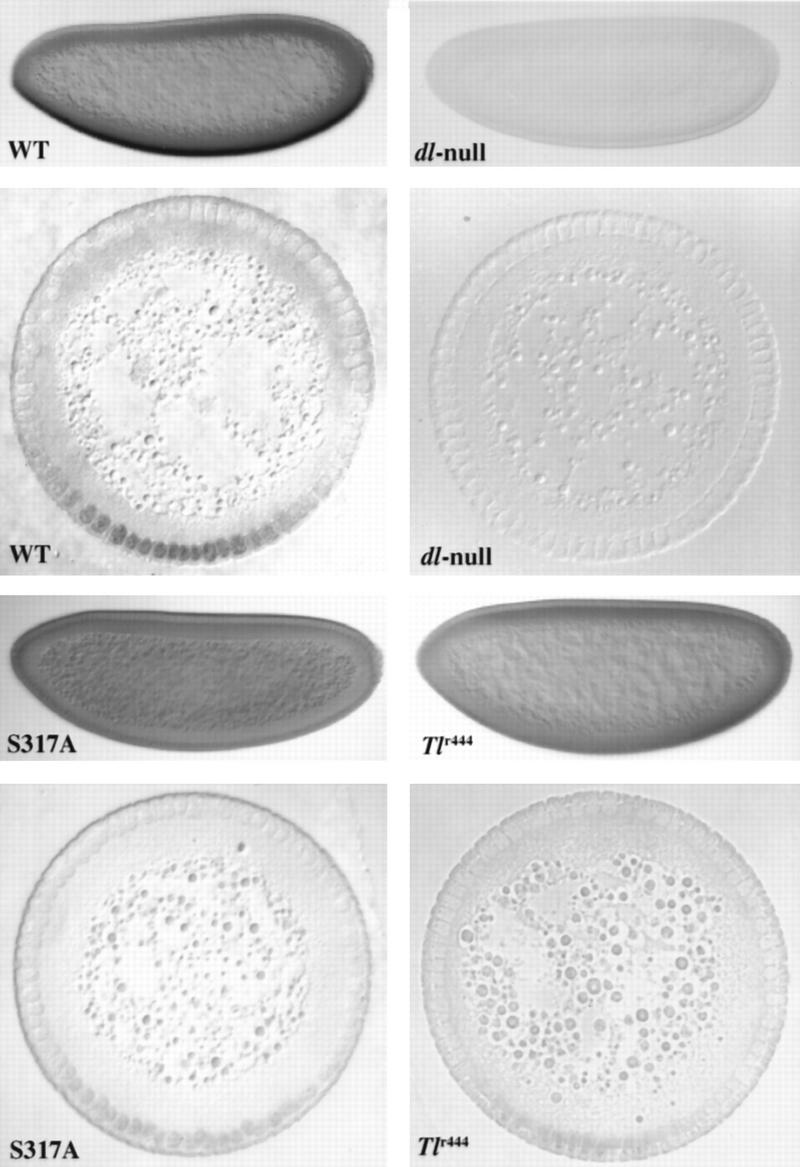
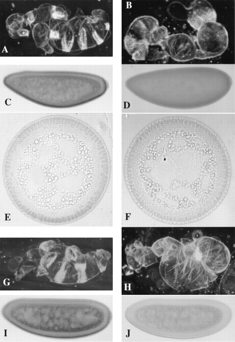
In Drosophila, dorsal-ventral polarity is determined by a maternally encoded signal transduction pathway that culminates in the graded nuclear localization of the Rel protein, Dorsal. Dorsal is retained in the cytoplasm by the IkappaB protein, Cactus. Signal-dependent phosphorylation of Cactus results in the degradation of Cactus and the nuclear targeting of Dorsal. We present an in-depth study of the functional importance of Dorsal phosphorylation. We find that Dorsal is phosphorylated by the ventral signal while associated with Cactus, and that Dorsal phosphorylation is essential for its nuclear import. In vivo phospholabeling of Dorsal is limited to serine residues in both ovaries and early embryos. A protein bearing mutations in six conserved serines abolishes Dorsal activity, is constitutively cytoplasmic, and appears to eliminate Dorsal phosphorylation, but still interacts with Cactus. Two individual serine-to-alanine mutations produce unexpected results. In a wild-type signaling background, a mutation in the highly conserved PKA site (S312) produces only a weak loss-of-function; however, it completely destabilizes the protein in a cactus mutant background. Significantly, the phosphorylation of another completely conserved serine (S317) regulates the high level of nuclear import found in ventral cells. We conclude that the formation of a wild-type Dorsal nuclear gradient requires the phosphorylation of both Cactus and Dorsal. The strong conservation of the serines suggests that phosphorylation of other Rel proteins is essential for their proper nuclear targeting.


Figures

Figure 1
Figure 1
Dorsal is subject to signal-dependent phosphorylation while associated with Cactus in the cytoplasm. Extracts from ∼20 embryos were loaded in each lane. The genotype of the mothers is indicated. (Arrow) Location of the hyperphosphorylated form of Dorsal in A and B. (A) Constitutively cytoplasmic ΔNLS–Dorsal is phosphorylated normally. (WT) Mothers are homozygous wild-type for dorsal and the dorsal–ventral signal transduction pathway. (Tl10) Mothers bear a dominant allele of the gene encoding the transmembrane receptor Toll, which results in constitutive signaling and causes Dorsal to translocate to all nuclei along the dorsal–ventral axis at a high, uniform level (Roth et al. 1989; Steward 1989). (ΔNLS) Mothers bear one copy of the ΔNLS–dorsal mutant minigene in an otherwise dorsal protein-null mutant background. (gd8) Mothers are homozygous for gd8 that completely disrupts signaling and causes Dorsal to remain cytoplasmic along the entire dorsal–ventral axis (Roth et al. 1989; Steward 1989). (B) Dorsal is phosphorylated when associated with Cactus. (WT) The same as in A. (dl-null) Mothers bear two different protein-null alleles of dorsal. (cactBQ) The mothers genotype is cactBQ/cactE10RN1. cactBQ is an allele of cactus that is completely recalcitrant to the ventral signal and thus constitutively maintains Dorsal in the cytoplasm (Bergmann et al. 1996). cactE10RN1 is a null allele of cactus (Roth et al. 1991). (gd8) The same as in A.
Figure 2
Figure 2
In vivo phospholabeling of Dorsal is limited to serine residues. (A) SDS-PAGE of labeled Dorsal from both ovaries (left) and early embryos (right). Extracts were made from labeled ovaries and embryos (see Materials and Methods). Dorsal was immunoprecipitated with pooled anti-Dorsal monoclonal antibodies that were conjugated to protein A–Sepharose beads. Parallel labelings and immunoprecipitations were done on ovaries and embryos from mothers bearing protein-null mutations in dorsal. These dorsal-null labelings had a similar level of total protein phosphorylation (data not shown). (B) Phosphoamino acid analysis of ovarian (left) and embryonic (right) Dorsal. The labeled Dorsal from A was transferred to Immobilon-P membrane and subjected to total hydrolysis in 5.7 n HCl (see Materials and Methods). The hydrolysates were separated by TLC. The ellipses indicate the locations of the ninhydrin-stained P-Thr and P-Tyr.
Figure 3
Figure 3
Conserved serines in the Rel homology domain of Dorsal. The Rel homology domain of Dorsal is compared with the other Drosophila Rel family members, Dif and Relish, as well as vertebrate forms, p65, c-Rel, RelB, and p50. The conserved serines are highlighted in bold. The conserved threonine and tyrosine residues we altered are indicated by shadowed characters. The bold numbers above the Dorsal sequence refer to the location of the residue within Dorsal.
Figure 4
Figure 4
Molecular and phenotypic analyses of S-to-A Dorsal mutants. Cuticle preparations of fully differentiated embryos and anti-Dorsal staining of blastoderm stage embryos. (A) An embryo from a mother bearing two copies of a wild-type dorsal cDNA. (B) An embryo from a dorsal-null mother without a dorsal minigene. (The dorsal-null background used throughout this work; see Materials and Methods). (C) An embryo from a mother bearing two copies of 6XS-A–dorsal in the dorsal-null background. (D) Anti-Dorsal staining of a blastoderm embryo as in C. (E) An embryo from a mother bearing one copy of S312A–dorsal in the dorsal-null background. (F) Anti-Dorsal staining of a blastoderm embryo as in E. (G,H) Embryos from mothers bearing one or two copies of S317A–dorsal in the dorsal-null background, respectively. See Fig. 6 for anti-Dorsal staining of wild-type Dorsal, dorsal-null, and S317A–Dorsal.
Figure 5
Figure 5
Western analyses of Dorsal serine-to-alanine mutations. (A) The 6XS-A mutant eliminates Dorsal phosphoforms. A low-bis Western blot probed with anti-Dorsal antibody. (Left) Embryos. Approximately thirty, 0 to 3 hr embryos were used for each lane. The lane markings indicate the origin of the Dorsal protein in each case. The 6XS-A comes from dorsal-null mothers bearing two copies of the 6XS-A–dorsal minigene. (WT de-P) Dephosphorylated wild-type Dorsal (see Materials and Methods). (Bact-Dl) Bacterially expressed Dorsal. (Right) Ovaries. One pair of ovaries was used for each lane. (B) The 6XS-A–Dorsal mutant protein interacts with Cactus. A Western blot probed with anti-Cactus antibody. The 6XS-A females carried only one copy of the 6XS-A–dorsal minigene, whereas the wild-type (WT) lane carries two endogenous dorsal copies. (C) The phosphoform distribution of single serine-to-alanine mutations. A representative low-bis Western blot probed with anti-Dorsal antibody. In each case, the embryos were from mothers bearing two copies of the respective transgenes. The subtle changes in phosphoform distribution between each serine-to-alanine mutant are somewhat variable. The S317A mutant differs consistently from both wild type and the other mutants.
Figure 6
Figure 6
Anti-Dorsal stainings of embryos show that S317A–Dorsal is defective for normal nuclear import. Whole mounts and cross sections of blastoderm stage embryos stained with pooled anti-Dorsal monoclonals (see Materials and Methods). In the whole mounts, anterior is to the left and dorsal is up; in the cross sections, dorsal is up. The cross sections were taken at roughly the anterior–posterior midpoint of the embryo. (WT) Embryos from mother bearing one copy of the wild-type dorsal minigene in the dorsal null background. (dl-null) Embryos from mothers bearing protein-null mutations in dorsal (see Materials and Methods) without any dorsal minigene. (S317A) Embryos from mothers bearing one copy of the S317A–dorsal mutant minigene instead of the wild-type minigene. (Tlr444) Embryos from a Tollr444 mother [genotype: Tlr444/Df(Tl); see Materials and Methods] raised at the restrictive temperature (29°C).
Figure 7
Figure 7
S317A–Dorsal is uniformly distributed and S312A–Dorsal in unstable in a cactusA2, dorsal-null mutant background. Cuticle preparations of fully differentiated embryos and blastoderm stage embryos stained with pooled anti-Dorsal monoclonals (see Materials and Methods). (A) A cuticle preparation of an embryo from a mother bearing one copy of the wild-type dorsal minigene in a cactA2, dorsal-null background. (B) The same as in A, except the mother carries one copy of the S317A–dorsal minigene instead of the wild-type minigene. (C) A whole mount antibody staining of an embryo from a mother bearing one copy of the wild-type dorsal minigene in a cactA2, dorsal-null background. (D) the same as in C, except the mother carries one copy of the S317A–dorsal minigene instead of the wild-type minigene. The nuclei are difficult to visualize in D because S317A–Dorsal does not make a nuclear gradient as seen in C, and stains the nuclei and cytoplasm at roughly equal levels. (E) A cross section of an embryo as in C. (F) A cross section of an embryo as in D, which more readily shows the uniform low level of S317A–Dorsal entering the nuclei. In this cross section, we cannot state that dorsal is up because the S317A–Dorsal shows no gradient to indicate dorsal–ventral polarity. (G,H) Cuticle preparations of embryos from a mother bearing one copy of S70A–dorsal. (G) One copy of S312A–dorsal in the cactusA2, dorsal-null background. (I,J) Whole mount anti-Dorsal stainings of blastoderm stage embryos as in G and H, respectively.

Similar articles

Cited by

References

    1. Anderson KV, Nüsslein-Volhard C. Genetic analysis of dorsal-ventral embryonic pattern in Drosophila. In: Molacinski GM, Bryant SV, editors. Pattern formation. New York, NY: MacMillan; 1984. pp. 269–289.
    1. Ashburner M. Drosophila: A laboratory manual. Cold Spring Harbor, NY: Cold Spring Harbor Laboratory Press; 1989.
    1. Baeuerle PA, Baltimore D. Activation of DNA-binding activity in an apparently cytoplasmic precursor of NF-κB transcription factor. Cell. 1998;53:211–217. - PubMed
    1. Belvin MP, Jin Y, Anderson KV. Cactus protein degradation mediates Drosophila dorsal–ventral signaling. Genes & Dev. 1995;9:783–793. - PubMed
    1. Bergmann A, Stein D, Geisler R, Hagenmaier S, Schmid B, Fernandez N, Schnell B, Nüsslein-Volhard C. A gradient of cytoplasmic Cactus degradation establishes the nuclear localization gradient of the dorsal morphogen in Drosophila. Mech Dev. 1996;60:109–123. - PubMed

Publication types

MeSH terms

LinkOut - more resources