. 1999 Mar 1;13(5):593-606.
doi: 10.1101/gad.13.5.593.

Binding of hnRNP H to an exonic splicing silencer is involved in the regulation of alternative splicing of the rat beta-tropomyosin gene

Affiliations

Binding of hnRNP H to an exonic splicing silencer is involved in the regulation of alternative splicing of the rat beta-tropomyosin gene

C D Chen et al. Genes Dev. .

Abstract

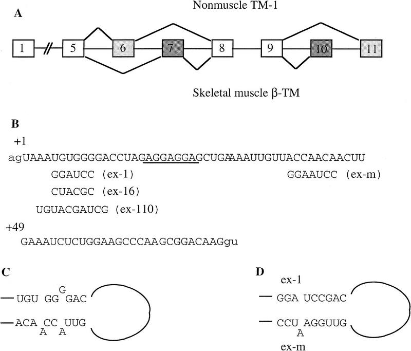
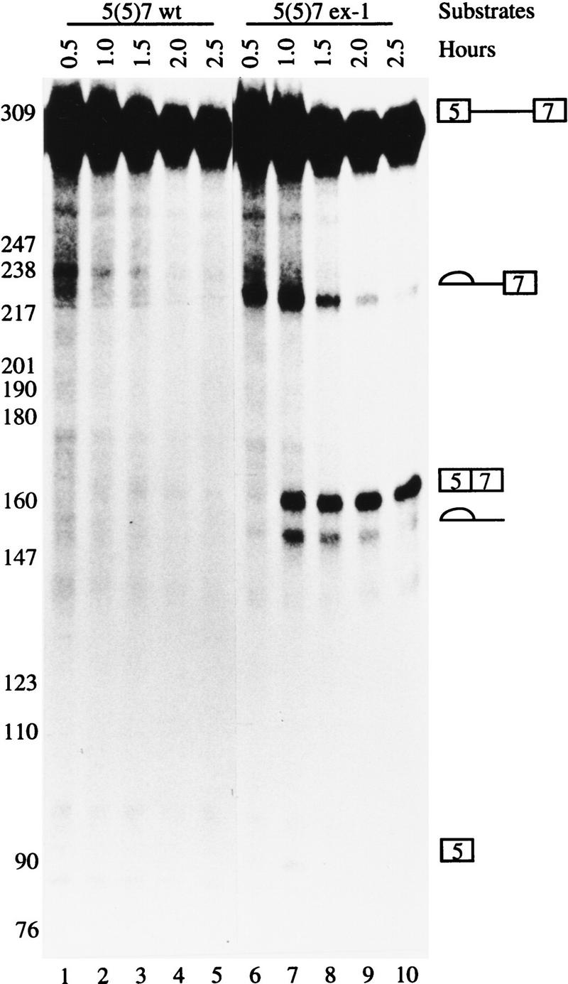
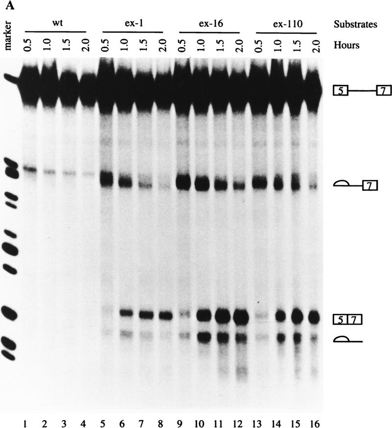
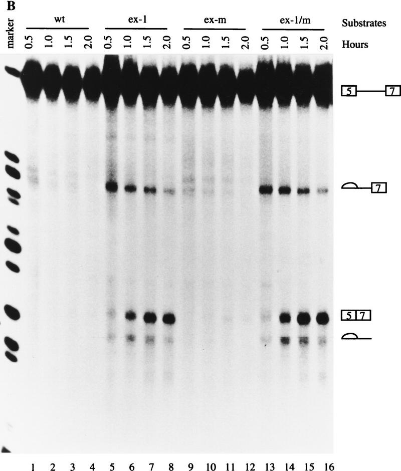
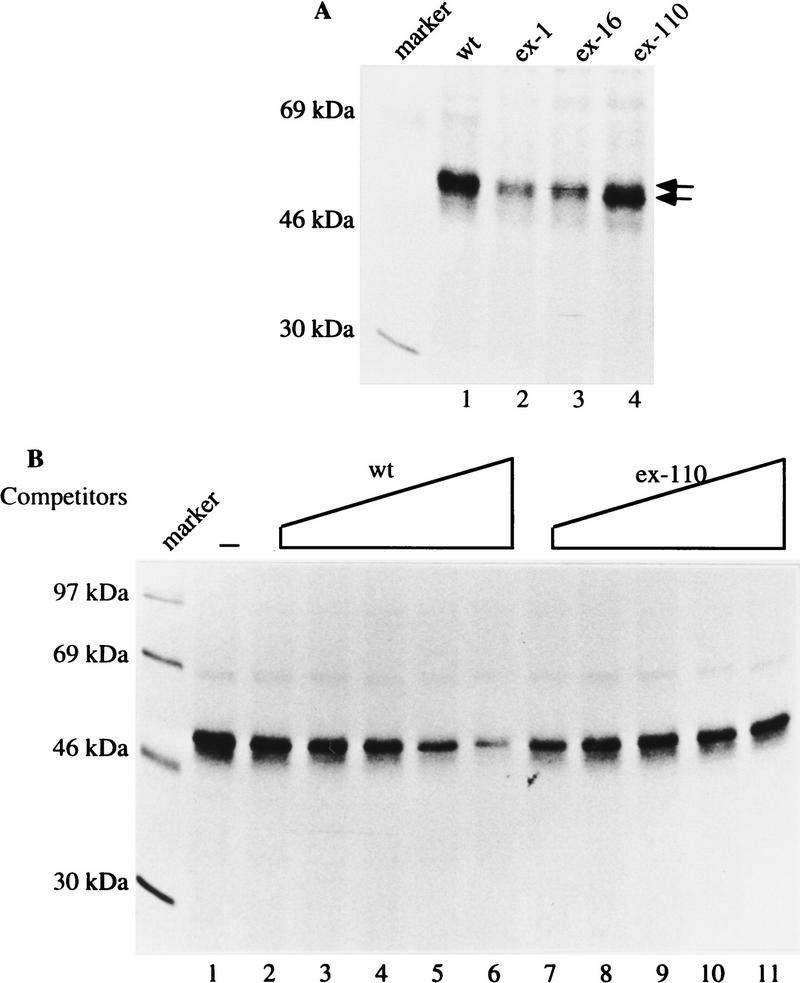
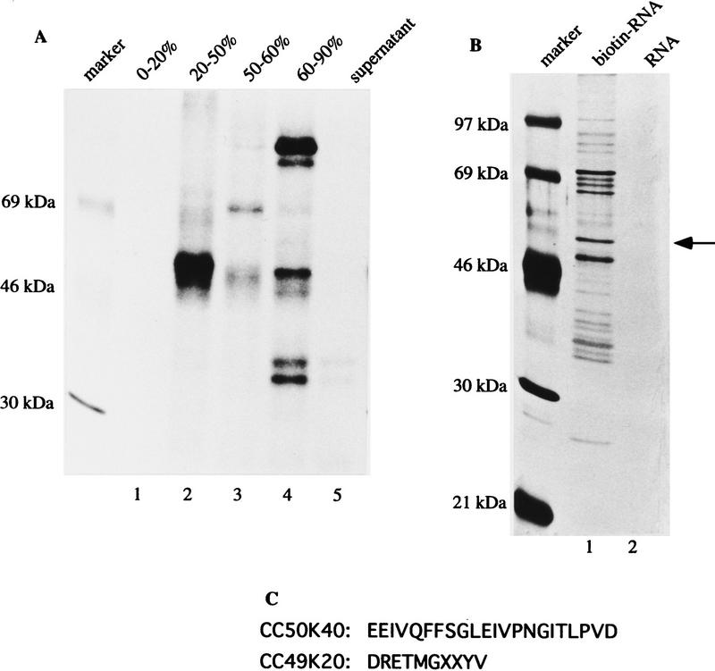
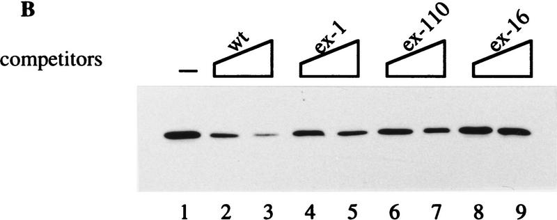
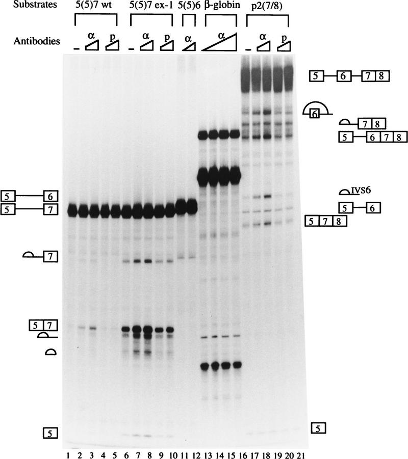
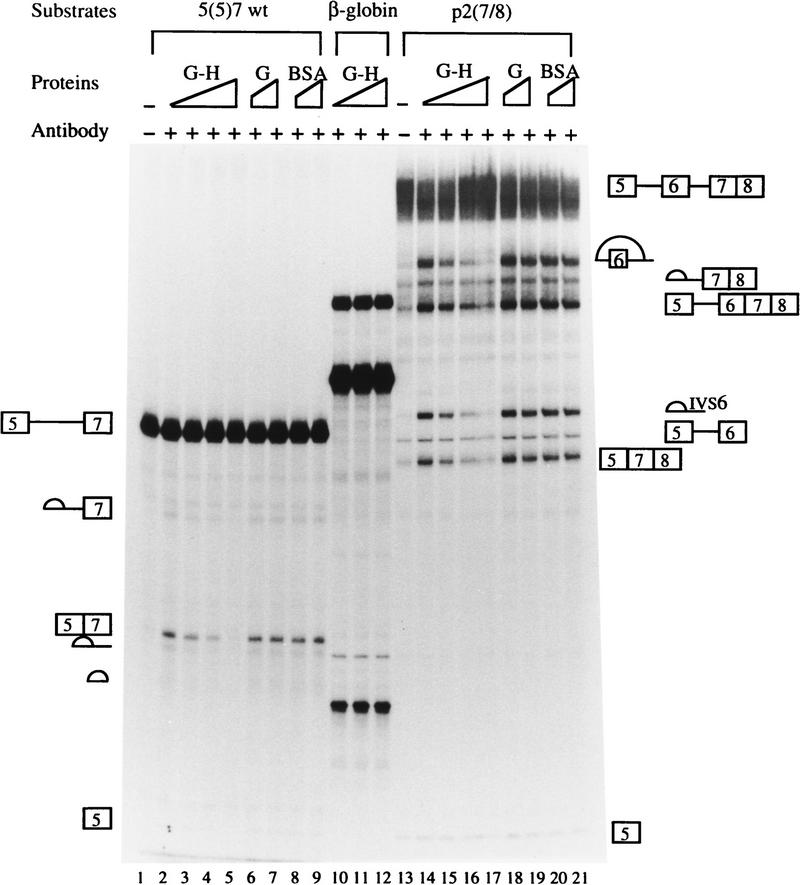
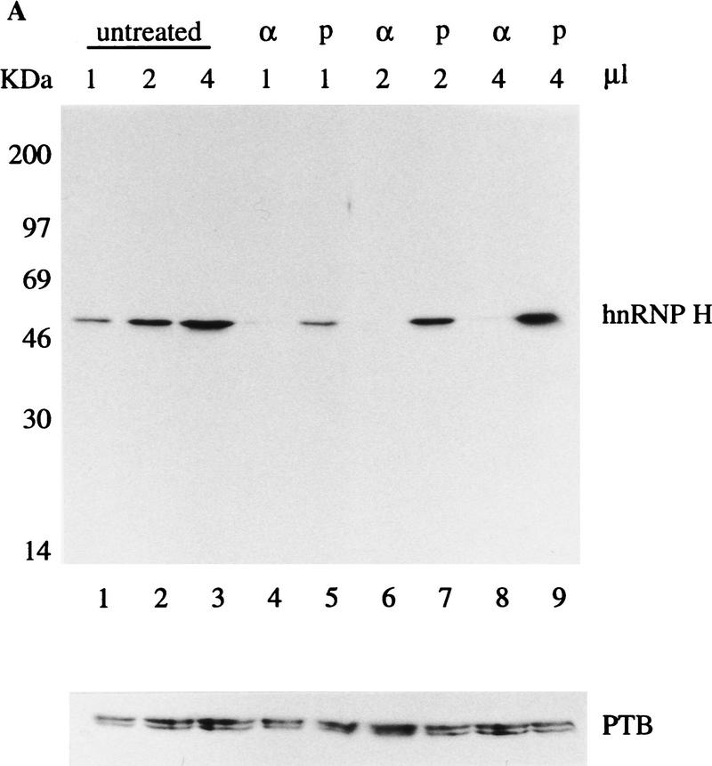
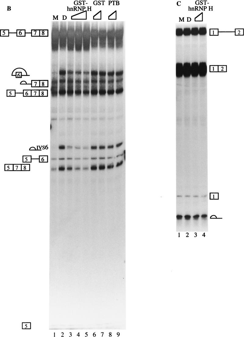
In the rat beta-tropomyosin (beta-TM) gene, exons 6 and 7 are spliced alternatively in a mutually exclusive manner. Exon 6 is included in mRNA encoding nonmuscle TM-1, whereas exon 7 is used in mRNA encoding skeletal muscle beta-TM. Previously, we demonstrated that a six nucleotide mutation at the 5' end of exon 7, designated as ex-1, activated exon 7 splicing in nonmuscle cells. In this study, we show that the activating effect of this mutation is not the result of creating an exonic splicing enhancer (ESE) or disrupting a putative secondary structure. The sequence in exon 7 acts as a bona fide exonic splicing silencer (ESS), which is bound specifically by a trans-acting factor. Isolation and peptide sequencing reveal that this factor is hnRNP H, a member of the heterogeneous nuclear ribonucleoprotein (hnRNP) family. Binding of hnRNP H correlates with the ESS activity. Furthermore, addition of antibodies that specifically recognizes hnRNP H to the splicing reactions or partial depletion of hnRNP H from nuclear extract activates exon 7 splicing in vitro and this effect can be reversed by addition of purified recombinant hnRNP H. These results indicate that hnRNP H participates in exclusion of exon 7 in nonmuscle cells. The involvement of hnRNP H in the activity of an ESS may represent a prototype for the regulation of tissue- and developmental-specific alternative splicing.


Figures

Figure 1
Figure 1
(A) A schematic diagram of rat β-TM gene. Boxes represent exons and lines introns; shaded boxes represent tissue-specific exons as indicated. (B) The wild-type and mutated sequences of exon 7. Lowercase letters represent sequences in adjacent introns, and +1 is the first nucleotide of exon 7. The mutated sequences are indicated below the wild-type sequences. (C) Stem I of the putative secondary structure in the wild-type substrate. (D) The restored stem I of the putative secondary structure when both complementary strands are mutated simultaneously.
Figure 2
Figure 2
An in vitro system to study the ex-1 mutation. The wild-type and ex-1 mutation 5(5)7 pre-mRNAs containing exon 5, intron 5, and exon 7 were synthesized and subjected to in vitro splicing reactions using the conditions indicated at the top. The RNAs were resolved on 4% polyacrylamide 8 m urea gel and visualized by autoradiography. The precursors, intermediate, and final products are indicated at left.
Figure 3
Figure 3
The 5′ end of exon 7 contains an ESS. Transcription and in vitro splicing were carried out as in Fig. 2, and the marker is the same as in Fig. 2. (A) Activation of ex-1 does not result from creation of an ESE. The wild-type and mutant 5(5)7 substrates are indicated at the top. (B) Activation of ex-1 mutation does not result from disruption of the putative secondary structure. The wild-type and mutant 5(5)7 substrates are indicated at the top. (C) Titration experiments. Splicing reactions were carried out in the absence (lanes 1,8,15,19) or presence of 10, 20, or 30 pmoles of the specific competitor ECS, or nonspecific competitor WU as indicated. The substrates are indicated at the top, the precursors and the products on either side. 5(5)6 contains exon 6 instead of exon 7 in 5(5)7; β-globin pre-mRNA consists of exon 1, intron 1, and exon 2 of the human β-globin gene.
Figure 3
Figure 3
The 5′ end of exon 7 contains an ESS. Transcription and in vitro splicing were carried out as in Fig. 2, and the marker is the same as in Fig. 2. (A) Activation of ex-1 does not result from creation of an ESE. The wild-type and mutant 5(5)7 substrates are indicated at the top. (B) Activation of ex-1 mutation does not result from disruption of the putative secondary structure. The wild-type and mutant 5(5)7 substrates are indicated at the top. (C) Titration experiments. Splicing reactions were carried out in the absence (lanes 1,8,15,19) or presence of 10, 20, or 30 pmoles of the specific competitor ECS, or nonspecific competitor WU as indicated. The substrates are indicated at the top, the precursors and the products on either side. 5(5)6 contains exon 6 instead of exon 7 in 5(5)7; β-globin pre-mRNA consists of exon 1, intron 1, and exon 2 of the human β-globin gene.
Figure 4
Figure 4
A protein cross-links to the ESS. (A) 32P-Labeled oligoribonucleotide that contains either the wild-type or mutated ESS as indicated at the top was cross-linked to HeLa cell nuclear extract and resolved on a 10% SDS-polyacrylamide gel. Two arrows indicate two cross-linked products in lane 4; the upper one migrated similarly to the products in other lanes. (B) 32P-Labeled oligoribonucleotide that contains the wild-type ESS was cross-linked to HeLa cell nuclear extract in the absence (lane 1) or presence of 10- , 20- , 40- , 60- , or 80-fold excess of the wild-type competitor or mutant competitor ex-110 as indicated.
Figure 5
Figure 5
Isolation and peptide-sequencing of hnRNP H. (A) HeLa cell nuclear extracts were fractionated by ammonium sulfate precipitation, and each fraction was UV cross-linked to an oligoribonucleotide containing the wild-type ESS. (B) The biotinylated RNA (lane 1) or RNA (lane 2) containing six tandem repeats of the wild-type ESS was incubated with the 20%–50% ammonium sulfate fraction. The RNA–protein complexes were pulled down by streptavidin–agarose. The proteins were eluted with wash buffer containing 0.5 m KCl, resolved on a 10% SDS-polyacrylamide gel, and visualized by silver staining. The arrow indicates the 50-kD protein of interest. (C) Two peptides from the 50-kD protein were obtained by peptide sequencing.
Figure 6
Figure 6
hnRNP H specifically binds to the ESS. (A) Biotin–streptavidin binding assay was carried out using the biotinylated wild-type and mutant 5(5)7, and 5(5)6 splicing substrates; the gels were probed with anti-hnRNP H antibody and visualized by ECL. (B) Competitive biotin–streptavidin binding assay. Biotin–streptavidin binding assay was carried out using the biotinylated wild-type 5(5)7 in the presence of 20 or 40 pmoles of oligoribonucleotide containing either the wild-type or mutated ESS, as indicated.
Figure 6
Figure 6
hnRNP H specifically binds to the ESS. (A) Biotin–streptavidin binding assay was carried out using the biotinylated wild-type and mutant 5(5)7, and 5(5)6 splicing substrates; the gels were probed with anti-hnRNP H antibody and visualized by ECL. (B) Competitive biotin–streptavidin binding assay. Biotin–streptavidin binding assay was carried out using the biotinylated wild-type 5(5)7 in the presence of 20 or 40 pmoles of oligoribonucleotide containing either the wild-type or mutated ESS, as indicated.
Figure 7
Figure 7
Anti-hnRNP H antibody specifically activates exon 7 splicing. In vitro splicing reactions were carried out using the substrates indicated at the top in the absence (lanes 1,5,17) or presence of 5 or 10 μg of anti-hnRNP H (α) or preimmune (p) rabbit sera as indicated below the substrates. The splicing reaction on lane 16 has 20 μg of anti-hnRNP H rabbit serum. Schematic representations of the precursors and products are shown on both sides. For clarity, the precursor and splicing products for the human β-globin pre-mRNA are not indicated (see Fig. 3C for reference).
Figure 8
Figure 8
Recombinant hnRNP H antagonizes the activation of anti-hnRNP H antibody. In vitro splicing reactions were carried out in the absence (lanes 1,13) or presence of 10 μg of anti-hnRNP H rabbit serum, and in the presence of 0.2, 0.4, or 0.6 μg of recombinant GST–hnRNP H (G-H), 0.3 or 0.6 μg of recombinant GST (G), and 0.3 or 0.6 μg of BSA as indicated. The substrates are indicated at the top, and schematic representations of the precursors and splicing products are shown on both sides. For clarity, the precursor and splicing products for the human β-globin pre-mRNA are not indicated (see Fig. 3C for reference).
Figure 9
Figure 9
Depletion of hnRNP H activates the splicing of exon 7. (A) Depletion of hnRNP H from HeLa cell nuclear extract. Western blot analysis was carried out with 1, 2, and 4 μg of untreated (lanes 1–3), antibody-depleted (lanes 4,6,8), and mock depleted extract (lanes 5,7,9), probed with anti-hnRNP or anti-PTB antibodies as indicated. (B) Splicing of p2(7/8) in mock-depleted extract (lane 1), or hnRNP H depleted extract without (lane 2) or with increasing amount (0.3, 0.6, and 0.9 μg) of recombinant GST–hnRNP H (lanes 3–5), recombinant GST (0.3 and 0.9 μg, lanes 6,7), or recombinant PTB (0.3 and 0.9 μg, lanes 8,9). (C) Splicing of the human β-globin pre-mRNA in mock-depleted extract (lane 1), hnRNP H-depleted extract without (lane 2), or with 0.3 and 0.9 μg of recombinant GST–hnRNP H (lanes 3,4).
Figure 9
Figure 9
Depletion of hnRNP H activates the splicing of exon 7. (A) Depletion of hnRNP H from HeLa cell nuclear extract. Western blot analysis was carried out with 1, 2, and 4 μg of untreated (lanes 1–3), antibody-depleted (lanes 4,6,8), and mock depleted extract (lanes 5,7,9), probed with anti-hnRNP or anti-PTB antibodies as indicated. (B) Splicing of p2(7/8) in mock-depleted extract (lane 1), or hnRNP H depleted extract without (lane 2) or with increasing amount (0.3, 0.6, and 0.9 μg) of recombinant GST–hnRNP H (lanes 3–5), recombinant GST (0.3 and 0.9 μg, lanes 6,7), or recombinant PTB (0.3 and 0.9 μg, lanes 8,9). (C) Splicing of the human β-globin pre-mRNA in mock-depleted extract (lane 1), hnRNP H-depleted extract without (lane 2), or with 0.3 and 0.9 μg of recombinant GST–hnRNP H (lanes 3,4).

Similar articles

Cited by

References

    1. Adams MD, Rudner DZ, Rio DC. Biochemistry and regulation of pre-mRNA splicing. Curr Opin Cell Biol. 1996;8:331–339. - PubMed
    1. Amendt BA, Si Z-H, Stoltzfus CM. Presence of exon splicing silencers within human immunodeficiency virus type 1 tat exon 2 and tat-rev exon 3: Evidence for inhibition mediated by cellular factors. Mol Cell Biol. 1995;15:4606–4615. - PMC - PubMed
    1. Ashiya M, Grabowski PJ. A neuron-specific splicing switch mediated by an array of pre-mRNA repressor sites: Evidence of a regulatory role for the polypyrimidine tract binding protein and a brain-specific PTB counterpart. Neuron. 1997;3:996–1015. - PMC - PubMed
    1. Black D. Activation of c-src neuron-specific splicing by an unusual RNA element in vitro and in vivo. Cell. 1992;69:795–807. - PubMed
    1. Boggs RT, Gregor P, Idriss S, Belote JM, McKeown M. Regulation of sexual differentiation in D. melanogaster via alternative splicing of RNA from the transformer gene. Cell. 1987;50:739–747. - PubMed

Publication types

LinkOut - more resources