. 1999 Mar 1;13(5):620-31.
doi: 10.1101/gad.13.5.620.

Mos positively regulates Xe-Wee1 to lengthen the first mitotic cell cycle of Xenopus

Affiliations

Mos positively regulates Xe-Wee1 to lengthen the first mitotic cell cycle of Xenopus

M S Murakami et al. Genes Dev. .

Abstract

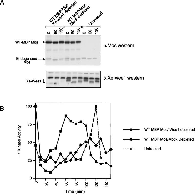
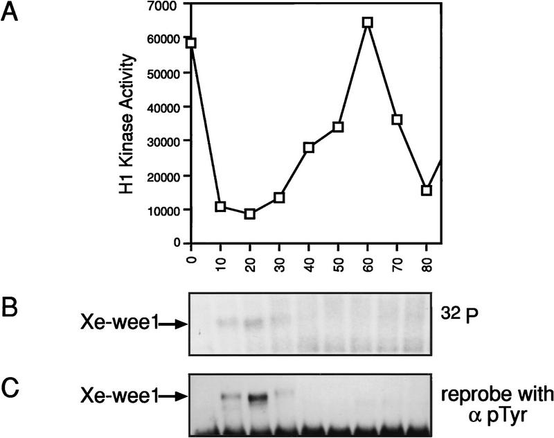
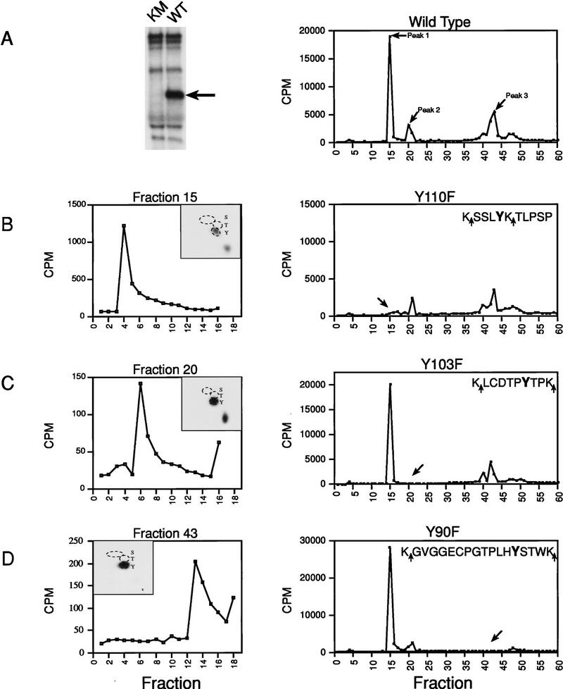
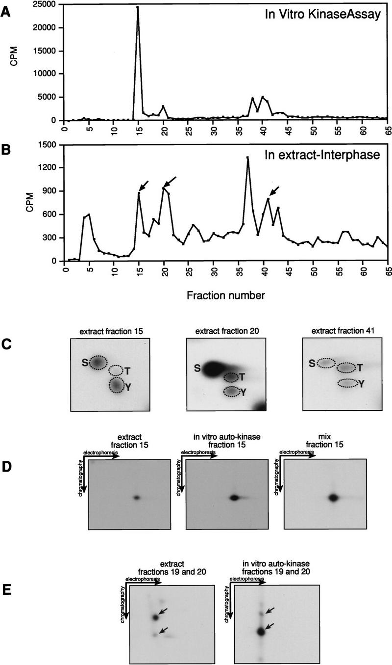
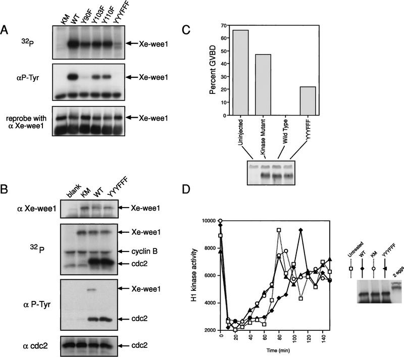
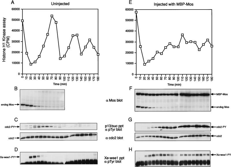
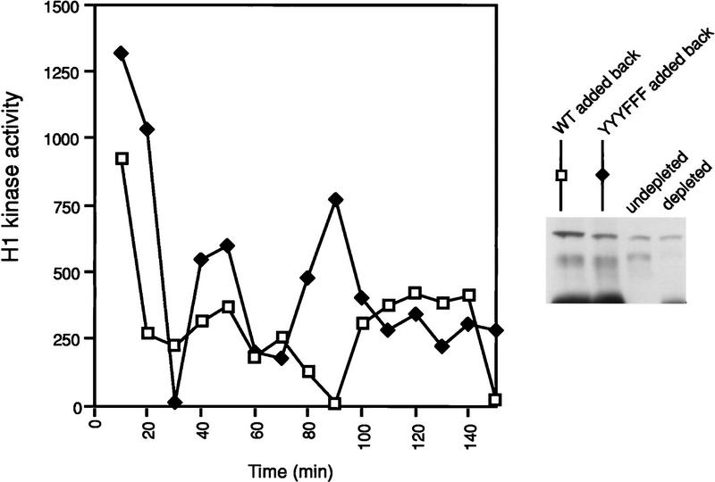
Several key developmental events occur in the first mitotic cell cycle of Xenopus; consequently this cycle has two gap phases and is approximately 60-75 min in length. In contrast, embryonic cycles 2-12 consist only of S and M phases and are 30 min in length. Xe-Wee1 and Mos are translated and degraded in a developmentally regulated manner. Significantly, both proteins are present in the first cell cycle. We showed previously that the expression of nondegradable Mos, during early interphase, delays the onset of M phase in the early embryonic cell cycles. Here we report that Xe-Wee1 is required for the Mos-mediated M-phase delay. We find that Xe-Wee1 tyrosine autophosphorylation positively regulates Xe-Wee1 and is only detected in the first 30 min of the first cell cycle. The level and duration of Xe-Wee1 tyrosine phosphorylation is elevated significantly when the first cell cycle is elongated with nondegradable Mos. Importantly, we show that the tyrosine phosphorylation of Xe-Wee1 is required for the Mos-mediated M-phase delay. These findings indicate that Mos positively regulates Xe-Wee1 to generate the G2 phase in the first cell cycle and establish a direct link between the MAPK signal transduction pathway and Wee1 in vertebrates.


Figures

Figure 1
Figure 1
G2 arrest mediated by Mos requires Xe-Wee1. (A) CSF extracts were prepared and supplemented with wild-type MBP-Mos (top). Xe-Wee1 was depleted from the CSF extract or the extract was mock depleted with preimmune sera (bottom). (B) The cell cycle was initiated by the addition of calcium, samples were taken at 10-min intervals and assayed for histone H1 kinase activity. Note that the endogenous Mos does not degrade efficiently in these extracts as compared to intact eggs (see Fig. 6B,F; Watanabe et al. 1991).
Figure 2
Figure 2
Xe-Wee1 has autophosphorylation activity in the first 30 min of the first mitotic cell cycle. The first mitotic cell cycle was initiated by treating unfertilized eggs with calcium ionophore (A23187). Ten eggs were collected at 10-min intervals and lysates were prepared in modified EB. Histone H1 kinase assays were used to monitor the progression through the cell cycle (A). Xe-Wee1 was immunoprecipitated (Ab 725:Ab 1532) from the remainder of each sample, and subjected to an immune complex autokinase assay with [γ-32P]ATP. The reactions were processed for SDS-PAGE, and the proteins were transferred to Immobilon membranes. After exposure to autoradiographic film (B), the filters were processed for immunoblotting with an anti-phosphotyrosine antibody (C).
Figure 3
Figure 3
Identification of Xe-Wee1 autophosphorylation sites. Wild type (WT) and kinase mutant (KM) Xe-Wee1 were translated in vitro and following immunoprecipitation, immune complex autokinase reactions were performed. After separation by SDS-PAGE (A, left), the wild-type 32P-labeled protein was eluted from the gel matrix, precipitated with TCA, and digested with either trypsin (which cleaves after Lys and Arg residues) or Lys-C (which cleaves after Lys residues). In the analysis of invitro-phosphorylated Xe-Wee1, both enzymes generated identical cleavage patterns, as the sites flanking the phosphopeptides were always lysine residues (B–D, right panels, sequences). The profile generated from the HPLC analysis of the wild-type Xe-Wee1 revealed three peaks of radioactivity (A, right). The peptides in fractions 15, 20, and 43, were subjected to phospho-amino acid analysis and two-dimensional thin-layer electrophoresis (B–D, left panels, insets), and Edman degradation (B–D, left panels). Each of the predicted tyrosine autophosphorylation sites was substituted with a phenylalanine residue, and the mutated versions of Xe-Wee1 were labeled with 32P in an autokinase assay and processed for HPLC analysis as described above; Y110F (B, right), Y103F (C, right), and Y90F (D, right). The amino acid sequence of the phosphopeptides is shown in the insets, arrows indicate LysC/trypsin cleavage sites.
Figure 4
Figure 4
Analysis of Xe-Wee1 phosphorylation in cell-free extracts. Interphase extracts were prepared as described in the Materials and Methods. [γ-32P]ATP (10 mCi) was added to 150–200 μl of these extracts, and endogenous Xe-Wee1 was isolated by immunoprecipitation (antibodies 725 and 1532) 30 min after the addition of calcium. The labeled proteins were isolated, digested with trypsin, and subjected to reverse-phase HPLC analysis (B; interphase Xe-Wee1). The HPLC profile for the wild-type Xe-Wee1 labeled in an in vitro autokinase assay (A) was generated as described in Fig. 3. The peptides in fractions 15, 20, and 41 were subjected to phospho-amino acid analysis. Tyrosine (as well as serine and threonine) phosphorylation was detected (4C). The peptide in fraction 15 from the Xe-Wee1 protein labeled in the interphase extracts (D, left) or from the Xe-Wee1 labeled in an in vitro autokinase assay (D, middle) was subjected to two-dimensional thin-layer electrophoresis and chromatography separately or as a mixture (D, right). In addition, the peptides from fraction 19 and 20 were also subjected to a two-dimensional thin-layer electrophoresis/chromatography analysis (E, left: extract; E, right: in vitro kinase). We were unable to perform a conclusive two-dimensional TLC analysis on the Y90 peptide. In our analysis, we compared the peptides isolated from the endogenous Xe-Wee1 labeled in an extract with those generated from our recombinant Xe-Wee1 labeled in an in vitro autophosphorylation reaction. Two sequences of Xe-Wee1 have been reported (Mueller et al. 1995a; Murakami and Vande Woude 1998). We believe that endogenous Xe-Wee1, in any given extract, is a mixture of these two isoforms. Importantly, the Xe-Wee1 sequence reported by the Dunphy laboratory (Mueller et al. 1995a) lacks the tryptic cleavage site just upstream of Y90 (which is present in our clone). Therefore, the tryptic digestion of the radiolabeled endogenous ‘Dunphy’ Xe-Wee1 would generate a peptide with a different mobility in both the HPLC analysis and two-dimensional TLC analysis, precluding comigration with the peptide generated from our recombinant Xe-Wee1.
Figure 5
Figure 5
Analysis of Xe-Wee1 mutant proteins. Tyrosine residues at position 90 (Y90F), 103 (Y103F), and 110 (Y110F), were substituted with phenylalanine residues singly or in combination (YYYFFF). The proteins were translated in vitro and immune complex autokinase assays were performed. Following separation by SDS-PAGE, the proteins were transferred to Immobilon membrane and exposed to autoradiographic film (A, top). The membranes were then processed for immunoblotting with anti-phosphotyrosine antibody 4G10 (A, middle) after which the membranes were stripped and re-probed for Xe-Wee1 (A, lower; antibody 725). The various forms of Xe-Wee1 were translated and immunoprecipitated as described above (B, top). The immune complexes were supplemented with recombinant cyclin B/cdc2 and [γ-32P]ATP. The reactions were subjected to SDS-PAGE and then transferred to Immobilon membrane and exposed to autoradiographic film (B; second panel). The filters were processed for Western analysis with antiphosphotyrosine antibody (B, third panel), then stripped and reprobed with anti-Xe-Wee1 antibody (B, first panel) and cdc2 (B, fourth panel). RNAs encoding the mutated versions of Xe-Wee1 were transcribed and capped in vitro. Approximately 40 ng of each RNA was injected into ∼75 stage-VI oocytes that were then left at 16°C overnight. Western analysis showed that equivalent amounts of Xe-Wee1 protein were present in the oocytes that had been injected with RNA (C, bottom). Progesterone was added at 10 μg/ml, and GVBD was scored by the appearance of a white spot in the animal hemisphere (C, top). CSF extracts were supplemented with exogenous Xe-Wee1 (D). CSF extracts were prepared and supplemented with wild-type or mutant Xe-Wee1. The cell cycle was initiated by the addition of calcium, aliquots were removed at 10-min intervals, and H1 kinase activity was assessed. The Western blot represents 1/13 of the exogenous Xe-Wee1 added to 60 μl of extract (right).
Figure 6
Figure 6
Analysis of Xe-Wee1 tyrosine phosphorylation in the first three mitotic cycles and in the first mitotic cycle with nondegradable Mos. Untreated eggs (A–D) and eggs that had been injected with wild-type nondegradable Mos (E–H) were activated with calcium ionophore and the progression through the cell cycle was monitored by histone H1 kinase activity (A,E). The levels of endogenous Mos and nondegradable Mos were detected by immunoblotting (B,F; one egg per lane). The level of tyrosine phosphorylated Cdc2 was determined following precipitation with p13/suc1 beads and immunoblotting with anti-phosphotyrosine antibody (C,G, top; 10 eggs per precipitate). The levels of Cdc2 in the p13 /suc1 precipitates are also shown (C,G, bottom). The levels of tyrosine-phosphorylated Xe-Wee1 were detected following immunoprecipitation with anti-Xe-Wee1 antibodies and immunoblotting with anti-phosphotyrosine antibody 4G10 (D,H; 10 eggs per precipitate).
Figure 7
Figure 7
CSF extracts were first depleted of endogenous Xe-Wee1 as described in Fig. 1. Recombinant wild-type and YYYFFF Xe-Wee1 were added back to the depleted extracts (legend). The extracts were also supplemented with one-tenth volume of MBP–Mos. The cell cycle was initiated by the addition of calcium and progression through the cell cycle was monitored by analysis of histone H1 activity.

Similar articles

Cited by

References

    1. Abrieu A, Lorca T, Labbe J-C, Morin N, Keyse S, Doree M. MAP kinase does not inactivate, but rather prevents the cyclin degradation pathway from being turned on in Xenopus egg extracts. J Cell Sci. 1996;109:239–246. - PubMed
    1. Abrieu A, Fisher D, Simon M-N, Doree M, Picard A. MAPK inactivation is required for the G2 to M-phase transition of the first mitotic cell cycle. EMBO J. 1997;16:6407–6413. - PMC - PubMed
    1. Aligue R, Wu L, Russell P. Regulation of Schizosaccharomyces pombe Wee1 tyrosine kinase. J Biol Chem. 1997;272:13320–13325. - PubMed
    1. Atherton-Fessler S, Liu F, Gabrielli B, Lee M S, Peng C-Y, Piwnica-Worms H. Cell cycle regulation of the p34cdc2 inhibitory kinases. Mol Biol Cell. 1994;5:989–1001. - PMC - PubMed
    1. Bitangcol JC, Chau AS-S, Stadnick E, Lohka MJ, Dicken B, Shibuya EK. Activation of the p42 mitogen-activated protein kinase pathway inhibits cdc2 activation and entry into M-phase in cycling Xenopus egg extracts. Mol Biol Cell. 1998;9:451–467. - PMC - PubMed

Publication types

LinkOut - more resources