. 1999 Jan;12(1):43-51.
doi: 10.1016/s0928-0197(98)00067-1.

Adaptation of human enteric coronavirus to growth in cell lines

Affiliations

Adaptation of human enteric coronavirus to growth in cell lines

J P Luby et al. J Clin Virol. 1999 Jan.

Abstract

Background: The existence of human enteric coronavirus (HEC) has been debated since its first description in stool by electron microscopy (EM) in 1975. Needed to resolve the issue is its cultivation in readily available cell lines.

Objectives: To grow HEC in cell lines. To describe its characteristics and to differentiate it from other human and animal coronaviruses.

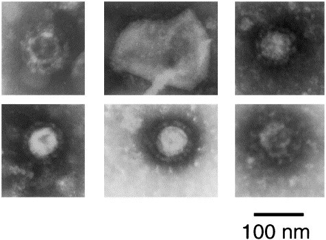
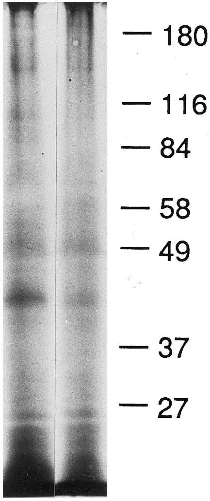
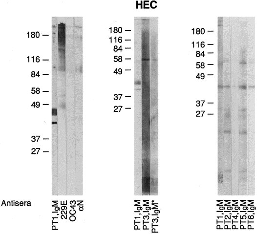
Study design: Originally grown in human fetal intestinal organ culture, HEC was passed in J774 cells (a mouse macrophage cell line) and C6/36 cells (a mosquito cell line). Its cytopathic effect (CPE) and pattern of immunofluorescence were described. Its appearance was ascertained by negative staining and transmission EM. Its structural proteins were delineated by polyacrylamide gel electrophoresis (PAGE) and Western blotting (WB). The antigenic character of the virus was determined by immunofluorescence and WB. Agglutination with mouse erythrocytes was performed.

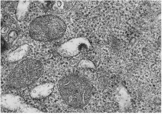
Results: In J774 cells, HEC induced the formation of giant cells and small syncytia. Immunofluorescence in both J774 and C6/36 cells was limited to the cytoplasm. Studies with transmission EM revealed the virus to have the typical appearance of other coronaviruses, to be 80-120 nm in diameter, and to bud into cysternae of the endoplasmic reticulum. By PAGE and WB, its major protein has an average molecular weight (MW) of 41 kilodaltons (kDa). Two other proteins had MWs of 190 and 24 kDa. By immunofluorescence and WB, HEC is antigenically distinct from human coronaviruses 0C43 and 229E and mouse hepatitis virus (A59 strain). Preparations of HEC did not agglutinate mouse erythrocytes.

Conclusion: We conclude that HEC is a human coronavirus that is antigenically unrelated to 0C43 and 229E viruses. Growth of HEC in readily available cell lines should aid in elucidating its role as a pathogen in human diarrheal illnesses.


Figures

Fig. 1
Fig. 1
Transmission electron microscopy of infected J774 cells. The picture is at a magnification of 112 500.
Fig. 2
Fig. 2
CVLPs from negatively stained resuspended pellets prepared from supernatants of infected cells. Row 1, frames 1 and 2: J774 cells. Row 1, frame 3, row 2, frame 1: C6/36 cells. Row 2, frames 2 and 3: pooled supernatants from J774 and C6/36 cells at 13 and 20 days after inoculation of virus seed stock.
Fig. 3
Fig. 3
PAGE of PEG precipitate centrifuged on a 20–60% sucrose gradient at 130 000 g for 18 h at 4°C and silver stained. The column on the right was from a band at mid-gradient; the column to the left was from a band just above mid-gradient.
Fig. 4
Fig. 4
Western blot of OC43 antigen and various antisera. Note that OC43 and A N antisera stain the major protein N at 49 kDa whereas Patient 1 (Ig and IgM) does not nor does antiserum to 229E.
Fig. 5
Fig. 5
Western blot of 229E antigen and various antisera. Note that 229E antiserum stains the major protein N at or around 49 kDa but this protein is not stained by antisera to OC43, A N or from Patient 1 (Ig).
Fig. 6
Fig. 6
Western blot of HEC preparations from J774 cells. Note patient reactions to the major protein centering at 41 kDa where antisera to 229E, OC43, and α N do not stain this protein. PT3, IgM* represents a serum specimen obtained from patient 3 at a later date.
Fig. 7
Fig. 7
Western blot of HEC purified from infected C6/36 cells and human immune IgM positive antisera. Sera from these patients (Ig) stained the cytoplasm of infected C6/36 cells but antisera to OC43 and 229E viruses did not.

Similar articles

Cited by

References

    1. Beards G.M, Green J, Hall C, Flewett T.H, Lamouliatte F, Du Pasquier P. An enveloped virus in stools of children and adults with gastroenteritis that resembles the Breda virus of calves. Lancet. 1984;1(8385):1050–1052. - PMC - PubMed
    1. Bencosme S.A, Tsutsumi V. A fast method for processing biologic material for electron microscopy. Lab Invest. 1970;23:447–450. - PubMed
    1. Caul E.O, Clarke S.K.R. Coronavirus propagated from patient with non-bacterial gastroenteritis. Lancet. 1975;2:953–954. - PMC - PubMed
    1. Caul E.O, Paver W.K, Clarke S.K.R. Coronavirus particles in faeces from patients with gastroenteritis. Lancet. 1975;1:1192. - PMC - PubMed
    1. Chaney C, Moscovici O, Lebon P, Rousset S. Association of coronavirus infection with neonatal necrotizing enterocolitis. Pediatrics. 1982;69:209–214. - PubMed

Publication types