Comparative Study
. 1999 Mar;181(6):1739-47.
doi: 10.1128/JB.181.6.1739-1747.1999.

Tyrosine aminotransferase catalyzes the final step of methionine recycling in Klebsiella pneumoniae

Affiliations
Comparative Study

Tyrosine aminotransferase catalyzes the final step of methionine recycling in Klebsiella pneumoniae

J Heilbronn et al. J Bacteriol. 1999 Mar.

Abstract

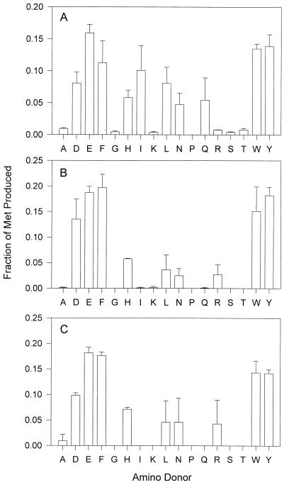
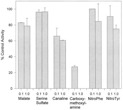
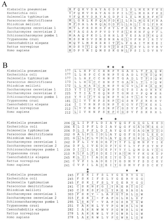
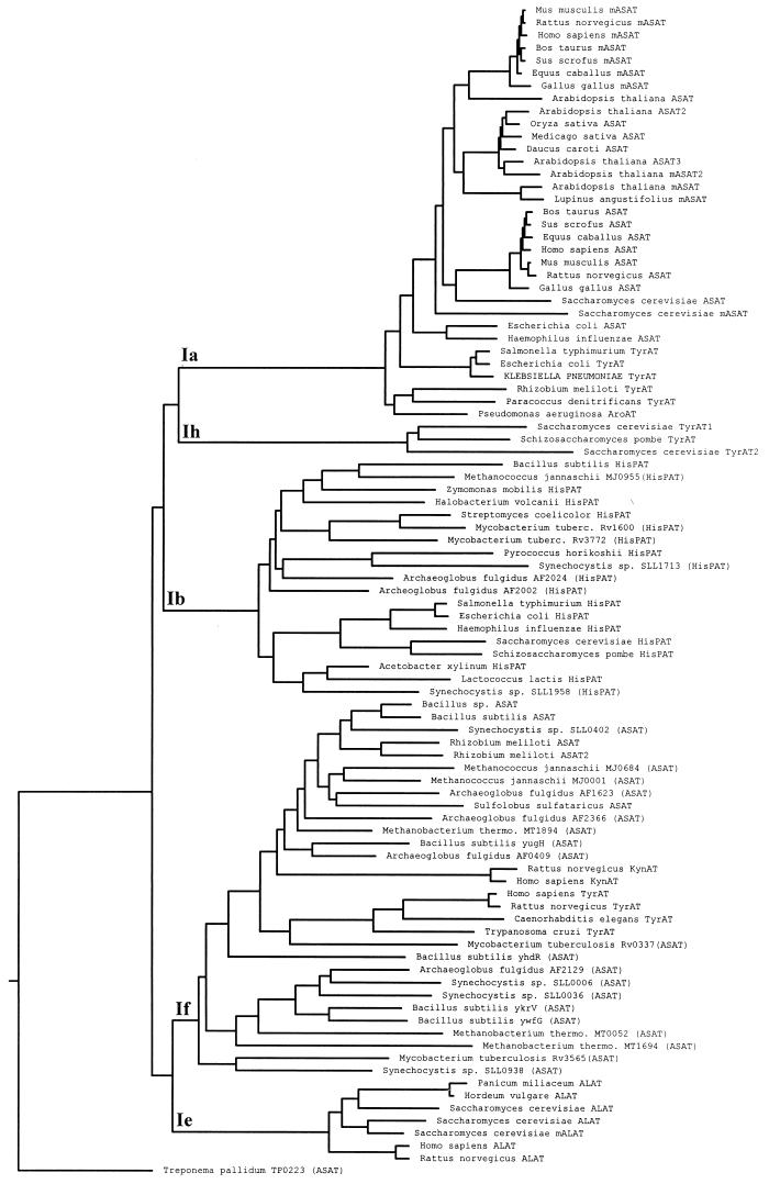
An aminotransferase which catalyzes the final step in methionine recycling from methylthioadenosine, the conversion of alpha-ketomethiobutyrate to methionine, has been purified from Klebsiella pneumoniae and characterized. The enzyme was found to be a homodimer of 45-kDa subunits, and it catalyzed methionine formation primarily using aromatic amino acids and glutamate as the amino donors. Histidine, leucine, asparagine, and arginine were also functional amino donors but to a lesser extent. The N-terminal amino acid sequence of the enzyme was determined and found to be almost identical to the N-terminal sequence of both the Escherichia coli and Salmonella typhimurium tyrosine aminotransferases (tyrB gene products). The structural gene for the tyrosine aminotransferase was cloned from K. pneumoniae and expressed in E. coli. The deduced amino acid sequence displayed 83, 80, 38, and 34% identity to the tyrosine aminotransferases from E. coli, S. typhimurium, Paracoccus denitrificans, and Rhizobium meliloti, respectively, but it showed less than 13% identity to any characterized eukaryotic tyrosine aminotransferase. Structural motifs around key invariant residues placed the K. pneumoniae enzyme within the Ia subfamily of aminotransferases. Kinetic analysis of the aminotransferase showed that reactions of an aromatic amino acid with alpha-ketomethiobutyrate and of glutamate with alpha-ketomethiobutyrate proceed as favorably as the well-known reactions of tyrosine with alpha-ketoglutarate and tyrosine with oxaloacetate normally associated with tyrosine aminotransferases. The aminotransferase was inhibited by the aminooxy compounds canaline and carboxymethoxylamine but not by substrate analogues, such as nitrotyrosine or nitrophenylalanine.


Figures

FIG. 1
FIG. 1
The Met regeneration pathway. The cycle functions to recycle Met (top of figure) for use as an aminopropyl donor in polyamine synthesis. The enzymes involved are as follows: 1, S-adenosylmethionine synthetase; 2, S-adenosylmethionine decarboxylase; 3, spermidine/spermine synthetase; 4, methylthioadenosine phosphorylase; 4a, methylthioadenosine nucleosidase; 4b, methylthioribose kinase; 5, undetermined isomerase; 6, undetermined dehydratase; 7a and 7b, bifunctional E-1 enolase-phosphatase; and 8a, E-2 dioxygenase. The reaction labelled 8 occurs nonenzymatically in K. pneumoniae and via the E-3 dioxygenase in rat liver (46). The reaction catalyzed by 4 occurs in eukaryotes, and those catalyzed by 4a and 4b occur in prokaryotes. The final step, labelled KMAT (KMTB to Met aminotransferase), is the subject of the present study.
FIG. 2
FIG. 2
The amino donor range for KMAT activity in K. pneumoniae. The enzyme source was incubated with 1 mM KMTB, 2 mM amino acid, and 50 μM pyridoxal phosphate for 30 min at 37°C before the Met produced was quantified by HPLC. Met production for each amino acid is shown as a relative percentage of activity for all amino acids. The enzyme sources were supernatants of cellular homogenates collected after centrifugation at 25,000 × g (A), purified aminotransferase from supernatants collected after centrifugation at 25,000 × g (B), and recombinant TyrAT (C).
FIG. 3
FIG. 3
The inhibition of Met production by the purified aminotransferase. The purified enzyme from K. pneumoniae was preincubated for 5 min with a single inhibitor at 0.1 mM (left column) or 1.0 mM (right column) before the addition of a reaction mixture containing 1 mM KMTB, certain amino acids (ADEFGHIKLNQRSTWY) at 2 mM each as amino donors, and 50 μM pyridoxal phosphate. After incubation at 37°C for 30 min, Met production was quantified by HPLC analysis. All inhibition is presented relative to a control value of 100%, which was determined by preincubating the enzyme with distilled water prior to adding the reagent mixture.
FIG. 4
FIG. 4
Alignment of the K. pneumoniae TyrAT with prokaryotic and eukaryotic analogues. In all cases, the boxed residues are identical to those found in the K. pneumoniae sequence. (A) A direct comparison of the N-terminal amino acid sequence obtained from the purified K. pneumoniae KMAT with known TyrATs. (B) Clustal alignment of the deduced amino acid sequence from the product of the K. pneumoniae tyrB gene with other known TyrATs. Regions around key residues which are conserved across the class I aminotransferase family are shown. In the top sequence, N-194, P-195, and G-197 have been marked with asterisks, in the middle sequence D-222 and Y-225 have been marked, and in the bottom sequence, K-258, R-266, and G-268 have been marked. K-258 (marked with a double asterisk) is the pyridoxal phosphate binding site. All conserved residues are numbered according to the nomenclature of Mehta et al. (31). Alignment was performed using the Megalign program of the DNAStar package (DNAStar, Madison, Wis.).
FIG. 5
FIG. 5
The class I family of aminotransferases. The dendrogram was constructed based on the clustal analysis of most aminotransferase members of the class I family, with nonaminotransferase members (subfamilies Ic and Id) and single member subfamilies (subfamily Ig) omitted. The K. pneumoniae TyrAT is shown capitalized, and subfamily groupings are presented according to the Iraqui et al. (22) revision of the nomenclature of Jensen and Gu (23). Enzyme abbreviations are as follows: ASAT, cytosolic aspartate aminotransferase; mASAT, mitochondrial aspartate aminotransferase; chASAT, chloroplast aspartate aminotransferase; TyrAT, tyrosine aminotransferase; ALAT, alanine aminotransferase; KynAT, kynurenine aminotransferase; and HisPAT, histidinol-phosphate aminotransferase. Putative aminotransferases identified from published whole genome sequences (, , , –16, 24, 25, 27, 39, 42) are shown with the appropriate identification code and with the annotation given in parentheses.

References

    1. Bacchi C J, Sufrin J R, Nathan H C, Spiess A J, Hannan T, Garafolo J, Alecia K, Katz L, Yarlett N. 5′-Alkyl-substituted analogs of 5′-methylthioadenosine as trypanocides. Antimicrob Agents Chemother. 1991;35:1315–1320. - PMC - PubMed
    1. Backlund P S, Chang C P, Smith R A. Identification of 2-keto-4-methylthiobutyrate as an intermediate compound in methionine synthesis from 5′-methylthioadenosine. J Biol Chem. 1982;257:4196–4202. - PubMed
    1. Backlund P S, Smith R A. Methionine synthesis from 5′-methylthioadenosine in rat liver. J Biol Chem. 1980;256:1533–1535. - PubMed
    1. Berger B J, Dai W W, Wang H, Stark R E, Cerami A. Aromatic amino acid transamination and methionine recycling in trypanosomatids. Proc Natl Acad Sci USA. 1996;93:4126–4130. - PMC - PubMed
    1. Berger B J, Dai W W, Wilson J. Methionine formation from α-ketomethiobutyrate in the trypanosomatid Crithidia fasciculata. FEMS Microbiol Lett. 1998;165:305–312. - PubMed

Publication types

MeSH terms

Associated data