. 1999 Mar;181(6):1801-10.
doi: 10.1128/JB.181.6.1801-1810.1999.

Sequence analysis of scaffolding protein CipC and ORFXp, a new cohesin-containing protein in Clostridium cellulolyticum: comparison of various cohesin domains and subcellular localization of ORFXp

Affiliations

Sequence analysis of scaffolding protein CipC and ORFXp, a new cohesin-containing protein in Clostridium cellulolyticum: comparison of various cohesin domains and subcellular localization of ORFXp

S Pagès et al. J Bacteriol. 1999 Mar.

Abstract

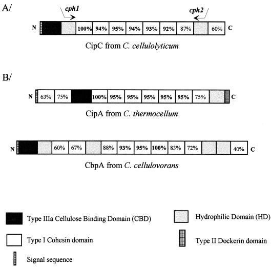
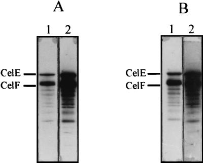
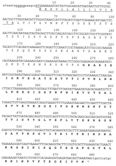
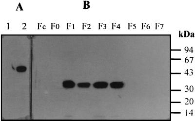
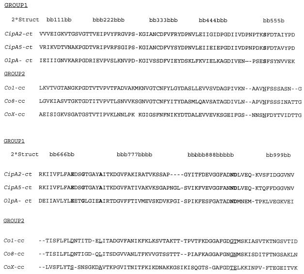
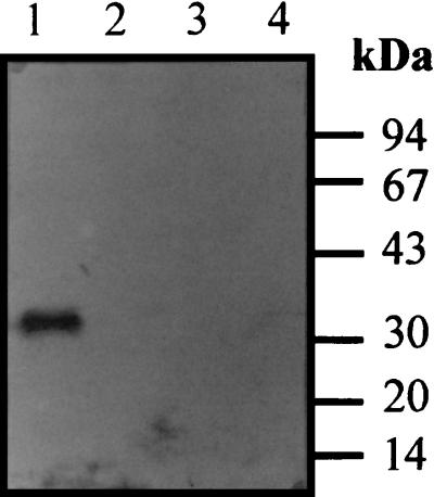
The gene encoding the scaffolding protein of the cellulosome from Clostridium cellulolyticum, whose partial sequence was published earlier (S. Pagès, A. Bélaïch, C. Tardif, C. Reverbel-Leroy, C. Gaudin, and J.-P. Bélaïch, J. Bacteriol. 178:2279-2286, 1996; C. Reverbel-Leroy, A. Bélaïch, A. Bernadac, C. Gaudin, J. P. Bélaïch, and C. Tardif, Microbiology 142:1013-1023, 1996), was completely sequenced. The corresponding protein, CipC, is composed of a cellulose binding domain at the N terminus followed by one hydrophilic domain (HD1), seven highly homologous cohesin domains (cohesin domains 1 to 7), a second hydrophilic domain, and a final cohesin domain (cohesin domain 8) which is only 57 to 60% identical to the seven other cohesin domains. In addition, a second gene located 8.89 kb downstream of cipC was found to encode a three-domain protein, called ORFXp, which includes a cohesin domain. By using antiserum raised against the latter, it was observed that ORFXp is associated with the membrane of C. cellulolyticum and is not detected in the cellulosome fraction. Western blot and BIAcore experiments indicate that cohesin domains 1 and 8 from CipC recognize the same dockerins and have similar affinity for CelA (Ka = 4.8 x 10(9) M-1) whereas the cohesin from ORFXp, although it is also able to bind all cellulosome components containing a dockerin, has a 19-fold lower Ka for CelA (2.6 x 10(8) M-1). Taken together, these data suggest that ORFXp may play a role in cellulosome assembly.


Figures

FIG. 1
FIG. 1
Schematic representation of CipC from C. cellulolyticum, CipA from C. thermocellum, and CbpA from C. cellulovorans. (A) The positions of the two oligonucleotides used for the amplification of the internal fragment of cipC are indicated by arrows. (B) The signal sequences, CBDs, hydrophilic domains, and cohesin domains are shown. The percent identity between cohesin domains is indicated in each box. Cohesin domains 1, 3, and 6 were used as the reference in CipC, CipA, and CbpA, respectively.
FIG. 2
FIG. 2
Phylogenetic tree of cohesin domains from CipC, CipA (C. thermocellum), and CbpA. The phylogenetic tree was constructed with the program AllAll accessible on the Computational Biochemestry Research Group server from E.T.H. Zurich, Switzerland. The length of each branch is proportional to the evolutionary distance between the nodes. The small black circle indicates the weighted centroid of the tree. The distances are expressed in pam (percent average mutation). For each scaffolding protein, the cohesin are numbered.
FIG. 3
FIG. 3
Alignment of CipC hydrophilic domains HD1 and HD2. The conserved amino acids are in gray boxes.
FIG. 4
FIG. 4
Diagram of the recombinant proteins used. Dark gray boxes indicate CBD, light gray boxes indicate hydrophilic domains, and white boxes represent cohesin domains. Small circles represent the His tag. The underlined residues are the first and last residues of the cohesin domain identified on the basis of the available crystal structures and sequence comparisons.
FIG. 5
FIG. 5
Recognition of SDS-PAGE-separated cellulosomal components by recombinant cohesin domains 1 and 8 of CipC. SDS-PAGE-separated samples were blotted onto nitrocellulose and probed with the biotinylated miniscaffolding protein desired. Blots were developed with streptavidin-peroxidase conjugate. Two different concentrations of cellulosomal preparation were used, 3 μg (lanes 1) and 6 μg (lanes 2). (A) SDS-PAGE-separated cellulosomal subunits probed with miniCipC1. (B) SDS-PAGE-separated cellulosomal subunits probed with miniCipC8. The presence of the two major cellulases of the C. cellulolyticum cellulosome, CelE and CelF, previously identified (17), are indicated.
FIG. 6
FIG. 6
Nucleotide and deduced amino acids sequences of the ORFX gene, encoding the predicted signal sequence (underlined), a putative linker domain, and a cohesin X domain (boldface type). The putative Shine-Dalgarno ribosome binding site is underlined upstream of the ATG codon.
FIG. 7
FIG. 7
Subcellular localization of ORFXp. (A) Two recombinant proteins, miniCipC1 (lane 1) and miniCipCX (lane 2), were separated by SDS-PAGE, transferred onto nitrocellulose, and probed with antibody raised against cohesin domain X. The blots were developed with anti immunoglobulin G-peroxidase conjugate. Incubation (lane 2) with miniCipCX indicates that antibodies (Ab α CX) specifically recognize cohesin domain X in the miniCipCX construction. (B) The different fractions obtained from the C. cellulolyticum cellulose growth culture (Fc to F7) were separated by SDS-PAGE and probed with antibody (Ab α CX) raised against cohesin domain X (see Materials and Methods for a discussion of fractionation of C. cellulolyticum cultures).
FIG. 8
FIG. 8
Sequence alignment of CipA cohesin domains 2 and 5 (CipA2-ct and CipA5-ct) and OlpA cohesin domain from C. thermocellum (OlpA-ct) (data from reference 4), and cohesin domains 1, 8, and X from C. cellulolyticum (Co1-cc, Co8-cc, and CoX-cc). For C. thermocellum, residues in boldface type are strictly conserved among cohesins of the same bacterium and expected to be involved in the interaction with the dockerin domain (group 1). In C. cellulolyticum, the five amino acids expected to be involved in the interaction with dockerin domain are underlined (group 2). The position of the β strands, based on the structure of cohesin domain 2 of CipA, are numbered and indicated by “b.”
FIG. 9
FIG. 9
Induction of ORFXp production by cellulose. A 500-μl volume of cellulose-grown culture (10 ml with 2 g of cellulose per liter) was used to encemence 10 ml of cellobiose medium (lane 1). This medium contains 0.1 g of residual cellulose per liter. A 500-μl volume of the first growth on cellobiose was used to encemence 10 ml of cellobiose medium (lane 2). This medium contains less than 0.005 g of residual cellulose per liter. Additional inocula were produced (lane 3 and 4) until the concentration of residual cellulose was so low that it could be neglected. In each case, cells were collected by centrifugation after 24 h. The pellet were resuspended in 100 μl of TBS buffer (50 mM Tris, 150 mM sodium chloride [pH 7.5]) containing 0.1% SDS and 100 mM mercaptoethanol. After being heated at 100°C for 15 min, the samples were centrifuged and 20 μl of the supernatant was subjected to SDS-PAGE. After transfer to nitrocellulose, the blots were incubated with antiserum raised against cohesin domain X.

References

    1. Bayer E, Lamed R. The cellulosome—a treasure-trove for biotechnology. Trends Biotechnol. 1994;12:379–386. - PubMed
    1. Bayer E A, Kenig R, Lamed R. Adherence of Clostridium thermocellum to cellulose. J Bacteriol. 1983;156:818–827. - PMC - PubMed
    1. Bayer E A, Lamed R. Ultrastructure of the cell surface cellulosome of Clostridium thermocellum and its interaction with cellulose. J Bacteriol. 1986;167:828–836. - PMC - PubMed
    1. Bayer E A, Morag E, Lamed R, Yaron S, Shoham Y. Cellulosome structure: four-pronged attack using biochemistry, molecular biology, crystallography and bioinformatics. In: Claeyssens M, Nerinckx W, Piens K, editors. Carbohydrates from Trichoderma reesei and other microorganisms. Structure, biochemistry, genetics and application. London, United Kingdom: The Royal Society of Chemistry; 1997. pp. 39–66.
    1. Bayer E A, Setter E, Lamed R. Organization and distribution of the cellulosome in Clostridium thermocellum. J Bacteriol. 1985;163:552–559. - PMC - PubMed

Publication types

MeSH terms

Associated data

LinkOut - more resources