. 1999 Apr;73(4):2569-75.
doi: 10.1128/JVI.73.4.2569-2575.1999.

Residues critical for duck hepatitis B virus neutralization are involved in host cell interaction

Affiliations

Residues critical for duck hepatitis B virus neutralization are involved in host cell interaction

C Sunyach et al. J Virol. 1999 Apr.

Abstract

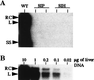
To date, no detailed analysis of the neutralization properties of duck hepatitis B virus (DHBV) has been reported, and it is not clear whether any of the known neutralization epitopes correspond to the viral receptor binding site or to sequences involved in the cell entry pathway. We demonstrate here that antibodies directed against two overlapping peptides (amino acids 83 to 97 and 93 to 107), covering the sequences of most DHBV pre-S neutralizing epitopes, both inhibit virus binding to primary duck hepatocytes and neutralize virus infectivity. An extensive mutagenesis of the motif 88WTP90, which is the shortest sequence of the epitope recognized by the virus-neutralizing monoclonal antibody (MAb) 900 was performed in order to define the amino acids involved in these interactions. Single point mutations within this epitope affected neither virus replication nor infectivity but abolished virus neutralization by MAb 900 completely. Interestingly, mutants with two and three consecutive residue replacements (SIP and SIH) within this epitope retained replication competence but were no longer infectious. The loss of infectivity of SIH and SIP mutant particles was associated with significantly reduced binding to primary duck hepatocytes and could be rescued by trans complementation with wild-type pre-S protein. Taken together, these results indicate that each amino acid of the DHBV pre-S sequence 88WTP90 is critical for recognition by the neutralizing MAb 900 and that replacement of the first two or all three residues strongly reduces virus interaction with hepatocytes and abrogates infectivity. These data imply that the motif 88WTP90 contains key residues which are critical for interaction with both the neutralizing MAb and the host cell.


Figures

FIG. 1
FIG. 1
Schematic representation of amino acid substitutions in the pre-S domain of the DHBV L envelope protein. The mutant names refer to the location and the nature of the amino acid changes within the 88WTP90 neutralization epitope recognized by MAb 900.
FIG. 2
FIG. 2
Binding of DHBV to PDHs is inhibited by antibodies to neutralization epitopes. PDH monolayers were incubated with concentrated DHBV-positive duck serum (DHBV) or uninfected duck serum (Mock). To test the capacity of DHBV pre-S antisera to inhibit binding, virus was preincubated with sera from a nonimmunized rabbit, DHBV pre-S polypeptide (aa 1 to 131) rabbit antiserum (DPS), or 83I-R97 (PS1), or/and 93D-R107 (PS2) peptide antisera. All antisera were diluted to similar anti-DHBV pre-S titers, as assessed by ELISA. Bound virus was detected with anti-S MAb 7C-12 and 125I-labeled goat anti-mouse IgG as described in Materials and Methods. The counts obtained for control DHBV are represented as 100%.
FIG. 3
FIG. 3
DHBV infectivity is neutralized in vitro by PS1 and PS2 peptide antisera. PDHs were infected with DHBV-positive duck serum (2 × 10⁸ VGE/well), preincubated with L15 medium (infection controls), unimmunized rabbit serum, DPS, or PS1 (83I-R97), PS2 (93D-R107), or PS1 and PS2 rabbit antisera. The release of viral particles into the supernatants was quantified by dot blot hybridization as described in Materials and Methods. The means of duplicate determinations are presented.
FIG. 4
FIG. 4
DHBV pre-S mutations do not affect virus replication capacity. pCMV-DHBV DNA plasmids containing each mutation presented in Fig. 1 were used to transfect the LMH hepatoma cell line. (A) Virus release into the transfected cell supernatants was monitored by dot blot hybridization as described in Materials and Methods. Lane 1, wild type; lane 2, W88L; lane 3, W88S; lane 4, T89I; lane 5, P90H; lane 6, P90T; lane 7, WIH; lane 8, SIP; lane 9, SIH. (B) Viral particles from the culture supernatants were concentrated and analyzed by immunoblotting. Proteins were probed with rabbit anti-DHBV pre-S antibody (DPS). Lane 1, wild type; lane 2, W88L; lane 3, W88S; lane 4, T89I; lane 5, P90H; lane 6, P90T; lane 7, WIH; lane 8, SIP; lane 9, SIH; lane 10, negative control. For comparison, circulating viral particles concentrated from DHBV-positive duck serum were loaded into lane 11.
FIG. 5
FIG. 5
SIP and SIH mutant viruses are not infectious in vivo. Viral particles produced by LMH cells transfected with pCMV-DHBV plasmids bearing the double (SIP) or triple (SIH) substitutions were used to infect ducklings. (A) Intrahepatic DHBV DNA replicative forms were analyzed by Southern blotting 3 weeks after inoculation in the liver from one duckling inoculated with wild-type DHBV (control) and two ducklings inoculated with either SIP or SIH mutant viruses. (B) Sensitivity of intrahepatic DHBV DNA detection in successive dilutions of the total liver DNA from a DHBV-positive liver. Arrows indicate the relaxed circular (RC), linear (L), and single-stranded (SS) DHBV DNA forms.
FIG. 6
FIG. 6
Mutations within the WTP epitope abolish in vitro neutralization of DHBV by MAb 900. PDH cultures were infected with wild-type DHBV or P90H mutant particles preincubated either with L15 culture medium (controls), with MAb 900 ascites fluid, or with DPS polyclonal antibody as described in Materials and Methods. Viral release into the culture medium was monitored from days 5 to 10 postinfection by dot blot hybridization.
FIG. 7
FIG. 7
Wild-type (WT) DHBV L protein trans-complements SIP and SIH mutant infectivity. LMH cells were transfected with pCMV-DHBV plasmids bearing the double (SIP) or triple (SIH) substitutions in the presence or absence of pCI–pre-S/S vector, which expresses wild-type L protein. PDH cultures were infected with viral particles produced by transfected LMH cells. (A) The viral release into cell supernatants was monitored from days 3 to 8 postinfection by dot blot hybridization. (B) Cells were harvested 8 days postinfection, and the L protein was detected in the cell lysate by immunoblotting with rabbit anti-DHBV pre-S antibody. For comparison, circulating viral particles concentrated from DHBV-positive duck serum were loaded in the right lane. The arrow shows the position of the 36-kDa DHBV L protein.
FIG. 8
FIG. 8
Binding of SIP and SIH mutant virus to PDH is reduced. PDH monolayers were incubated with wild type (WT), SIP, or SIH viral particles produced by transfected LMH cells as described in Materials and Methods. The results are expressed as the percentages of counts obtained with wild-type DHBV particles, after substraction of the values obtained with concentrated medium from mock-transfected LMH cells.

Similar articles

Cited by

References

    1. Baowei C, Przyla A E. An efficient site-directed mutagenesis method based on PCR. BioTechniques. 1994;17:657–659. - PubMed
    1. Borel C, Sunyach C, Hantz O, Trépo C, Kay A. Phosphorylation of DHBV pre-S: identification of the major site of phosphorylation and effects of mutations on the virus life cycle. Virology. 1998;242:90–98. - PubMed
    1. Breiner K, Urban S, Schaller H. Carboxypeptidase D (gp180), a Golgi-resident protein, functions in the attachement and entry of avian hepatitis B viruses. J Virol. 1998;72:8098–8104. - PMC - PubMed
    1. Chassot S, Lambert V, Kay A, Godinot C, Roux B, Trepo C, Cova L. Fine mapping of neutralization epitopes on duck hepatitis B virus (DHBV) pre-S protein using monoclonal antibodies and overlapping peptides. Virology. 1993;192:217–223. - PubMed
    1. Cheung R C, Robinson W, Marion P L, Greenberg H B. Epitope mapping of neutralization monoclonal antibodies against duck hepatitis B virus. J Virol. 1989;63:2445–2451. - PMC - PubMed

Publication types

LinkOut - more resources