. 1999 Apr;73(4):2641-9.
doi: 10.1128/JVI.73.4.2641-2649.1999.

The transmembrane domain of hepatitis C virus glycoprotein E1 is a signal for static retention in the endoplasmic reticulum

Affiliations

The transmembrane domain of hepatitis C virus glycoprotein E1 is a signal for static retention in the endoplasmic reticulum

L Cocquerel et al. J Virol. 1999 Apr.

Abstract

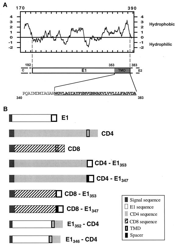
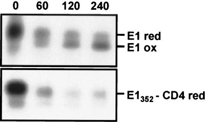
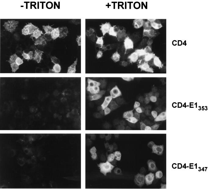
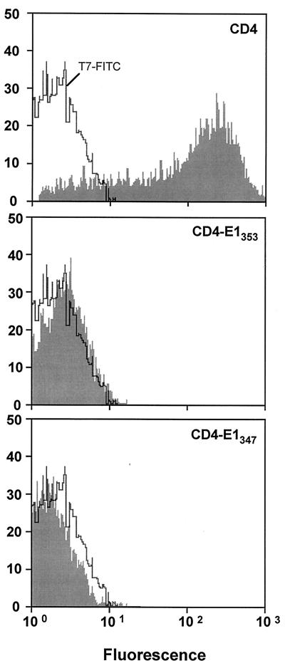
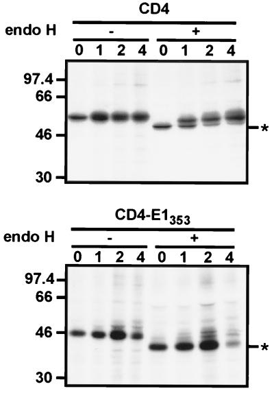
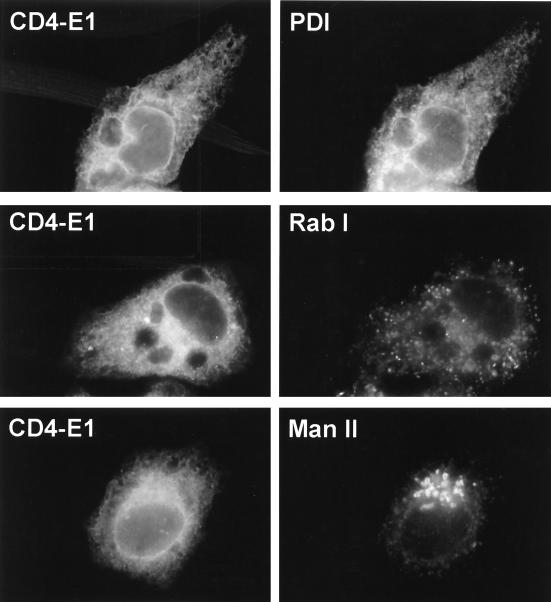
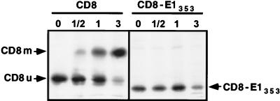
Hepatitis C virus (HCV) glycoproteins E1 and E2 assemble to form a noncovalent heterodimer which, in the cell, accumulates in the endoplasmic reticulum (ER). Contrary to what is observed for proteins with a KDEL or a KKXX ER-targeting signal, the ER localization of the HCV glycoprotein complex is due to a static retention in this compartment rather than to its retrieval from the cis-Golgi region. A static retention in the ER is also observed when E2 is expressed in the absence of E1 or for a chimeric protein containing the ectodomain of CD4 in fusion with the transmembrane domain (TMD) of E2. Although they do not exclude the presence of an intracellular localization signal in E1, these data do suggest that the TMD of E2 is an ER retention signal for HCV glycoprotein complex. In this study chimeric proteins containing the ectodomain of CD4 or CD8 fused to the C-terminal hydrophobic sequence of E1 were shown to be localized in the ER, indicating that the TMD of E1 is also a signal for ER localization. In addition, these chimeric proteins were not processed by Golgi enzymes, indicating that the TMD of E1 is responsible for true retention in the ER, without recycling through the Golgi apparatus. Together, these data suggest that at least two signals (TMDs of E1 and E2) are involved in ER retention of the HCV glycoprotein complex.


Figures

FIG. 1
FIG. 1
Schematic representation of the proteins used in this study. (A) Hydropathy plot (28) of HCV glycoprotein E1. The putative ectodomain and TMD of E1 are from amino acids 192 to 352 and amino acids 353 to 383 (position on the polyprotein), respectively. The amino acid sequence of the C-terminal region of E1 is shown, indicated by the single-letter amino acid code. The TMD of E1 is underlined. (B) Schematic representation of the parental (E1, CD4, and CD8) and chimeric proteins. CD4-E1353 and CD8-E1353, ectodomains of CD4 and CD8 fused to the TMD of E1; CD4-E1347 and CD8-E1347, same as CD4-E1353 and CD8-E1353 with an additional 6-amino-acid spacer from the ectodomain of E1 at the N terminus of the TMD of E1; E1352-CD4, ectodomain of E1 fused to the TMD and cytoplasmic domain of CD4; E1346-CD4, same as E1352-CD4 with a 6-amino-acid deletion at the C terminus of the ectodomain of E1. The ectodomain of CD4 is from amino acid 1 to 373, its TMD is from 374 to 395, and its cytosolic domain is from 396 to 435. The ectodomain of CD8 is from amino acid 1 to 160, its TMD is from 161 to 187, and its cytosolic domain is from 188 to 214. Details on the constructions are described in Materials and Methods.
FIG. 2
FIG. 2
Analysis of intramolecular disulfide bond formation in E1352-CD4. HepG2 cells were coinfected with vTF7-3 and a vaccinia virus recombinant expressing E1352-CD4 at a multiplicity of infection of 5 PFU/cell. Cells coinfected with vTF7-3 and a vaccinia virus recombinant expressing E1, E2, and p7 were used as a control. At 4.5 h postinfection, infected cells were pulse-labeled for 10 min and chased for the indicated times (in minutes). Cell lysates were immunoprecipitated with MAb A4. Immunoprecipitates were analyzed under nonreducing condition by SDS-PAGE (10% acrylamide). red, reduced; ox, oxidized.
FIG. 3
FIG. 3
Absence of cell surface expression of CD4-E1353 and CD4-E1347. HepG2 cells were coinfected with vTF7-3 and the appropriate vaccinia virus recombinant at a multiplicity of infection of 3 PFU/cell. At 8 h postinfection, cells were treated for indirect immunofluorescence light microscopy. Cells were fixed with paraformaldehyde, permeabilized or not with Triton X-100, and immunostained with anti-CD4 MAb OKT4 (secondary donkey anti-mouse IgG-Cy2).
FIG. 4
FIG. 4
Expression of chimeric proteins analyzed by flow cytometry. HeLa cells were coinfected with vTF7-3 and the appropriate vaccinia virus recombinant at a multiplicity of infection of 10 PFU/cell. At 8 h postinfection, cells were immunostained with FITC-conjugated anti-CD4 MAb 13B8.2. Stained cells were fixed in PBS–1% paraformaldehyde before flow-cytometric analysis. The level of cell surface expression is indicated by the shift of the solid histogram to the right from the open control histogram (T7-FITC, MAb 13B3.2 on cells infected with vTF7-3 alone).
FIG. 5
FIG. 5
Sensitivity of CD4-E1353 to endo H treatment. HepG2 cells were coinfected with vTF7-3 and the appropriate vaccinia virus recombinant at a multiplicity of infection of 5 PFU/cell. At 4.5 h postinfection, infected cells were pulse-labeled for 10 min and chased for the indicated times (in hours). Cell lysates were immunoprecipitated with MAb OKT4 and then treated or not with endo H. Samples were separated by SDS-PAGE (10% polyacrylamide). Deglycosylated proteins are indicated by asterisks. Sizes (in kilodaltons) of protein molecular-mass markers are indicated on the left.
FIG. 6
FIG. 6
Indirect double-label immunofluorescence characterization of the organelle containing CD4-E1353. Subconfluent HepG2 cells grown on coverslips were infected with vTF7-3 and vCD4-E1353 at a multiplicity of infection of 3 PFU/cell. At 8 h postinfection, cells were fixed with paraformaldehyde, permeabilized with Triton X-100, and labeled with anti-CD4 MAb OKT4 (secondary donkey anti-mouse IgG-Cy2) and antibodies to PDI, Rab I, or mannosidase II (Man II) (secondary donkey anti-rabbit IgG-rhodamine red-X).
FIG. 7
FIG. 7
Expression of CD8 and CD8-E1353 analyzed in pulse-chase experiments. HepG2 cells were coinfected with vTF7-3 and the appropriate vaccinia virus recombinant at a multiplicity of infection of 5 PFU/cell. At 4.5 h postinfection, infected cells were pulse-labeled for 10 min and chased for the indicated times (in hours). Cell lysates were immunoprecipitated with MAb OKT8 (anti-CD8). Samples were separated by SDS-PAGE (10% polyacrylamide). CD8u, unglycosylated precursor of CD8; CD8m, mature form of CD8.
FIG. 8
FIG. 8
Analysis of the oligosaccharides bound to CD4-E1353. HepG2 cells were coinfected with vTF7-3 and vCD4-E1353. At 4.5 h postinfection, infected cells were pulse-labeled for 30 min with [2-3H]mannose, chased for an additional 4 h, and lysed with Triton X-100. Cell lysates were used for immunoprecipitation with MAb OKT4, and labeled glycans were removed by PNGase F treatment as described in Materials and Methods. Panel A represents concanavalin A-Sepharose chromatography of glycan fractions obtained after PNGase F treatment with the equilibration buffer alone (a), with 10 mM methyl-α-d-glucoside (b), or with 100 mM methyl-α-d-mannoside (c). Panel B shows HPLC analysis of glycans bound to immunoprecipitated glycoproteins after PNGase F treatment. M7, M8, and M9 indicate the oligosaccharide species possessing two GlcNAc residues at their reducing end and 7, 8, or 9 mannose residues, respectively.

References

    1. Ahn K, Szczesna-Skorupa E, Kemper B. The amino-terminal 29 amino acids of cytochrome P450 2C1 are sufficient for retention in the endoplasmic reticulum. J Biol Chem. 1993;268:18726–18733. - PubMed
    1. Andersson A M, Melin L, Bean A, Pettersson R F. A retention signal necessary and sufficient for Golgi localization maps to the cytoplasmic tail of a Bunyaviridae (Uukuniemi virus) membrane glycoprotein. J Virol. 1997;71:4717–4727. - PMC - PubMed
    1. Armstrong J, Patel S. The Golgi sorting domain of coronavirus E1 protein. J Cell Sci. 1991;98:567–575. - PubMed
    1. Barlowe C, Orci L, Yeung T, Hosobuchi M, Hamamoto S, Salama N, Rexach M F, Ravazzola M, Amherdt M, Schekman R. COPII: a membrane coat formed by Sec proteins that drive vesicle budding from the endoplasmic reticulum. Cell. 1994;77:895–907. - PubMed
    1. Bause E, Breuer W, Schweden J, Roeser R, Geyer R. Effect of substrate structure on the activity of Man9-mannosidase from pig liver involved in N-linked oligosaccharide processing. Eur J Biochem. 1992;208:451–457. - PubMed

Publication types

Substances