. 1999 Apr;73(4):2650-7.
doi: 10.1128/JVI.73.4.2650-2657.1999.

Bacterial lipopolysaccharide inhibits dengue virus infection of primary human monocytes/macrophages by blockade of virus entry via a CD14-dependent mechanism

Affiliations

Bacterial lipopolysaccharide inhibits dengue virus infection of primary human monocytes/macrophages by blockade of virus entry via a CD14-dependent mechanism

Y C Chen et al. J Virol. 1999 Apr.

Abstract

Monocytes/macrophages (MO/Mphi) are the major target cells for both dengue virus (DV) and bacterial lipopolysaccharide (LPS), and the aim of this study was to define their interactions. We had found that LPS markedly suppressed DV infection of primary human MO/Mphi when it was added to cultures prior to or together with, but not after, viral adsorption. The inhibitory effect of LPS was direct and specific and was not mediated by LPS-induced secretion of cytokines and chemokines such as tumor necrosis factor alpha, interleukin-1beta (IL-1beta), IL-6, IL-8, IL-12, alpha interferon, MIP-1alpha, and RANTES. In fact, productive DV infection was not blocked but was just postponed by LPS, with a time lag equal to one viral replication cycle. Time course studies demonstrated that LPS was only effective in suppressing DV infection of MO/Mphi that had not been previously exposed to the virus. At various time points after viral adsorption, the level of unbound viruses that remained free in the culture supernatants of LPS-pretreated cultures was much higher than that of untreated controls. These observations suggest that the LPS-induced suppression of DV replication was at the level of virus attachment and/or entry. Blockade of the major LPS receptor, CD14, with monoclonal antibodies MY4 or MoS39 failed to inhibit DV infection but could totally abrogate the inhibitory effect of LPS. Moreover, human serum could significantly enhance the LPS-induced DV suppression in a CD14-dependent manner, indicating that the "binding" of LPS to CD14 was critical for the induction of virus inhibition. Taken together, our results suggest that LPS blocked DV entry into human MO/Mphi via its receptor CD14 and that a CD14-associated cell surface structure may be essential for the initiation of a DV infection.


Figures

FIG. 1
FIG. 1
Effect of LPS treatment on the production of infectious DV by young monocytes (A) and differentiated macrophages (B) cultured for 1 and 7 days, respectively. For each infection experiment, three comparison groups were included. (i) MO/Mφ were infected with DV without any stimulation (DV alone), (ii) LPS (5 or 10 μg/ml) was added to the cultures and incubated for 22 h before DV infection (LPS-DV), and (iii) LPS (5 μg/ml) was added to the cultures 6 h (A) or 12 h (B) after viral adsorption (DV-LPS). The cells were infected with DV at an MOI of 3 PFU per cell. After 42 to 46 h of infection, cells and culture supernatants were collected and assayed for both intracellular and extracellular infectious virus production. For each experimental point, triplicate wells were performed, and the results are given as the mean ± the standard error (SE).
FIG. 2
FIG. 2
Dose-response inhibition of DV infection of MO/Mφ by LPS. (A) Seven-day-old cells were left untreated or were treated with increasing concentrations of LPS (10 pg/ml to 25 μg/ml) for 24 h and then infected with DV at an MOI of 3 PFU per cell. (B) Six-day-old MO/Mφ were left untreated or were treated with 5 μg of LPS per ml for 22 h and then infected with DV at an MOI of 5, 0.5, 0.05, or 0.005 PFU per cell. After viral adsorption, cells were incubated with fresh medium without LPS. Culture supernatants were harvested after 44 to 46 h of infection and assayed to determine the infectious-virus titers. Each experimental point is presented as the mean ± the SE of results obtained from three separate wells.
FIG. 3
FIG. 3
Effects of MO/Mφ activators and stimulatory cytokines on DV infection of MO/Mφ. One-week-old MO/Mφ were left untreated or were treated with LPS (5 μg/ml), zymosan (50 μg/ml), PHA-CM (20% [vol/vol]), rhGM-CSF (20,000 U/ml), or rhTNF-α (20 ng/ml), either alone or in combination, for 20 to 24 h. The cells were then washed and infected with DV at an MOI of 3 PFU per cell. Culture supernatants were harvested after 42 to 46 h of infection and assayed for infectious-virus titers. Each experimental point is expressed as the mean ± the SE.
FIG. 4
FIG. 4
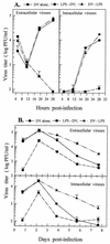
Kinetics of DV replication in primary human MO/Mφ. Infection experiments on 7-day-old (A) and 6-day-old (B) MO/Mφ cultures were performed with three comparison groups (DV alone, LPS-DV, and DV-LPS) as described in the legend to Fig. 1. For LPS-DV, LPS at a concentration of 6 μg/ml was incubated with the cells for 22 h prior to infection and for DV-LPS, LPS was added to the cultures immediately after viral adsorption. All cultures were infected with DV at an MOI of 3 PFU per cell and then incubated to follow the short-term (A) and long-term (B) kinetics of extracellular and intracellular infectious virus production. For each time point, three separate wells were prepared and analyzed, and the results are presented as the mean ± the SE.
FIG. 5
FIG. 5
Time course of the effectiveness of LPS treatment on reducing DV yields. (A) LPS (5 μg/ml) was added to the 7-day-old MO/Mφ cultures 15 or 1.5 h prior to or 1.5 h after viral adsorption, which was performed in 0.25 ml of serum-free α-MEM. (B) LPS was added to MO/Mφ cultures 20 h before, together with, or immediately after viral adsorption, which was performed in α-MEM supplemented with 3% ΔAHS. The inoculated MOI was 3 PFU per cell. After 44 h of infection, culture supernatants were harvested and assayed to determine the infectious-virus titers. Each experimental point is expressed as the mean ± the SE of results obtained from three independent wells. Control, no LPS treatment.
FIG. 6
FIG. 6
Blockade of LPS-induced suppression of DV infection by two anti-CD14 MAbs, MY4 and MoS39. One-week-old MO/Mφ were infected with DV at an MOI of 3 PFU per cell without any treatment (a) or were treated with LPS (5 μg/ml) for 20 h prior to DV infection (b). In addition, MO/Mφ were preincubated with 10 or 20 μg of MY4 per ml (c and d, respectively) or with 20 or 40 μg of MoS39 per ml (e and f, respectively) for 2 h before the addition of LPS (i.e., 22 h prior to DV infection). Some cultures were incubated with 10 μg of MY4 per ml for 2 h (g) or 20 μg of MoS39 per ml for 22 h (h) prior to DV infection, without a subsequent LPS treatment. In some cultures, MY4 was added after 2 h of viral adsorption (i). Each experimental point represents the mean ± the SE of results obtained from three separate wells.
FIG. 7
FIG. 7
Proposed model for DV entry into human MO/Mφ. The LPS receptor on the cell surface of human MO/Mφ is a heterodimeric complex containing a GPI-anchored CD14 and an unidentified transmembrane coreceptor, pRt. (A) In the absence of LPS, DV utilizes the pRt for infection. (B) In the presence of LPS, occupancy of the pRt by LPS blocks the access for DV entry. (C) Blockage of CD14 with anti-CD14 MAbs precludes the LPS from occupying the pRt and makes it accessible for DV. pRt, putative transmembrane receptor.

Similar articles

Cited by

References

    1. Anderson R, Wang S, Osiowy C, Issekutz A C. Activation of endothelial cells via antibody-enhanced dengue virus infection of peripheral blood monocytes. J Virol. 1997;71:4226–4232. - PMC - PubMed
    1. Bagasra O, Wright S D, Seshamma T, Oakes J W, Pomerantz R J. CD14 is involved in control of human immunodeficiency virus type 1 expression in latently infected cells by lipopolysaccharide. Proc Natl Acad Sci USA. 1992;89:6285–6289. - PMC - PubMed
    1. Bergelson J M, Cunningham J A, Droguett G, Kurt-Jones E A, Krithivas A, Hong J S, Horwitz M S, Crowell R L, Finberg R W. Isolation of a common receptor for coxsackie B viruses and adenoviruses 2 and 5. Science. 1997;275:1320–1323. - PubMed
    1. Bernstein M S, Tong-Starksen S E, Locksley R M. Activation of human monocyte-derived macrophages with lipopolysaccharide decreases human immunodeficiency virus replication in vitro at the level of gene expression. J Clin Invest. 1991;88:540–545. - PMC - PubMed
    1. Biswas P, Poli G, Kinter A L, Justement J S, Stanley S K, Maury W J, Bressler P J M, Orenstein J M, Fauci A S. Interferon γ modulates the expression of human immunodeficiency virus in persistently infected promonocytic cells by redirecting the production of virions to intracytoplasmic vacuoles. J Exp Med. 1992;176:739–750. - PMC - PubMed

Publication types

Substances