. 1999 Apr;73(4):2667-74.
doi: 10.1128/JVI.73.4.2667-2674.1999.

Vif and the p55(Gag) polyprotein of human immunodeficiency virus type 1 are present in colocalizing membrane-free cytoplasmic complexes

Affiliations

Vif and the p55(Gag) polyprotein of human immunodeficiency virus type 1 are present in colocalizing membrane-free cytoplasmic complexes

J H Simon et al. J Virol. 1999 Apr.

Abstract

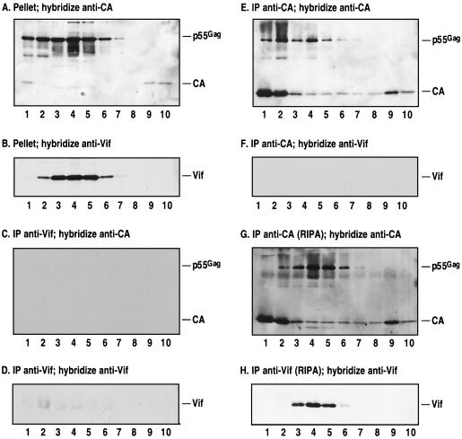
The Vif protein of human immunodeficiency virus type 1 (HIV-1) is a potent regulator of viral infectivity. Current data posit that Vif functions late in replication to modulate assembly, budding, and/or maturation. Consistent with this model, earlier indirect immunofluorescence analyses of HIV-1-infected cells demonstrated that Vif and Gag colocalize to a substantial degree (J. H. M. Simon, R. A. M. Fouchier, T. E. Southerling, C. B. Guerra, C. K. Grant, and M. H. Malim, J. Virol. 71:5259-5267, 1997). Here, we describe a series of subcellular fractionation studies which indicate that Vif and the p55(Gag) polyprotein are present in membrane-free cytoplasmic complexes that copurify in sucrose density gradients and are stable in nonionic detergents. Both Vif and Gag are targeted to these complexes independent of each other, and their association with them appears to be mediated by protein-protein interactions. We propose that these complexes may represent viral assembly intermediates and that Vif is appropriately localized to influence the final stages of the viral life cycle and, therefore, the infectivity of progeny virions.


Figures

FIG. 1
FIG. 1
Fractionation of HIV-1-infected T cells. HIV-1-infected cells were lysed in PBS–1% TX-100 and fractionated by differential centrifugation into nuclear (N), TX-100-soluble (S), and TX-100-insoluble (I) fractions. Aliquots of each fraction corresponding to equivalent numbers of cells were resolved on SDS-polyacrylamide gels, transferred to nitrocellulose, and analyzed by Western blotting using antibodies specific for hnRNP C1/C2, vimentin, α-tubulin, calreticulin, HIV-1 CA, or HIV-1 Vif.
FIG. 2
FIG. 2
Solubilization of Vif and Gag. HIV-1-infected H9 cells were lysed in PBS–1% TX-100, and the nuclei were pelleted by low-speed centrifugation. Various detergents were added to aliquots of the postnuclear supernatant to a final concentration of 1%, or salt was added to a final concentration of 1 M. These were then centrifuged at 100,000 × g to separate soluble (S) from insoluble (I) fractions. Aliquots of the fractions corresponding to equal numbers of cells were examined by Western blot analysis using antibodies specific for calreticulin, HIV-1 CA, or HIV-1 Vif.
FIG. 3
FIG. 3
Fractionation of cytoplasmic extracts of HIV-1-infected H9 cells by using sucrose density gradients. (A) Densities of fractions from a typical sucrose gradient, in this case the gradient analyzed in panel B. H9 cells transiently infected with HIV-1 (B) or HIV-1/Δvif (C) and uninfected H9/hVif cells (D) were lysed with PBS–1% TX-100. In addition, H9/hVif cells were also lysed in the absence of detergent by nitrogen cavitation (E). The postnuclear supernatant from each lysate was loaded onto a 20 to 60% (wt/vol) continuous sucrose gradient and centrifuged at 150,000 × g for 2 h. Ten fractions were harvested (1 = top; 10 = bottom), diluted and centrifuged to pellet the high-molecular-mass complexes. Equivalent amounts from each fraction were resolved on SDS-polyacrylamide gels and analyzed by Western blotting using antibodies specific for CA or Vif.
FIG. 4
FIG. 4
Coimmunoprecipitation analysis of Vif-Gag interactions, using density gradient-purified fractions of infected cell lysates. The postnuclear supernatant of HIV-1-infected H9 cells was fractionated on a sucrose density gradient, and equal portions of each fraction were either pelleted at 100,000 × g for 60 min (A and B) or incubated with protein A-conjugated agarose beads bound to rabbit polyclonal antibodies specific for HIV-1 Vif (C, D, and H) or HIV-1 CA (E, F, G, and H) for immunoprecipitation (IP). Aliquots of each sample equivalent to equal numbers of cells were resolved on SDS-polyacrylamide gels and examined by Western blot analysis using murine monoclonal antibodies specific for CA (A, C, E, and G) or Vif (B, D, F, and H).
FIG. 5
FIG. 5
Quantitation of Vif and Gag recovered by high-speed centrifugation and immunoprecipitation. The fraction 4 (Fig. 4) samples that had been either pelleted, or immunoprecipitated (IP) with anti-CA or preimmune (pre-imm.) serum were resolved on SDS-polyacrylamide gels together with dilution series of purified recombinant His6-tagged Vif and CA (which served as standard curves). The proteins were visualized by using monoclonal antibodies, chemiluminescence, and autoradiography, and the bands were quantitated by densitometry. Short (lanes 1 to 8) and long (lanes 9 and 10) exposures of the final filters are shown. Note that three times as much sample was loaded on this gel as for the gels shown in Fig. 4.

Similar articles

Cited by

References

    1. Bolognesi D P, Montelaro R C, Frank H, Schafer W. Assembly of type C oncornaviruses: a model. Science. 1978;199:183–186. - PubMed
    1. Borman A M, Quillent C, Charneau P, Dauguet C, Clavel F. Human immunodeficiency virus type 1 Vif− mutant particles from restrictive cells: role of Vif in correct particle assembly and infectivity. J Virol. 1995;69:2058–2067. - PMC - PubMed
    1. Bouyac M, Courcoul M, Bertoia G, Baudat Y, Gabuzda D, Blanc D, Chazal N, Boulanger P, Sire J, Vigne R, Spire B. Human immunodeficiency virus type 1 Vif protein binds to the pr55Gag precursor. J Virol. 1997;71:9358–9365. - PMC - PubMed
    1. Bouyac M, Rey F, Nascimbeni M, Courcoul M, Sire J, Blanc D, Clavel F, Vigne R, Spire B. Phenotypically Vif− human immunodeficiency virus type 1 is produced by chronically infected restrictive cells. J Virol. 1997;71:2473–2477. - PMC - PubMed
    1. Brown D A, Rose J K. Sorting of GPI-anchored proteins to glycolipid-enriched membrane subdomains during transport to the apical cell surface. Cell. 1992;68:533–544. - PubMed

Publication types

Substances

LinkOut - more resources