. 1999 Apr;73(4):2994-3003.
doi: 10.1128/JVI.73.4.2994-3003.1999.

HMG protein family members stimulate human immunodeficiency virus type 1 and avian sarcoma virus concerted DNA integration in vitro

Affiliations

HMG protein family members stimulate human immunodeficiency virus type 1 and avian sarcoma virus concerted DNA integration in vitro

P Hindmarsh et al. J Virol. 1999 Apr.

Abstract

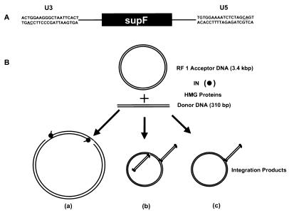
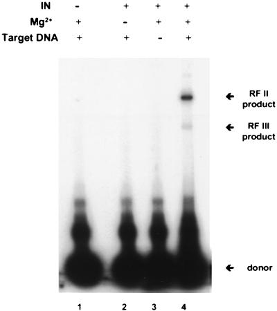
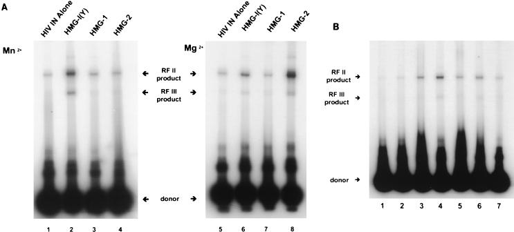
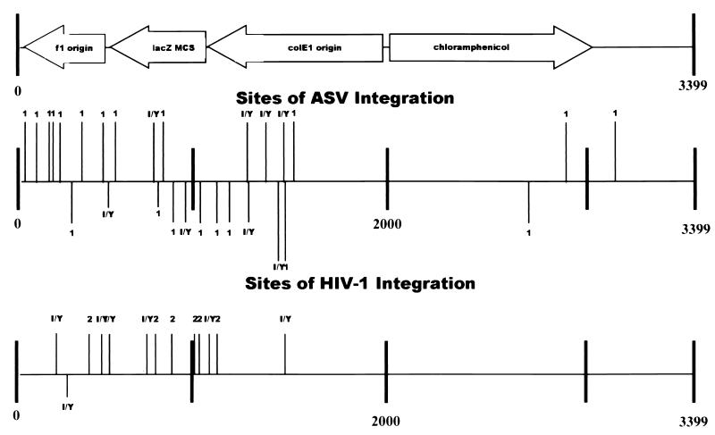
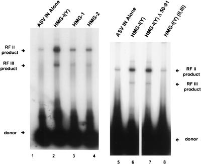
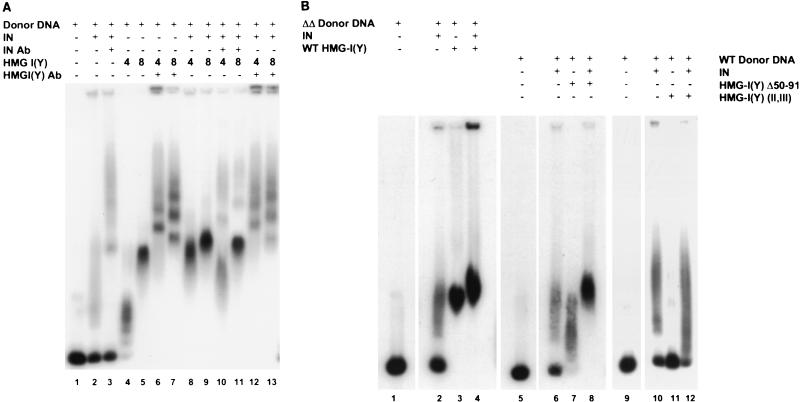
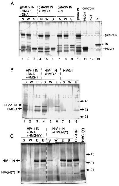
We have reconstituted concerted human immunodeficiency virus type 1 (HIV-1) integration in vitro with specially designed mini-donor HIV-1 DNA, a supercoiled plasmid acceptor, purified bacterium-derived HIV-1 integrase (IN), and host HMG protein family members. This system is comparable to one previously described for avian sarcoma virus (ASV) (A. Aiyar et al., J. Virol. 70:3571-3580, 1996) that was stimulated by the presence of HMG-1. Sequence analyses of individual HIV-1 integrants showed loss of 2 bp from the ends of the donor DNA and almost exclusive 5-bp duplications of the acceptor DNA at the site of integration. All of the integrants sequenced were inserted into different sites in the acceptor. These are the features associated with integration of viral DNA in vivo. We have used the ASV and HIV-1 reconstituted systems to compare the mechanism of concerted DNA integration and examine the role of different HMG proteins in the reaction. Of the three HMG proteins examined, HMG-1, HMG-2, and HMG-I(Y), the products formed in the presence of HMG-I(Y) for both systems most closely match those observed in vivo. Further analysis of HMG-I(Y) mutants demonstrates that the stimulation of integration requires an HMG-I(Y) domain involved in DNA binding. While complexes containing HMG-I(Y), ASV IN, and donor DNA can be detected in gel shift experiments, coprecipitation experiments failed to demonstrate stable interactions between HMG-I(Y) and ASV IN or between HMG-I(Y) and HIV-1 IN.


Figures

FIG. 1
FIG. 1
Reconstitution of integration in vitro. (A) Diagrammatic representation of a mini-HIV-1 donor DNA substrate of 310 bp in length. The 20 bp shown are from the U3 and U5 HIV-1 LTRs. The underlined nucleotides denote the highly conserved CA dinucleotide found near the ends of the LTR sequences. The solid box represents an expression cassette for the supF suppressor tRNA. (B) Diagrammatic representation of reconstituted integration with an acceptor DNA, purified IN, HMG proteins, and mini-donor DNA. Possible products include those that result from concerted donor integration (majority product) (a) and from nonconcerted integration by two (b) or one (c) donor DNA(s) via one-ended insertion events.
FIG. 2
FIG. 2
Requirements for reconstitution of HIV-1 IN-dependent integration in vitro. Integration reactions with wild-type HIV-1 donor DNA (0.3 pmol), acceptor DNA (0.02 pmol), and bacterially expressed HIV-1 IN (6.4 μmol) were incubated and analyzed as described in Materials and Methods. The arrows indicate the labeled donor, RF II, and RF III integration products. Lane 1, IN omitted; lane 2, Mg2+ omitted; lane 3, acceptor DNA omitted; lane 4, complete reaction with HMG-I(Y) present. Migration on the gel is from top to bottom.
FIG. 3
FIG. 3
(A) HMG protein family members stimulate reconstituted DNA integration catalyzed by HIV-1 IN. Reaction conditions were as described in Materials and Methods. Wild-type donor DNA was incubated in the presence of MnCl2 (lanes 1 to 4) or MgCl2 (lanes 5 to 8) with HMG protein family members as follows: lanes 1 and 4, no HMG protein; lanes 2 and 6, 20 and 4 pmol of HMG-I(Y), respectively; lanes 3 and 7, 20 pmol of HMG-1; lanes 4 and 8, 20 and 10 pmol of HMG-2, respectively. (B) Effect of varying the concentration of HMG-I(Y) on HIV-1 IN-dependent integration in vitro. HIV-1 IN-dependent integration reactions were reconstituted with MgCl2 in the presence of increasing concentrations of HMG-I(Y), and the products were analyzed by gel electrophoresis as described in Materials and Methods. HMG-I(Y) was added as follows: lane 1, 20 pmol; lane 2, 10 pmol; lane 3, 4 pmol; lane 4, 2 pmol; lane 5, 1 pmol; lane 6, 0.4 pmol. Lane 7, no added HMG-I(Y). All other notations are as in legend to Fig. 2.
FIG. 4
FIG. 4
Sites of concerted integration of donor ASV or HIV-1 DNA. The locations and lengths of flanking duplications of cell DNA for integrants using wild-type ASV or HIV-1 donor DNA as described in the tables are presented. Data for wild-type ASV from Aiyar et al. (1) are also included. The plasmid acceptor data is drawn in a linear representation to scale. The genes of the plasmid and origin of replication are indicated by the open arrows. MCS, multiple cloning site. The thick vertical lines represent numbers of nucleotide in the plasmid DNA. Each thin vertical line represents a separate sequenced integration event. The length of the thin vertical line above or below each bar represents either 4-, 5-, 6-, or 7-bp duplication of the acceptor DNA. The thin lines above the bar are in vitro duplications of acceptor DNA that match duplications observed in vivo, six for ASV and five for HIV-1. The thin lines below the bar represents base pair duplications of the acceptor DNA different from duplications observed in vivo. The presence of HMG-I(Y), HMG-1, or HMG-2 in the reaction is indicated for ASV or HIV-1 integrants above or below the thin vertical lines.
FIG. 5
FIG. 5
Reconstituted avian IN-dependent concerted DNA integration is stimulated by several HMG protein family members. Integration reactions with the wild-type ASV 32P-labeled donor DNA (0.3 pmol), acceptor DNA (0.02 pmol), and bacterially expressed IN (6 pmol) were incubated as described in Materials and Methods and analyzed by agarose gel electrophoresis. The arrows indicate the labeled donor, RF II, and RF III integration products. Different HMG protein family members or altered HMG proteins were added to the reaction as follows: lanes 1 and 5, no HMG protein added; lanes 2 and 6, wild-type HMG-I(Y) (4 pmol); lane 3, HMG-1 (8 pmol); lane 4, HMG-2 (8 pmol); lane 7, HMG-I(Y) (Δ50-91) derivative with an N-terminal truncation (4 pmol); lane 8, HMG-I(Y) (II, III) with amino acid substitutions in the A-T hook regions of the protein (4 pmol).
FIG. 6
FIG. 6
Complexes of donor DNA, ASV IN, and HMG-I(Y) detected by gel shift assays. (A) 32P-labeled donor DNA was incubated with ASV IN and/or HMG-I(Y) protein in the presence or absence of antibodies (Ab) to IN or HMG-I(Y) as described in Materials and Methods. The complexes formed were analyzed by agarose gel electrophoresis. Lane 1, labeled donor DNA alone; lane 2, donor DNA and 4 pmole ASV IN; lanes 3, donor DNA, 4 pmol ASV IN and anti-IN serum; lane 4, donor DNA and 4 pmol of wild-type HMG-I(Y); lane 5, donor DNA and 8 pmol of HMG-I(Y); lane 6, donor DNA, 4 pmol of HMG-I(Y), and anti-HMG-I(Y) serum; lane 7, donor DNA, 8 pmol of HMG-I(Y) and anti-HMG-I(Y); lane 8, donor DNA, 4 pmol each of ASV IN and HMG-I(Y); lane 9, donor DNA, 4 pmol of ASV IN, and 8 pmol of HMG-I(Y); lane 10, donor DNA, 4 pmol of ASV IN, 4 pmol HMG-I(Y), and anti-IN serum; lane 11, donor DNA, 4 pmol of ASV IN, 8 pmol of HMG-I(Y), and anti-IN serum; lane 12, donor DNA, 4 pmol of ASV IN, 4 pmol HMG-I(Y), and anti-HMG-I(Y) serum; lane 13, donor DNA, 4 pmol of ASV IN, 8 pmol HMG-I(Y), and anti-HMG-I(Y) serum. (B) Gel shifts using wild-type ASV donor DNA or donor DNA with random sequences at each end (ΔΔ), ASV IN, and HMG-I(Y) protein or mutants HMG-I(Y) (II, III) and HMG-I(Y) Δ50-91. Lanes 1 to 4 use the ΔΔ donor DNA. Lane 1, no IN or HMG protein; lane 2, 1 pmol of ASV IN; lane 3, 2 pmol of HMG-I(Y); lane 4, 1 pmol of ASV IN and 2 pmol of HMG-I(Y). Lanes 5 to 12 use the wild-type ASV donor DNA and HMG-I(Y) Δ50-91 where indicated. Lane 5, no IN or mutant HMG protein; lane 6, 1 pmol of ASV IN; lane 7, 2 pmol of HMG-I(Y) Δ50-91; lane 8, 1 pmol of ASV IN and 2 pmol of HMG-I(Y) Δ50-91. Lanes 9-12 use the wild-type ASV donor DNA and 1 pmol of HMG-I(Y) (II, III). Lane 9, no IN or mutant HMG protein; lane 10, 1 pmol of ASV IN; lane 11, 2 pmol of HMG-I(Y) (II, III); lane 12, 1 pmol of ASV IN and 2 pmol of HMG-I(Y) (II, III).
FIG. 7
FIG. 7
Analysis of complexes of HMG proteins and IN in the presence or absence of DNA. (A) GST-ASV IN was incubated with HMG-1 in the presence (lanes 1 to 3) or absence (lanes 4 to 6) of donor DNA as described in Materials and Methods. As a positive control for protein-protein interactions, GST-ASV IN was incubated with wild-type ASV IN and these complexes were treated in a identical fashion (lanes 7 to 9). Complexes were pelleted with glutathione-agarose, and supernatants (lanes S) were removed. Half of the pelleted complexes were washed (lanes W), and the other half were directly analyzed (lanes N) by SDS-PAGE followed by silver staining. Lanes 10 to 13 show starting amounts of each component loaded directly on the gel in separate lanes; arrows and dashed lines show the migration positions of each component. (B) His-tagged HIV-1 IN was incubated with HMG-1 in the presence (lanes 1 to 3) or absence (lanes 4 to 6) of donor DNA as described in Materials and Methods. Complexes were pelleted by interaction with Ni2+-charged chelating Sepharose, and the unbound supernatant fraction was removed (S). Pellets were washed, and this fraction (W) was removed and subjected to elution with buffer containing Imidazole and EDTA (E). Each of these three fractions (S, W, and E) were TCA precipitated and analyzed by SDS-PAGE followed by silver staining. Lanes 7 to 9 show the partitioning of HMG-1 alone with no His-tagged IN or DNA present and show a minor level nonspecific binding of HMG-1 to the Sepharose beads (lane 9). Positions of proteins are marked at the left, and positions of molecular weight markers are shown in kilodaltons on the right. (C) His-tagged HIV-1 IN was incubated with HMG-I(Y) in the presence (lanes 1 to 4) or absence (lanes 5 to 8) of donor DNA and analyzed as described for panel B. Any protein remaining with the beads following removal of the elution fraction was released by boiling in SDS sample buffer (fraction B) and also analyzed by SDS-PAGE. Lanes 9 and 10 show starting amounts of each protein loaded directly on the gel in separate lanes.

References

    1. Aiyar A, Hindmarsh P, Skalka A M, Leis J. Concerted integration of linear retroviral DNA by the avian sarcoma virus integrase in vitro: dependence on both long terminal repeat termini. J Virol. 1996;70:3571–3580. - PMC - PubMed
    1. Andrake M D, Skalka A M. Multimerization determinants reside in both the catalytic core and C terminus of avian sarcoma virus integrase. J Biol Chem. 1995;270:29299–29306. - PubMed
    1. Asante-Appiah E, Skalka A M. A metal-induced conformational change and activation of HIV-1 integrase. J Biol Chem. 1997;272:16196–16205. - PubMed
    1. Ausubel F M, Brent R, Kingston R E, Moore D D, Seidman J G, Smith J A, Struhl K, editors. Short protocols in molecular biology. 2nd ed. New York, N.Y: Greene Publishing Associates and John Wiley & Sons; 1992.
    1. Bustin M, Lehn D A, Landsman D. Structural features of the HMG chromosomal proteins and their genes. Biochim Biophys Acta. 1990;1049:231–243. - PubMed

Publication types

LinkOut - more resources