. 1999 Apr;73(4):3095-101.
doi: 10.1128/JVI.73.4.3095-3101.1999.

Yellow fever/Japanese encephalitis chimeric viruses: construction and biological properties

Affiliations

Yellow fever/Japanese encephalitis chimeric viruses: construction and biological properties

T J Chambers et al. J Virol. 1999 Apr.

Abstract

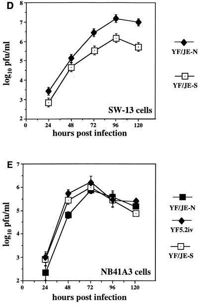
A system has been developed for generating chimeric yellow fever/Japanese encephalitis (YF/JE) viruses from cDNA templates encoding the structural proteins prM and E of JE virus within the backbone of a molecular clone of the YF17D strain. Chimeric viruses incorporating the proteins of two JE strains, SA14-14-2 (human vaccine strain) and JE Nakayama (JE-N [virulent mouse brain-passaged strain]), were studied in cell culture and laboratory mice. The JE envelope protein (E) retained antigenic and biological properties when expressed with its prM protein together with the YF capsid; however, viable chimeric viruses incorporating the entire JE structural region (C-prM-E) could not be obtained. YF/JE(prM-E) chimeric viruses grew efficiently in cells of vertebrate or mosquito origin compared to the parental viruses. The YF/JE SA14-14-2 virus was unable to kill young adult mice by intracerebral challenge, even at doses of 10(6) PFU. In contrast, the YF/JE-N virus was neurovirulent, but the phenotype resembled parental YF virus rather than JE-N. Ten predicted amino acid differences distinguish the JE E proteins of the two chimeric viruses, therefore implicating one or more residues as virus-specific determinants of mouse neurovirulence in this chimeric system. This study indicates the feasibility of expressing protective antigens of JE virus in the context of a live, attenuated flavivirus vaccine strain (YF17D) and also establishes a genetic system for investigating the molecular basis for neurovirulence determinants encoded within the JE E protein.


Figures

FIG. 1
FIG. 1
Structure of cDNA templates for chimeric YF/JE viruses (truncated within the NS1 protein for clarity). YF/JE-S and YF/JE-N refer to the two chimeric viruses as described in the text. The 5′ nontranslated region is derived from YF5.2iv (6). Hatched regions are from JE SA14-14-2, and solid regions are from JE-N. The remainder of the nonstructural region is derived from YF5.2iv. The amount of virus recovered is indicated as the titer of virus (log10 PFU per milliliter) in the media at the time of harvest at 96 h after RNA transfection of Vero cells.
FIG. 2
FIG. 2
Immunoprecipitation of viral proteins from chimeric YF/JE viruses. (A) Parental or chimeric viruses were grown in LLC-MK2 cells, labelled with [35S]methionine, and harvested from the media at 48 to 60 h postinfection. Viral E proteins were immunoprecipitated from the media as described in Materials and Methods, and proteins were analyzed on an SDS–9% polyacrylamide gel. The E protein of YF5.2iv virus was immunoprecipitated with rabbit polyclonal antiserum against YF. The E proteins of the JE-S (JE SA14-14-2), YF/JE-S, YF/JE-N, and JE-N viruses were immunoprecipitated with mouse hyperimmune ascitic fluid against JE virus. (B) Viral proteins produced in Vero cells. Proteins were labelled with [35S]methionine for 6 h, and lysates were prepared at 48 h postinfection as described in Materials and Methods. Virus in the media was harvested and immunoprecipitated with hyperimmune ascitic fluid to either YF or JE, as described for panel A. Mock-infected lysates were immunoprecipitated with a mixture of YF and JE hyperimmune ascitic fluids. Proteins were analyzed on 13% SDS–polyacrylamide gels. Tunicamycin was added (+) or not added (−) at the time of labelling at a concentration of 7.5 μg/μl. Molecular mass markers, indicated by small lines in the right margin, represent 220, 97.4, 66, 46, 30, and 14.3 kDa, respectively.
FIG. 3
FIG. 3
Immunoprecipitation of YF and JE proteins with monoclonal and polyclonal antisera. LLC-MK2 cells were infected and labelled as described for Fig. 2A. Viral proteins were immunoprecipitated from the media with monoclonal antibody to JE (A) or polyclonal antisera to the YF and JE viruses (B and C), as described in Materials and Methods. In panel A, YF, YF/JE-N, and YF/JE-S refer to media from cells infected with these respective viruses. In panel B, YF-infected cells were used. In panel C, YF/JE-N-infected cells were used. NI refers to nonimmune ascites fluid. YF-HI and JE-HI refer to hyperimmune ascites fluid to the YF and JE viruses, respectively. Proteins were analyzed on 10% SDS gels.
FIG. 4
FIG. 4
Growth curves of chimeric viruses in cell culture. Cells were infected at a multiplicity of 0.5 PFU/cell, and media were collected at 12- or 24-h intervals, followed by plaque titration on LLC-MK2 cells. (A) YF/JE-S compared to its parental viruses on LLC-MK2 cells. (B) YF/JE-N compared to its parental viruses on LLC-MK2 cells. The titers represent averages of triplicate samples for both experiments. (C) Chimeric virus growth on C6/36 cells, with values representing an average of two samples for each virus. (D) Growth of YF/JE-S and YF/JE-N viruses on SW-13 cells. (E) Growth of YF5.2iv, YF/JE-S, and YF/JE-N on NB41A3 cells. The titers represent averages of triplicate samples, except for the last time point, which was determined in duplicate.
FIG. 4
FIG. 4
Growth curves of chimeric viruses in cell culture. Cells were infected at a multiplicity of 0.5 PFU/cell, and media were collected at 12- or 24-h intervals, followed by plaque titration on LLC-MK2 cells. (A) YF/JE-S compared to its parental viruses on LLC-MK2 cells. (B) YF/JE-N compared to its parental viruses on LLC-MK2 cells. The titers represent averages of triplicate samples for both experiments. (C) Chimeric virus growth on C6/36 cells, with values representing an average of two samples for each virus. (D) Growth of YF/JE-S and YF/JE-N viruses on SW-13 cells. (E) Growth of YF5.2iv, YF/JE-S, and YF/JE-N on NB41A3 cells. The titers represent averages of triplicate samples, except for the last time point, which was determined in duplicate.
FIG. 5
FIG. 5
Mouse neurovirulence assay. A fixed-dose intracerebral challenge with 104 PFU was carried out in 4-week-old ICR mice. (A) YF/JE-S compared with its parental viruses. (B) YF/JE-N compared with its parental viruses. Differences in mortality between YF/JE-S and YF5.2iv were significant (P < .005), based on χ2 analysis of the proportion of survivors.

References

    1. Bray M, Men R, Lai C-J. Monkeys immunized with intertypic chimeric dengue viruses are protected against wild-type virus challenge. J Virol. 1996;70:4162–4166. - PMC - PubMed
    1. Calisher C H, Karabatsos N, Dalrymple J M, Shope R E, Porterfield J S, Westaway E G, Brandt W E. Antigenic relationships between flaviviruses as determined by cross-neutralization tests with polyclonal antisera. J Gen Virol. 1989;70:37–43. - PubMed
    1. Cecilia D, Gould E A. Nucleotide changes responsible for loss of neuroinvasiveness in Japanese encephalitis virus neutralization-resistant mutants. Virology. 1991;181:70–77. - PubMed
    1. Chambers T J, Hahn C S, Galler R, Rice C M. Flavivirus genome organization, expression and replication. Annu Rev Microbiol. 1990;44:649–688. - PubMed
    1. Chambers T J, McCourt D W, Rice C M. Production of yellow fever virus proteins in infected cells: identification of discrete polyprotein species and analysis of cleavage kinetics using region-specific polyclonal antisera. Virology. 1990;177:159–174. - PubMed

Publication types

LinkOut - more resources