. 1999 Apr;73(4):3125-33.
doi: 10.1128/JVI.73.4.3125-3133.1999.

Sendai virus and simian virus 5 block activation of interferon-responsive genes: importance for virus pathogenesis

Affiliations

Sendai virus and simian virus 5 block activation of interferon-responsive genes: importance for virus pathogenesis

L Didcock et al. J Virol. 1999 Apr.

Abstract

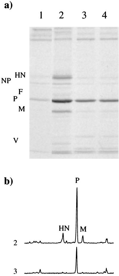
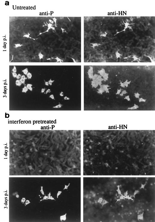
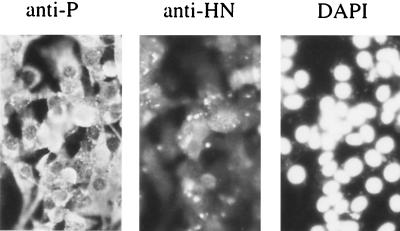
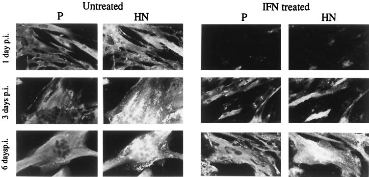
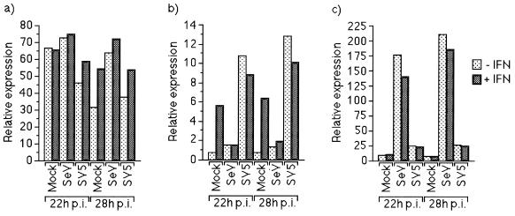
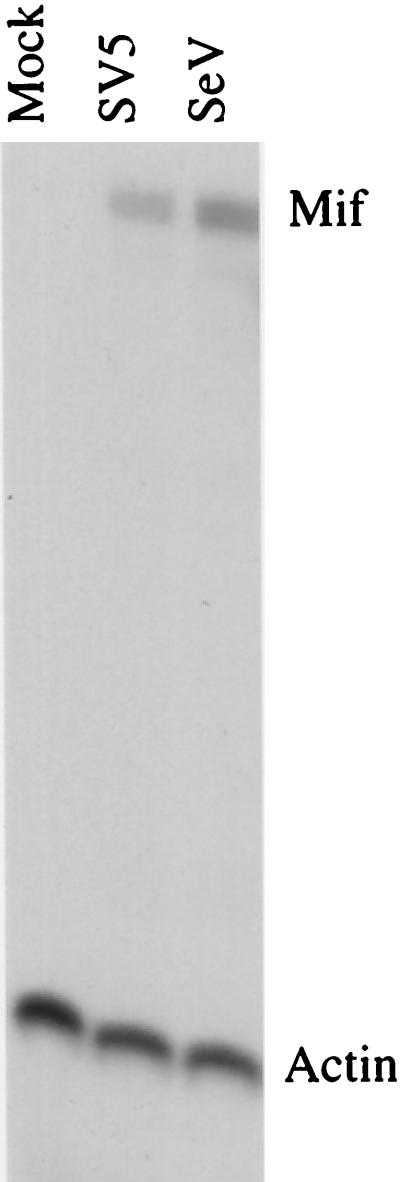
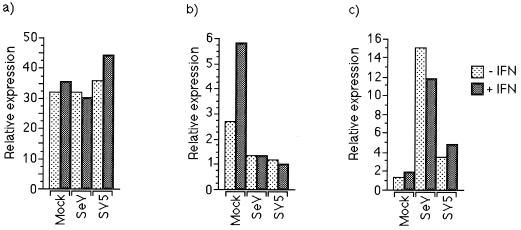
Sendai virus (SeV) is highly pathogenic for mice. In contrast, mice (including SCID mice) infected with simian virus 5 (SV5) showed no overt signs of disease. Evidence is presented that a major factor which prevented SV5 from productively infecting mice was its inability to circumvent the interferon (IFN) response in mice. Thus, in murine cells that produce and respond to IFN, SV5 protein synthesis was rapidly switched off. In marked contrast, once SeV protein synthesis began, it continued, even if the culture medium was supplemented with alpha/beta IFN (IFN-alpha/beta). However, in human cells, IFN-alpha/beta did not inhibit the replication of either SV5 or SeV once virus protein synthesis was established. To begin to address the molecular basis for these observations, the effects of SeV and SV5 infections on the activation of an IFN-alpha/beta-responsive promoter and on that of the IFN-beta promoter were examined in transient transfection experiments. The results demonstrated that (i) SeV, but not SV5, inhibited an IFN-alpha/beta-responsive promoter in murine cells; (ii) both SV5 and SeV inhibited the activation of an IFN-alpha/beta-responsive promoter in human cells; and (iii) in both human and murine cells, SeV was a strong inducer of the IFN-beta promoter, whereas SV5 was a poor inducer. The ability of SeV and SV5 to inhibit the activation of IFN-responsive genes in human cells was confirmed by RNase protection experiments. The importance of these results in terms of paramyxovirus pathogenesis is discussed.


Figures

FIG. 1
FIG. 1
Autoradiogram of a Western blot used to detected the P and V proteins of SV5 in extracts of BF cells infected with SV5 for 24 h (lane 1) or in lung extracts of mock-infected SCID mice (lane 2) or SCID mice that were infected with SV5 for 1, 4, 9, 13, or 21 days (lanes 3 to 7, respectively).
FIG. 2
FIG. 2
Analysis of 35S-labelled polypeptides present in immune precipitates (a) formed by the reaction of a pool of MAbs specific for the HN, NP, F, M, and P or V proteins of SV5 with soluble antigen extracts made from BF cells infected with SV5 for 1 day (lanes 1 and 2) or 3 days (lanes 3 and 4). The cells were pretreated with IFN-α/β (100 IU/ml) 24 h prior to infection (lane 1) or left untreated (lanes 2 to 4). At 24 h p.i., exogenous rHuIFN-alphaA/D (100 IU/ml) was added to the culture medium of cells used to make the extract shown in lane 4. The amount of 35S label in the precipitated polypeptides was quantitated by phosphoimage analysis, and the profiles of lanes 2 and 3 are shown in panel b.
FIG. 3
FIG. 3
Analysis of 35S-labelled polypeptides present in immune precipitates formed by the reaction of MAbs specific for the P (a), HN (b), and F (c) proteins of SeV with soluble antigen extracts made from BF cells infected with SeV for 1 day (lanes 1 and 2) or 3 days (lanes 3 and 4). The cells were pretreated with IFN 24 h (100 IU/ml) prior to infection (lane 1) or left untreated (lanes 2 to 4). At 24 h p.i., exogenous IFN (100 IU/ml) was added to the culture medium of cells used to make the extract shown in lane 4.
FIG. 4
FIG. 4
Photographs showing the localization of the P and HN proteins in monolayers of BF cells untreated (a) or treated with IFN (b) 24 h prior to infection with SeV. Monolayers were fixed at 1 and 3 days p.i. prior to staining with the appropriate MAbs.
FIG. 5
FIG. 5
Photographs showing the localization of the P and HN proteins of SeV in BF cells infected at a high MOI with SeV and passaged twice over a 2-week period. The cells were also stained with DAPI. As can be seen, all the cells remained infected with SeV.
FIG. 6
FIG. 6
Photograph showing the localization of the P and HN proteins of SV5 in human MRC-5 cells pretreated with IFN (100 IU/ml) 24 h prior to infection or left untreated. Cells were fixed at 1, 3, and 6 days p.i. prior to staining with the appropriate MAbs.
FIG. 7
FIG. 7
SeV, but not SV5, can block activation of the Type I IFN-responsive promoter in BF cells. BF cells were transfected with 0.3 and 0.1 μg of control plasmids, pUC13 and pJATlacZ, respectively, and 0.1 μg of one of the HSV TK promoter containing-plasmid (a), the IFN-α/β-responsive plasmid (b), or the IFN-β promoter-containing plasmid (c). At 16 h posttransfection, the cells were infected with SeV or SV5. Eighteen or twenty-four hours postinfection, the culture medium was supplemented with IFN or left untreated as indicated. Four hours later, luciferase and β-galactosidase activities in cellular lysates were measured. Luciferase activity, expressed in relative light units, was normalized to β-galactosidase activity.
FIG. 8
FIG. 8
SeV and SV5 induce IFN-β mRNA in murine BF cells. BF cells were infected with either SeV or SV5 for 24 h. Twenty micrograms of total cellular RNA from cells infected with SeV or SV5 was mapped with RNase protection probes corresponding to mouse IFN-β (Mif) or γ-actin mRNAs, and the protected fragments are indicated at the right.
FIG. 9
FIG. 9
SeV and SV5 block activation of the IFN-α/β-responsive promoter in 2fTGH cells. Cells were transfected with 0.3 and 0.1 μg of control plasmids pUC13 and pJATlacZ, respectively, and 0.1 μg of one of the HSV TK promoter-containing plasmid (a), the IFN-α/β-responsive plasmid (b), or the IFN-β promoter-containing plasmid (c). At 16 h postinfection, cells were infected with either SeV or SV5, and at 18 or 24 h p.i. the culture medium was supplemented with IFN or left untreated as indicated. Four hours later, luciferase and β-galactosidase activities in cellular lysates were measured. Luciferase activity, expressed in relative light units, was normalized to β-galactosidase activity.
FIG. 10
FIG. 10
SeV and SV5 block the induction of IFN-responsive gene mRNAs in MG-63 cells. MG-63 cells were infected with either SeV or SV5, and at 24 h p.i. the culture medium was supplemented with IFN for 4 h or left untreated. Twenty micrograms of total cellular RNA from cells was mapped with RNase protection probes corresponding to human IFN-β (5′IF), 6-16, IRF-1, or γ-actin mRNAs, and the protected fragments are indicated at the right.

Similar articles

Cited by

References

    1. Bosma G C, Custer R P, Bosma M J. A severe combined immunodeficiency mutation in mice. Nature. 1983;301:479–482. - PubMed
    1. Choppin P W. Multiplication of a myxovirus (SV5) with minimal cytopathic effects and without interference. Virology. 1964;23:224–233. - PubMed
    1. Darnell J E, Jr, Kerr I M, Stark G R. JAK-STAT pathways and transcriptional activation in response to interferon and other extracellular signalling proteins. Science. 1994;264:1415–1421. - PubMed
    1. de Wet J R, Wood K V, DeLuca M, Helinski D R, Subramani S. Firefly luciferase gene: structure and expression in mammalian cells. Mol Cell Biol. 1987;7:725–737. - PMC - PubMed
    1. Didcock, L., and R. E. Randall. Unpublished data.

Publication types

LinkOut - more resources