. 1999 Apr;73(4):3284-91.
doi: 10.1128/JVI.73.4.3284-3291.1999.

The U69 gene of human herpesvirus 6 encodes a protein kinase which can confer ganciclovir sensitivity to baculoviruses

Affiliations

The U69 gene of human herpesvirus 6 encodes a protein kinase which can confer ganciclovir sensitivity to baculoviruses

A Ansari et al. J Virol. 1999 Apr.

Abstract

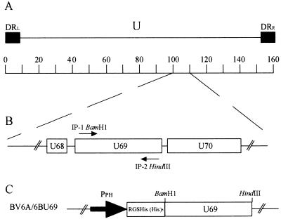
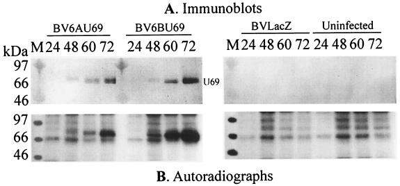
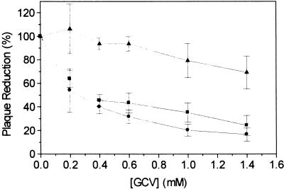
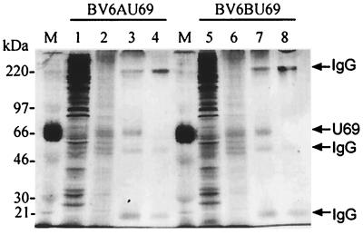
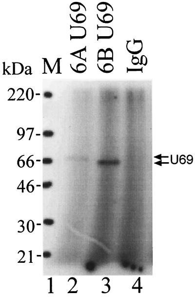
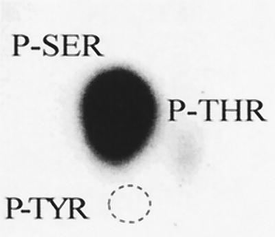
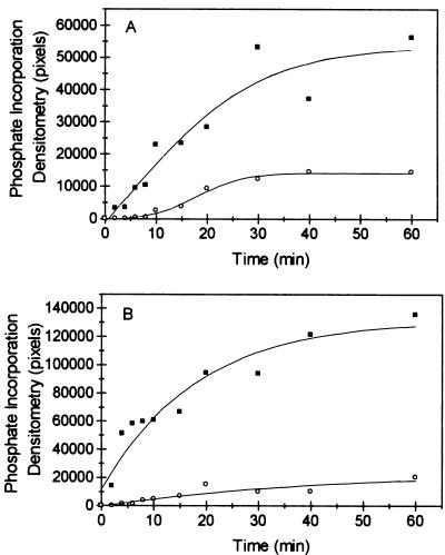
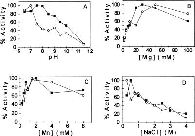
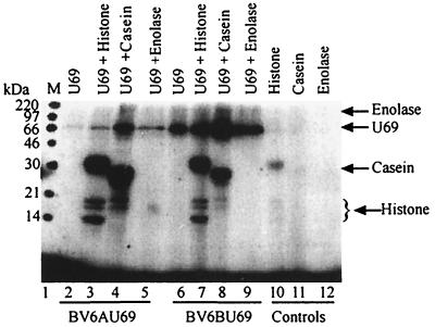
The protein encoded by the U69 open reading frame (ORF) of human herpesvirus 6 (HHV-6) has been predicted to be a protein kinase. To investigate its functional properties, we have expressed the U69 ORFs from both HHV-6 variants, A and B, by using recombinant baculoviruses (BV6AU69 and BV6BU69). Nickel agarose and antibody affinity chromatography was used to purify the proteins to homogeneity and when incubated with [gamma-32P]ATP, both U69 proteins became phosphorylated on predominantly serine residues. These data strongly suggest that U69 is a protein kinase which autophosphorylates. The phosphorylation reaction was optimal at physiological pH and low NaCl concentrations. It required the presence of Mg2+ or Mn2+, and Mg2+ was able to support phosphorylation over a wider range of concentrations than Mn2+. Both ATP and GTP could donate phosphate in the protein kinase assay and the former was more efficient. U69 was capable of phosphorylating histone and casein (serine/threonine kinase substrates) but not enolase (a tyrosine kinase substrate). For the autophosphorylation reaction, the Michaelis constants for ATP of baculovirus-expressed HHV-6A and HHV-6B U69 were calculated to be 44 and 11 microM, respectively. U69 is a homologue of the UL97 gene encoded by human cytomegalovirus which has been shown to phosphorylate the antiviral drug ganciclovir (GCV). We analyzed whether the U69 ORF alone was capable of conferring GCV sensitivity on baculoviruses BV6AU69 and BV6BU69. In plaque reduction experiments, these baculoviruses displayed a GCV-sensitive phenotype compared to a control baculovirus (BVLacZ). The 50% inhibitory concentrations (IC50) of BV6AU69 and BV6BU69 were calculated to be 0.35 and 0.26 mM, respectively, whereas the control baculovirus had an IC50 of >1.4 mM. This shows that the U69 gene product is the only one required to confer GCV sensitivity on baculovirus.


Figures

FIG. 1
FIG. 1
Cloning of the HHV-6A and HHV-6B U69 proteins. (A) Schematic organization of the HHV-6 genome with terminal direct repeats (■) and a unique region (U). The scale is marked in kilobase pairs. (B) PCR primers IP1-BamHI and IP2-HindIII generated HHV-6A and HHV-6B U69 amplicons which had restriction endonuclease recognition sites at either end to allow subsequent cloning of the amplicons into appropriately digested baculovirus transfer vector pBluBacHis. (C) Recombinant baculoviruses (BV6AU69 and BV6BU69) were constructed containing the HHV-6 U69 gene driven by the polyhedrin promoter (➞). The U69 protein was expressed as an N-terminal antibody epitope (RGSHis) and polyhistidine [(His)5] fusion.
FIG. 2
FIG. 2
Detection and phosphorylation of baculovirus-expressed U69. Insect cells were infected with recombinant baculovirus BV6AU69, BV6BU69, or BVLacZ and harvested at 24, 48, 60, and 72 h p.i. Protein extracts were prepared, incubated in a standard protein kinase assay (see Materials and Methods), and subjected to SDS-PAGE. The gels were then either analyzed by Western blotting (A) or exposed to autoradiography (B). The major phosphorylated species accumulated with polyhedrin promoter kinetics and comigrated with recombinant U69 as detected by Western blotting. The images shown here and in other figures are continuous two-tone images obtaining by scanning with a UMAX Astra 600S and Adobe Photoshop 4.0.
FIG. 3
FIG. 3
Baculovirus plaque reduction assays. Baculoviruses expressing the U69 ORFs of HHV-6A and HHV-6B (BV6AU69, ■; BV6BU69, ●) were individually cultured in increasing concentrations of GCV (0 to 1.4 mM), and the effect of the drug on virus replication was measured by plaque assay. The inhibitory effect of GCV was greater for the U69-expressing baculoviruses than for the control (BVlacZ, ▴). A slight reduction in plaque formation was observed for BvlacZ; however, this was at GCV concentrations near the cytotoxic concentration. The IC50s were calculated to be 0.35 and 0.26 mM for BV6AU69 and BV6BU69, respectively. The IC50 for the control virus could not be determined within this data set. The values shown are means of three independent experiments, each performed in duplicate.
FIG. 4
FIG. 4
Purification of BV6AU69- and BV6BU69-expressed U69. Aliquots of protein samples were taken from each step of the purification procedure and analyzed by SDS-PAGE followed by silver staining. Lanes 1 and 5 represent crude lysates of insect cells infected with BV6AU69 or BV6BU69, respectively. Eluates of the Ni2+-NTA affinity purification step (lanes 2 and 6) were then immobilized by immunoaffinity chromatography, and samples were loaded on lanes 3 and 7. Lanes 4 and 8 represent aliquots of immobilized IgG. The molecular sizes of the protein markers (lane M) are indicated on the left.
FIG. 5
FIG. 5
Phosphorylation of purified recombinant U69. Equal amounts of U69, purified from BV6AU69- or BV6BU69-infected insect cells, were incubated with [γ-32P]ATP in a protein kinase assay and subjected to SDS-PAGE (lanes 2 and 3, respectively). The gel was dried and exposed to film. The resulting autoradiograph is shown. Lane 4 is immobilized U69-specific IgG used for immunoprecipitation of BVLacZ-infected insect cells and subjected to a protein kinase assay as described above. The molecular sizes of the protein markers (lane M) are indicated on the left.
FIG. 6
FIG. 6
Phosphoamino acid analysis of recombinant U69. U69 was partially purified with Ni2+-NTA and phosphorylated in vitro in the presence of [γ-32P]ATP. The proteins were separated by SDS-PAGE and transferred electrophoretically onto a PVDF membrane, and the band corresponding to phosphorylated U69 was excised and acid hydrolyzed. The hydrolysate was mixed with unlabelled phosphoamino acids, phosphoserine (P-SER), phosphothreonine (P-THR), and phosphotyrosine (P-TYR) and subjected to two-dimensional electrophoretic thin-layer chromatography. The unlabelled phosphoamino acids were visualized with ninhydrin, and the autoradiograph of the thin-layer chromatography plate for the phosphoaminoacid analysis of HHV-6A U69 is shown. Phosphoamino acid analysis of HHV-6B U69 revealed a similar autoradiograph. The labelled phosphoamino acids comigrated with P-SER and P-THR, and the migration of P-TYR is indicated by the dotted circle.
FIG. 7
FIG. 7
Velocity of autophosphorylation of recombinant HHV-6A (A) and HHV-6B (B) U69 with respect to time. Protein kinase assays were performed for the times indicated by using either ATP (■) or GTP (○) as the radiolabelled phosphate donor. The reaction was terminated and subjected to SDS-PAGE. The amount of radiolabelled phosphate incorporation was measured by densitometry of the autoradiographs.
FIG. 8
FIG. 8
Biochemical characteristics of HHV-6A (■) and HHV-6B (○) U69. Baculovirus-expressed U69 was subjected to a standard protein kinase assay using radiolabelled ATP as the phosphate donor, except that the pH (A), divalent cation concentration (B and C), and NaCl concentration (D) were varied as indicated. In order to measure initial rates, 2-min reactions were performed. Autophosphorylation was measured for each condition by densitometry.
FIG. 9
FIG. 9
Phosphorylation of exogenous substrates. Purified U69 was coincubated in a protein kinase assay with exogenous proteins. The reaction was terminated, samples were subjected to SDS-PAGE, and the gel was exposed to film. The autoradiograph shows purified U69 from BV6AU69-infected insect cells incubated either alone (lane 2) or with histone casein or enolase (lanes 3 to 5) and purified U69 from BV6BU69-infected insect cells incubated either alone (lane 6) or with the exogenous substrates indicated (lanes 7 to 9). The positions and molecular sizes of marker proteins (lane M) are shown on the left side, and the migration of the exogenous substrates is shown on the right side. Lanes 10 to 12 are the exogenous substrates incubated in the absence of U69 in a standard protein kinase assay.

Similar articles

Cited by

References

    1. Ablashi D V, Salahuddin S Z, Josephs S F, Imam F, Lusso P, Gallo R C, Hung C, Lemp J, Markham P D. HBLV (or HHV-6) in human cell lines. Nature. 1987;329:207. - PubMed
    1. Agut H, Huraux J M, Collandre H, Montagnier L. Susceptibility of human herpesvirus 6 to acyclovir and ganciclovir. Lancet. 1989;ii:626. - PubMed
    1. Asano Y, Yoshikawa T, Suga S, Yazaki T, Kondo K, Yamanishi K. Fatal fulminant hepatitis in an infant with human herpesvirus-6 infection. Lancet. 1990;335:862–863. - PubMed
    1. Asano Y, Yoshikawa T, Kajita Y, Ogura R, Suga S, Yazaki T, Nakashima T, Yamada A, Kurata T. Fatal encephalitis/encephalopathy in primary human herpesvirus-6 infection. Arch Dis Child. 1992;67:1484–1485. - PMC - PubMed
    1. Baboonian C, Venables P J, Maini R N, Kangro H O, Osman H K. Antibodies to human herpesvirus-6 in Sjogren’s syndrome. Arthritis Rheum. 1990;33:1749–1750. - PubMed

Publication types

MeSH terms

LinkOut - more resources