. 1999 Apr;73(4):3359-65.
doi: 10.1128/JVI.73.4.3359-3365.1999.

Late domain function identified in the vesicular stomatitis virus M protein by use of rhabdovirus-retrovirus chimeras

Affiliations

Late domain function identified in the vesicular stomatitis virus M protein by use of rhabdovirus-retrovirus chimeras

R C Craven et al. J Virol. 1999 Apr.

Abstract

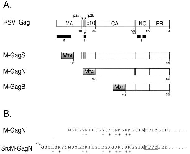
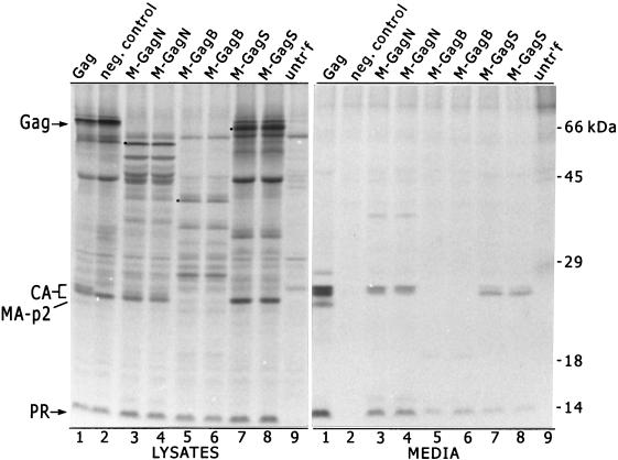
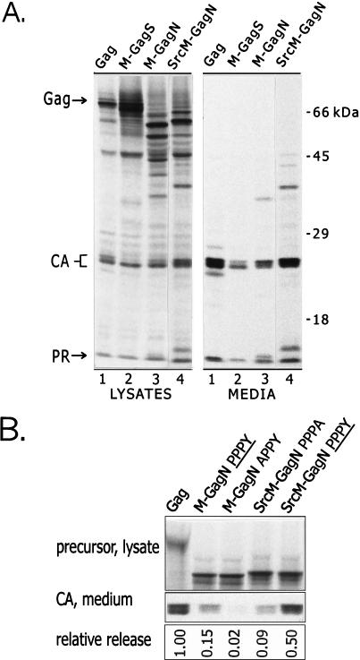
Little is known about the mechanisms used by enveloped viruses to separate themselves from the cell surface at the final step of budding. However, small sequences in the Gag proteins of several retroviruses (L domains) have been implicated in this process. A sequence has been identified in the M proteins of rhabdoviruses that closely resembles the PPPPY motif in the L domain of Rous sarcoma virus (RSV), an avian retrovirus. To evaluate whether the PPPY sequence in vesicular stomatitis virus (VSV) M protein has an activity analogous to that of the retroviral sequence, M-Gag chimeras were characterized. The N-terminal 74 amino acids of the VSV (Indiana) M protein, including the PPPY motif, was able to replace the L domain of RSV Gag and allow the assembly and release of virus-like particles. Alanine substitutions in the VSV PPPY motif severely compromised the budding activity of this hybrid protein but not that of another chimera which also contained the RSV PPPPY sequence. We conclude that this VSV sequence is functionally homologous to the RSV L domain in promoting virus particle release, making this the first example of such an activity in a virus other than a retrovirus. Both the RSV and VSV motifs have been shown to interact in vitro with certain cellular proteins that contain a WW interaction module, suggesting that the L domains are sites of interaction with unknown host machinery involved in virus release.


Figures

FIG. 1
FIG. 1
Structure of the Gag protein and M-Gag chimeras. (A) Structure of the wild-type Gag protein of RSV. The cleavage sites that are cut by the viral protease (PR) to generate the mature viral proteins are marked by the residue numbers. Black bars mark the positions of the three assembly domains in RSV Gag: M, membrane-binding domain; L, late domain; I, interaction domains. The PPPPY sequence that defines the core of the L domain lies within the p2b peptide (gray). The M-GagS, M-GagN, and M-GagN proteins are chimeras formed by the fusion of 74 amino acids from the N terminus of the VSV M protein (M74, shaded box) to different lengths of the Gag protein. The residue in Gag to which the M74 fragment is fused is indicated below each molecule. Foreign amino acids were inserted at the fusion junction in M-GagS (A-L-V) and in M-GagN (A-P-R-Q) but not in M-GagS. (B) Amino-terminal sequence of the M-GagN chimera and its Src derivative. The terminal sequence of M-GagN is identical to that of the VSV M protein. Eight lysine residues which have been implicated in membrane binding are indicated by plus signs. The PPPY motif is boxed. In the SrcM-GagN molecule, the amino-terminal sequence of the M protein is preserved. The addition of a sequence containing the first 10 codons of v-src to the chimeric M-gag gene in pSV.M-GagN is expected to yield the protein shown. The N-terminal methionine is predicted to be removed cotranslationally as myristic acid (indicated by the zigzag) is linked to the subterminal glycine in the Src peptide.
FIG. 2
FIG. 2
Release of particles from cells expressing M-Gag chimeras. Wild-type Gag protein and the three M-Gag chimeras were expressed in COS-1 cells (31). Two independent clones were tested for each of the chimeras. Gag-related proteins present in the cell lysates and in the medium of expressing cells were analyzed by radioimmunoprecipitation, using an anti-RSV serum which recognizes primarily the Gag protein and its cleavage products. The labelled proteins were resolved by sodium dodecyl sulfate-polyacrylamide gel electrophoresis on a 12% polyacrylamide gel. The positions of full-length Gag protein and the CA, MA, and PR proteins in cell lysates are marked on the left. The full-length M-Gag chimeras are marked by dots. A mutant Gag protein which has a defective membrane-binding domain serves as the negative control for budding (lanes 2). Samples from untransfected cells were loaded in lanes 9.
FIG. 3
FIG. 3
Enhancement of the budding activity of the M-GagN chimera by the addition of the Src membrane-binding signal. (A) Particle assembly and release by the SrcM-GagN protein were compared to those of the wild-type Gag protein and the M-GagS and M-GagN chimeras. Anti-RSV serum was used for immunoprecipitation. (B) The relative levels of particle release by the various proteins were compared. Expression and labelling were carried out as described in the legend for Fig. 2, except that an anti-CA serum was used for immunoprecipitation. The CA protein that accumulated in the medium during a 2.5-h labelling period (bottom panel) was measured by PhosphorImager analysis of the dried gel. These counts were normalized for the amount of Gag or M-Gag precursor present in lysates of parallel plates labelled for only 5 min (top panel) and for the number of methionine residues in the precursors. The release of each chimera was then expressed relative to that of the wild-type Gag protein. The gel shown represents one typical experiment. The chimeras which contain a wild-type VSV L domain are indicated by PPPY in the lane heading; the other chimeras carry the indicated mutant sequences. In three independent experiments, the average relative release (± standard error) was as follows: Gag, 1.00; M-GagN PPPY, 0.157 ± 0.052; M-GagN APPY, 0.014 ± 0.004; SrcM-GagN PPPA, 0.057 ± 0.016; SrcM-GagN PPPY, 0.348 ± 0.075.
FIG. 4
FIG. 4
Alterations to the PPPY motif of M-GagN cause a drastic reduction in budding activity. Particle release by the M-GagN chimera bearing the wild-type PPPY motif (distinguished by PPPY in the lane heading) was compared to those of mutants in which the peptide was altered by an alanine substitution at either the tyrosine (PPPA; two independent clones shown) or the first proline (APPY; two clones). Similar mutations in M74 were also evaluated in the context of the M-GagS protein. The negative control for budding is a mutant Gag protein that contains a large deletion spanning the p2b sequence (lanes 2). Samples from untransfected cells were loaded in lanes 1.

References

    1. Bennett R P, Nelle T D, Wills J W. Functional chimeras of the Rous sarcoma virus and human immunodeficiency virus Gag proteins. J Virol. 1993;67:6487–6498. - PMC - PubMed
    1. Chen H I, Einbond A, Kwak S-J, Linn H, Koepf E, Peterson S, Kelly J W, Sudol M. Characterization of the WW domain of human Yes-associated protein and its polyproline-containing ligands. J Biol Chem. 1997;272:17070–17077. - PubMed
    1. Chen H I, Sudol M. The WW domain of Yes-associated protein binds a proline-rich ligand that differs from the consensus established for Src homology 3-binding modules. Proc Natl Acad Sci USA. 1995;92:7819–7823. - PMC - PubMed
    1. Chong L D, Rose J K. Interactions of normal and mutant vesicular stomatitis virus matrix proteins with the plasma membrane and nucleocapsids. J Virol. 1994;68:441–447. - PMC - PubMed
    1. Craven R C, Bennett R P, Wills J W. Role of the avian retroviral protease in the activation of reverse transcriptase during virion assembly. J Virol. 1991;65:6205–6217. - PMC - PubMed

Publication types

Substances

LinkOut - more resources