. 1999 Apr;73(4):3386-403.
doi: 10.1128/JVI.73.4.3386-3403.1999.

Mutagenic analysis of the 3' cis-acting elements of the rubella virus genome

Affiliations

Mutagenic analysis of the 3' cis-acting elements of the rubella virus genome

M H Chen et al. J Virol. 1999 Apr.

Abstract

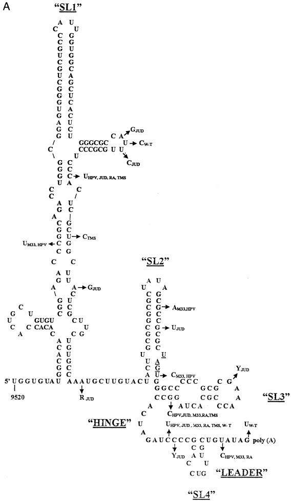
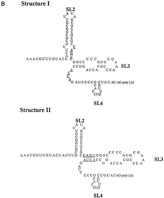
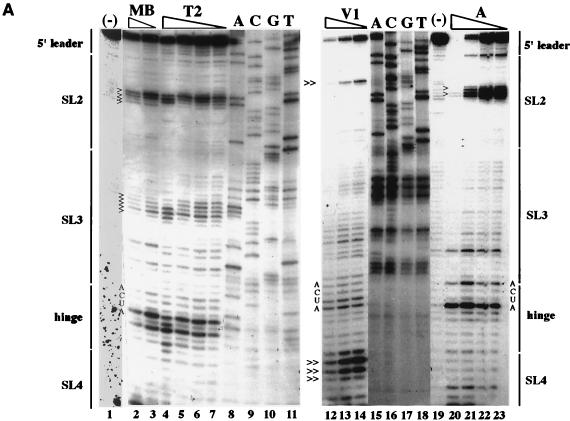
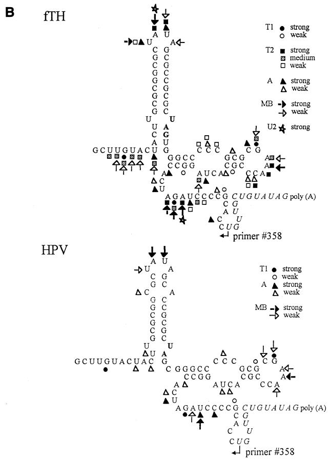
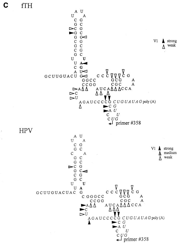
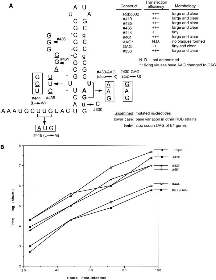
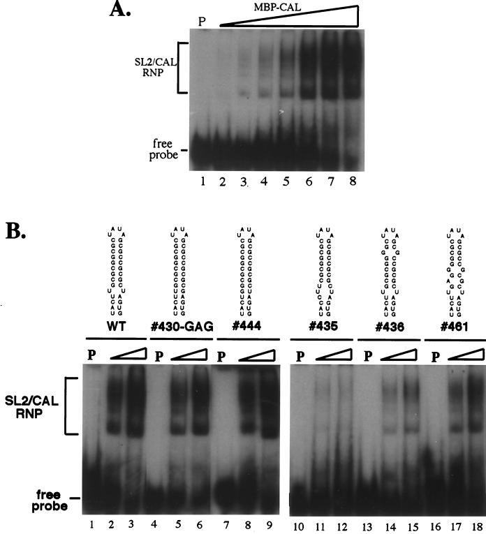
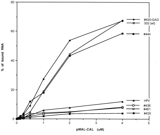
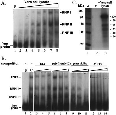
Thermodynamically predicted secondary structure analysis of the 3'-terminal 305 nucleotides (nt) of the rubella virus (RUB) genome, a region conserved in all RUB defective interfering RNAs, revealed four stem-loop (SL) structures; SL1 and SL2 are both located in the E1 coding region, while SL3 and SL4 are within the 59-nt 3' untranslated region (UTR) preceding the poly(A) tract. SL2 is a structure shown to interact with human calreticulin (CAL), an autoantigen potentially involved in RUB RNA replication and pathogenesis. RNase mapping indicated that SL2 and SL3 are in equilibrium between two conformations, in the second of which the previously proposed CAL binding site in SL2, a U-U bulge, is not formed. Site-directed mutagenesis of the 3' UTR with a RUB infectious clone, Robo302, revealed that most of the 3' UTR is required for viral viability except for the 3'-terminal 5 nt and the poly(A) tract, although poly(A) was rapidly regenerated during subsequent replication. Maintenance of the overall SL3 structure, the 11-nt single-stranded sequence between SL3 and SL4, and the sequences forming SL4 were all important for viral viability. Studies on the interaction between host factors and the 3' UTR showed the formation of three RNA-protein complexes by gel mobility shift assay, and UV-induced cross-linking detected six host protein species, with molecular masses of 120, 80, 66, 55, 48, and 36 kDa, interacting with the 3' UTR. Site-directed mutagenesis of SL2 by nucleotide substitutions showed that maintenance of SL2 stem rather than the U-U bulge was critical in CAL binding since mutants having the U-U bulge base paired had a similar binding activity for CAL as the native structure whereas mutants having the SL2 stem destabilized had much lower binding activity. However, all of these mutations gave rise to viable viruses when introduced into Robo302, indicating that binding of CAL to SL2 is independent of viral viability.


Figures

FIG. 1
FIG. 1
(A) Thermodynamically predicted secondary structure of the 3′-terminal 240 nt of the RUB genome (W-Therien strain). A structure with four prominent SLs (SL1 to SL4) and a ΔG of ∼−99.8 kcal/mol was predicted; SL2 is the structure shown to interact with CAL. Both SL1 and SL2 are within the E1 coding region; the UAG stop codon, located at the base of SL2, is underlined. SL3 and SL4 are within the 3′ UTR and are connected by a single-stranded hinge region. SL4 is followed by a 7-nt leader and the poly(A) tract. Nucleotide variations found in other RUB strains are indicated by arrows. fTH, F-Therien (clear-plaque variant derived from W-Therien) (accession no. D00156); M33, M33 (X05259); HPV, HPV77 (vaccine strain derived from M33) (M30776); JUD, Judith (L19420); THM, Thomas (L19421); RA, RA27/3 (vaccine strain currently used in the United States) (L78917). (B) Alternative structures of SL2 and SL3. Structure I (top) was predicted for the W- and F-Therien strains (ΔG ∼−30 kcal/mol); structure II (bottom) was predicted for the M33/HPV77, Judith, RA27/3, and Thomas strains (both are drawn by reference to the W-Therien sequence) (ΔG ∼−25 Kcal/mol in the RA27/3 strain). The complementary UAGU and ACUA stretches which pair to form the longer SL3 in the second structure are underlined.
FIG. 1
FIG. 1
(A) Thermodynamically predicted secondary structure of the 3′-terminal 240 nt of the RUB genome (W-Therien strain). A structure with four prominent SLs (SL1 to SL4) and a ΔG of ∼−99.8 kcal/mol was predicted; SL2 is the structure shown to interact with CAL. Both SL1 and SL2 are within the E1 coding region; the UAG stop codon, located at the base of SL2, is underlined. SL3 and SL4 are within the 3′ UTR and are connected by a single-stranded hinge region. SL4 is followed by a 7-nt leader and the poly(A) tract. Nucleotide variations found in other RUB strains are indicated by arrows. fTH, F-Therien (clear-plaque variant derived from W-Therien) (accession no. D00156); M33, M33 (X05259); HPV, HPV77 (vaccine strain derived from M33) (M30776); JUD, Judith (L19420); THM, Thomas (L19421); RA, RA27/3 (vaccine strain currently used in the United States) (L78917). (B) Alternative structures of SL2 and SL3. Structure I (top) was predicted for the W- and F-Therien strains (ΔG ∼−30 kcal/mol); structure II (bottom) was predicted for the M33/HPV77, Judith, RA27/3, and Thomas strains (both are drawn by reference to the W-Therien sequence) (ΔG ∼−25 Kcal/mol in the RA27/3 strain). The complementary UAGU and ACUA stretches which pair to form the longer SL3 in the second structure are underlined.
FIG. 2
FIG. 2
Analysis of SL2/SL3 conformations by RNase probing. RNA probes consisting of the 3′-terminal 90 nt plus a poly(A) tract from the fTH or HPV77 (HPV) strain were digested with a battery of single- or double-stranded RNases, and the digestion pattern was resolved by primer extension. (A) Results of primer extension from pUC3′RUB110-fTH transcripts digested in a 20-μl reaction with no RNase (−; lanes 1 and 19); mung bean nuclease (MB; cleaves single-stranded RNA with no nucleotide specificity), 10 (lane 2) and 5 (lane 3) U; RNase T2 (cleaves single-stranded RNA with no nucleotide specificity), 0.5 (lane 4), 0.25 (lane 5), 0.1 (lane 6), and 0.05 (lane 7) U; RNase V1 (cleaves double-stranded RNA with no nucleotide specificity), 0.1 (lane 12), 0.05 (lane 13), and 0.025 (lane 14) U; and RNase A (cleaves single-stranded RNA with preference for C and U), 0.1 (lane 20), 0.05 (lane 21), 0.025 (lane 22), and 0.01 (lane 23) U. In lanes 8 to 11 and 15 to 18 are the sequencing ladders for orientation produced by using the primer extension primer and plasmid pUCRUB3′110-fTH as a template. Structural regions within the probe are shown on both margins. Digestion landmarks highlighted include the loops of SL2 and SL3 (>), both of which are sensitive to the single-stranded RNases but not RNase V1 (the SL3 loop is not sensitive to RNase A because it contains a GAAA sequence), and the single efficient RNase V1 digestion site in SL2 and the digestion of the 5′ side of the SL4 stem by RNase V1 (≫). The overall results of RNase probing on both probes are summarized in panels B (single-stranded RNases) and C (double-stranded RNase V1). Because the primer was complementary to the 11 nt preceding the poly(A) tract (italics), digestion within these nucleotides could not be resolved. Single-stranded RNases: T1, RNase T1 (G residue); T2, RNase T2 (no specificity); A, RNase A (C and U residues); MB, mung bean nuclease (no specificity); U2, RNase U2 (A residue).
FIG. 2
FIG. 2
Analysis of SL2/SL3 conformations by RNase probing. RNA probes consisting of the 3′-terminal 90 nt plus a poly(A) tract from the fTH or HPV77 (HPV) strain were digested with a battery of single- or double-stranded RNases, and the digestion pattern was resolved by primer extension. (A) Results of primer extension from pUC3′RUB110-fTH transcripts digested in a 20-μl reaction with no RNase (−; lanes 1 and 19); mung bean nuclease (MB; cleaves single-stranded RNA with no nucleotide specificity), 10 (lane 2) and 5 (lane 3) U; RNase T2 (cleaves single-stranded RNA with no nucleotide specificity), 0.5 (lane 4), 0.25 (lane 5), 0.1 (lane 6), and 0.05 (lane 7) U; RNase V1 (cleaves double-stranded RNA with no nucleotide specificity), 0.1 (lane 12), 0.05 (lane 13), and 0.025 (lane 14) U; and RNase A (cleaves single-stranded RNA with preference for C and U), 0.1 (lane 20), 0.05 (lane 21), 0.025 (lane 22), and 0.01 (lane 23) U. In lanes 8 to 11 and 15 to 18 are the sequencing ladders for orientation produced by using the primer extension primer and plasmid pUCRUB3′110-fTH as a template. Structural regions within the probe are shown on both margins. Digestion landmarks highlighted include the loops of SL2 and SL3 (>), both of which are sensitive to the single-stranded RNases but not RNase V1 (the SL3 loop is not sensitive to RNase A because it contains a GAAA sequence), and the single efficient RNase V1 digestion site in SL2 and the digestion of the 5′ side of the SL4 stem by RNase V1 (≫). The overall results of RNase probing on both probes are summarized in panels B (single-stranded RNases) and C (double-stranded RNase V1). Because the primer was complementary to the 11 nt preceding the poly(A) tract (italics), digestion within these nucleotides could not be resolved. Single-stranded RNases: T1, RNase T1 (G residue); T2, RNase T2 (no specificity); A, RNase A (C and U residues); MB, mung bean nuclease (no specificity); U2, RNase U2 (A residue).
FIG. 3
FIG. 3
Site-directed mutagenesis of the 3′ UTR. The Robo302 sequence of the 3′ UTR beginning with the UAG stop codon of the E1 gene is given on the top line, and the structural features of the UTR are delineated [in addition to those shown in Fig. 1, a UG(U/C) triplet is marked in bold]. Among the mutations created, deletions are indicated by dashes and substitutions are indicated by lowercase letters. Viability is indicated as (nonviable), + (viable with plaques or CPE apparent in transfected cultures), +1 (CPE apparent after one passage), and +3 (CPE after three passages). Sequences were confirmed on all viable mutants after one amplification in Vero cells; those designated +a contained alterations in the mutant sequence (Fig. 4), while those designated +r contained regenerated poly(A) tracts and formed opaque plaques in transfected cells but clear plaques in the subsequent passages. Mutants which formed plaques similar to those of Robo302 on transfection plaques are inicated as “WT,” while those that formed tiny plaques on transfection plaques or no plaques are indicated as “tiny” and “N.P.,” respectively. Plaque morphologies indicated in parentheses are those formed after subsequent passaging for amplification.
FIG. 4
FIG. 4
Characterization of viruses with mutations in SL4, the leader, and the poly(A) tract. Sequences determined from 15 viable mutants are given (a and b indicate results of independent transfections); altered nucleotides not present in the original transcripts are in lowercase. For comparison, sequences of six nonviable mutants are also given at the bottom. To determine replication ability, mutant viruses isolated from transfection plaques (p) or medium (m) were amplified by one passage in Vero cells, and the titers of the amplified stocks were determined. These stocks were then used for infection of Vero cells (in the first experiment [superscript “1”], which compared the replication of Robo302 and eight mutants, the multiplicity of infection [MOI] was 5; in the second experiment [superscript “2”], which compared Robo302 and six additional mutants, the MOI was 0.01). The infected culture media were harvested at 2 (MOI = 5) or 3 (MOI = 0.01) days postinfection, and the titer (PFU per milliliter) was determined. The relative titer produced by each mutant is shown in comparison with the Robo302 titer (set at 100%) produced in the same experiment (N.D., not determined). The proposed critical stretch of UG(U/C) triplets is underlined.
FIG. 5
FIG. 5
Thermodynamically predicted secondary structure of SL3 in virus transcripts with mutations in this region. The native SL3 structure (WT) is at the top. Point mutations are underlined. The entropy of each mutant is also given.
FIG. 6
FIG. 6
(A) Mutagenesis of SL2. Because SL2 is located in the E1 coding region, analysis was done by creating point mutations that resulted in silent changes (436) or synonymous codons (435 and 461). However, to eliminate the U-U bulge, we created two missense mutations (444 and 430-AAG/GAG) which led to changes in coding as indicated. Mutation 419 was made to create an NsiI site for cloning, and 330 was made to conform to HPV77 sequence. The bold characters are the stop codon ending the E1 gene; the nucleotide variations found in other RUB strains are in lowercase. Transfection efficiencies of mutant transcripts are shown as +++ (equivalent to that of Robo302); ++ (about 30 to 50% of that of Robo302) +, (about 5 to 10% of that of Robo302), and N.D., not determinable, but transfected cells inoculated with medium showed CPE. The plaque morphology described is that produced following transfection. Following amplification of 430-AAG from transfection medium, the sequence was found to have changed to CAG. (B) Growth curves of SL2 mutants. Vero cells were infected with viruses amplified from transfection plaques at an MOI of 0.01 PFU/cell. At indicated times postinfection, aliquots of the infected culture media were harvested and titered on Vero cells.
FIG. 7
FIG. 7
Binding of MBP-CAL to SL2 probes. (A) Robo302 SL2 probe (<1 ng) was incubated in a binding reaction with various amounts of MBP-CAL prior to resolution of binding by electrophoresis in a nondenaturing polyacrylamide gel. Lane 1, control (0 μM MBP-CAL); lanes 2 to 7, 0.125, 0.25, 0.5, 1, 2, and 4 μM MBP-CAL. (B) SL2 probes containing sequences of WT Robo302 (lane 1 to 3) and mutations 430-GAG (lanes 4 to 6), 444 (lanes 7 to 9), 435 (lanes 10 to 12), 436 (lanes 13 to 15), and 461 (lanes 16 to 18) were incubated with 0 μM (lanes 1, 4, 7, 10, 13, and 16), 0.25 μM (lanes 2, 5, 8, 11, 14, and 17), or 1 μM (lane 3, 6, 9, 12, 15, and 18) MBP-CAL. After binding, formation of RNP complexes was determined by electrophoresis in polyacrylamide gels under nondenaturing conditions. P, probe only.
FIG. 8
FIG. 8
Comparison of binding activities of Robo302 and mutated SL2s to MBP-CAL. Robo302 and mutant SL2 probes were incubated with increasing amounts of MBP-CAL, and formation of RNP complexes was determined by gel mobility shift (an example of binding of MBP-CAL and Robo302 SL2 is shown in Fig. 6A). The binding was quantitated as the percentage of total radioactivity (bound plus unbound) in RNP complexes.
FIG. 9
FIG. 9
Binding of cellular factors to the 3′ UTR. (A) 32P-labeled UTR probe (lacking both the poly(A) tract and the 3′-terminal 5 nt was incubated with 0 μg (lane 1), 0.2 μg (lane 2), 0.375 μg (lane 3), 0.75 μg (lane 4), 1.5 μg (lane 5), 3 μg (lane 6), 6 μg (lane 7), and 12 μg (lane 8) of cytoplasmic lysate from uninfected Vero cells, and formation of complexes was detected by electrophoresis in nondenaturing polyacrylamide gels. The three RNP complexes detected are indicated on the right as RNP I, II, and III. (B) Increasing amounts of different competitor RNAs, including both nonspecific [SL2, poly(I)-poly(C), and yeast tRNA] and specific (3′ UTR RNA transcripts) competitors, were incubated with 4 μg of Vero cell lysate prior to the addition of the 32P-labeled probe. Lane 5: 1 (P), probe alone; 2 (C), positive control without any competitor; 3 to 5, SL2 at 20× (lane 3), 50× (lane 4), or 150× (lane 5) molar excess; 6 to 8, poly(I)-poly(C) at 50 ng (lane 6), 100 ng (lane 7), or 200 ng (lane 8) [the standard binding assay buffer containing contains 100 ng of poly(I)-poly(C)]; 9 to 11, yeast tRNA at 20× (lane 9), 50× (lane 10), or 150× (lane 11) molar excess; 12 to 14, 3′ UTR RNA at 20× (lane 12), 50× (lane 13), or 150× (lane 14) molar excess. (C) 3′ UTR probe in the absence (lane 2, P [probe only]) or presence (lane 3) of 30 μg of Vero cell lysates was exposed to UV light and digested with RNase. Cross-linked proteins were revealed by SDS-polyacrylamide gel electrophoresis. Approximate positions of size markers (lane M) are given on the right.

References

    1. Andino R, Rieckhof G E, Achacoso P L, Baltimore D. Poliovirus RNA synthesis utilizes and RNP complex formed around the 5′-end of viral RNA. EMBO J. 1993;12:3587–3598. - PMC - PubMed
    1. Atreya, C. D. Personal communication.
    1. Atreya C D, Singh N K, Nakhasi H L. The rubella virus RNA binding activity of human calreticulin is localized to the N-terminal domain. J Virol. 1995;69:3848–3851. - PMC - PubMed
    1. Barr J N, Whelan S P J, Wertz G W. cis-acting signals involved in termination of vesicular stomatitis virus mRNA synthesis include the conserved AUAC and the U7 signal for polyadenylation. J Virol. 1997;71:8718–8725. - PMC - PubMed
    1. Belsham G J, Sonenberg N. RNA protein interactions in regulation of picornavirus RNA translation. Microbiol Rev. 1996;60:499–511. - PMC - PubMed

Publication types