. 1999 Apr;73(4):3484-90.
doi: 10.1128/JVI.73.4.3484-3490.1999.

Infectious pancreatic necrosis virus: identification of a VP3-containing ribonucleoprotein core structure and evidence for O-linked glycosylation of the capsid protein VP2

Affiliations

Infectious pancreatic necrosis virus: identification of a VP3-containing ribonucleoprotein core structure and evidence for O-linked glycosylation of the capsid protein VP2

A Hjalmarsson et al. J Virol. 1999 Apr.

Abstract

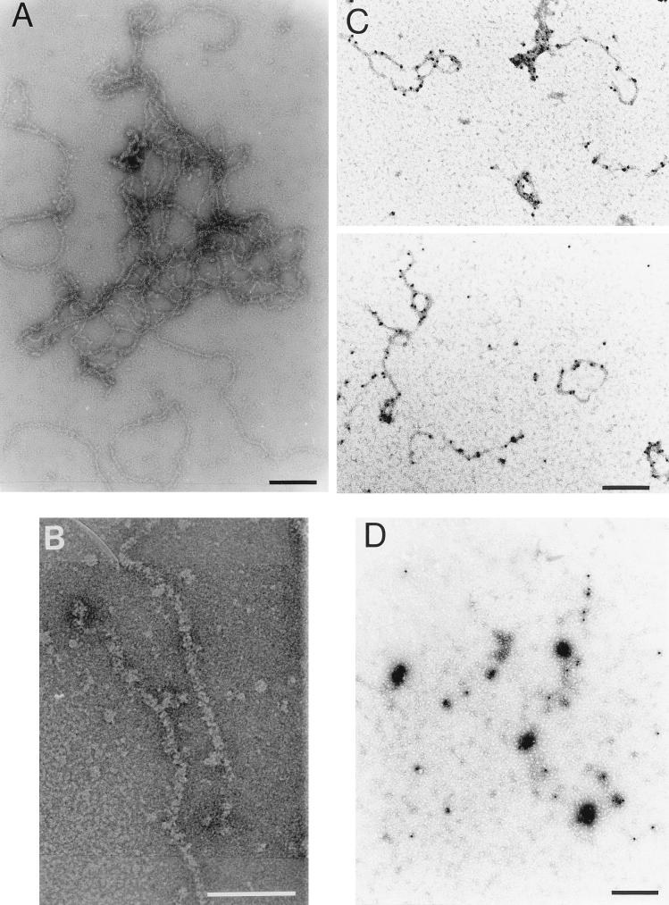
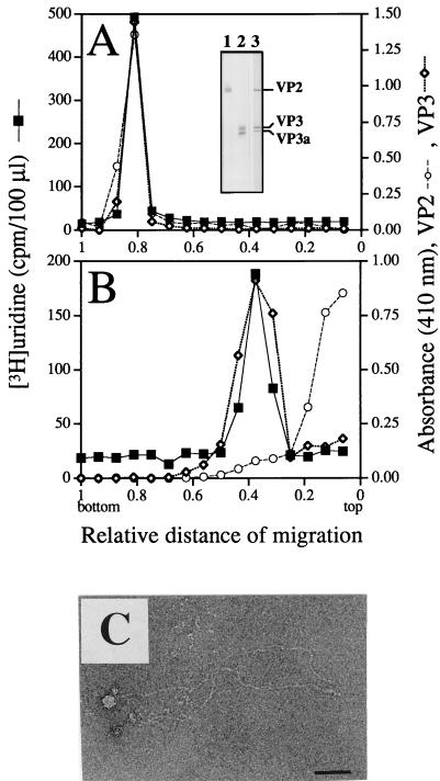
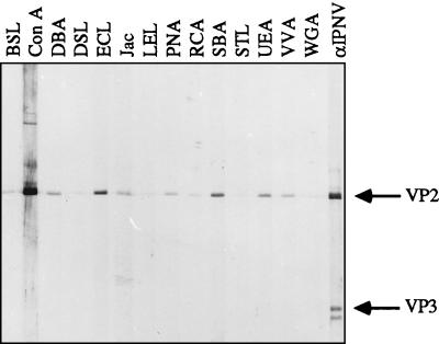
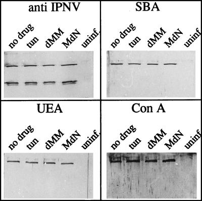
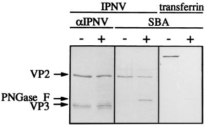
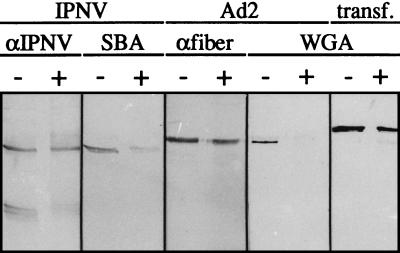
Virions of infectious pancreatic necrosis virus (IPNV) were completely disintegrated upon dialysis against salt-free buffers. Direct visualization of such preparations by electron microscopy revealed 5.0- to 6.5-nm-thick entangled filaments. By using a specific colloidal gold immunolabeling technique, these structures were shown to contain the viral protein VP3. Isolation by sucrose gradient centrifugation of the filaments, followed by serological analysis, demonstrated that the entire VP3 content of the virion was recovered together with the radiolabeled genomic material forming the unique threadlike ribonucleoprotein complexes. In a sensitive blotting assay, the outer capsid component of IPNV, i.e., the major structural protein VP2, was shown to specifically bind lectins recognizing sugar moieties of N-acetylgalactosamine, mannose, and fucose. Three established metabolic inhibitors of N-linked glycosylation did not prevent addition of sugar residues to virions, and enzymatic deglycosylation of isolated virions using N-glycosidase failed to remove sugar residues of VP2 recognized by lectins. However, gentle alkaline beta elimination clearly reduced the ability of lectins to recognize VP2. These results suggest that the glycosylation of VP2 is of the O-linked type when IPNV is propagated in RTG-2 cells.


Figures

FIG. 1
FIG. 1
Subviral components of IPNV. Visualization by EM of filamentous structures released from virions dialyzed against a low-ionic-strength buffer (A) and an enlargement of a section of a tentative ribonucleoprotein complex (B) are shown. The horizontal bars indicate 100 (A) and 50 (B) nm. Subviral components labeled with colloidal-gold-conjugated MAbs against VP3 (C) or VP2 (D) and stained with 2% uranyl acetate before examination by EM are shown. Under the present conditions, the specimens in panels C and D were stained positively. Bars in panels C and D, 200 nm.
FIG. 2
FIG. 2
Rate zonal sedimentation of intact and disintegrated virions. IPNV stored in TNE (A) and IPNV dialyzed against low-ionic-strength buffer (B) were sedimented by sucrose gradient centrifugation. The RNA was measured as trichloroacetic acid-precipitable, [3H]uridine-labeled material, and viral proteins VP2 and VP3 were identified by MAbs in an ELISA and detected as A410. The specificities of the MAbs are shown in an immunoblot (inset in panel A). IPNV-infected RTG-2 cells were run in lanes 1 and 2, and purified IPNV was run in lane 3. Proteins in lane 1 were displayed by a MAb against VP2 (14/2d, also recognizing pVP2), those in lane 2 by a MAb against VP3 (1/2f-3), and the proteins in lane 3 were revealed by an anti-IPNV specific serum. Material of the fraction from the sucrose gradient sedimentation of disintegrated virions containing the highest concentrations of VP3 and RNA was observed by negative staining and EM (C). Bar, 50 nm.
FIG. 3
FIG. 3
Lectin blot of virion polypeptides. IPNV was purified by an additional step of isodensity centrifugation after detergent treatment. Upon SDS-PAGE of the recovered virus and transblotting of the polypeptides to a nitrocellulose membrane, carbohydrates were monitored with 14 different lectins. The total amount of virion proteins subjected to SDS-PAGE was 67 ng per channel (42). The specificities of the different lectins are explained in Table 1. The positions of the viral proteins were detected with a polyclonal serum against IPNV (αIPNV). For definitions of abbreviations, see the text and Table 1, footnote a.
FIG. 4
FIG. 4
Effects of inhibitors of glycosylation. Virus-infected cells were treated with the following glycosylation inhibitors added at 1 h p.i.: 10-μg/ml tunicamycin (tun), 3 mM deoxymannojirimycin (dMM), and 4 mM N-methyl-1-deoxynojirimycin (MdN). The two controls were virus replicated in the absence of any drug (no drug) and mock-infected cells treated in the same way as the infected cells, without any drug (uninf.). The latter material was recovered from positions in a CsCl gradient corresponding to the viral bands in gradients separating virus-containing material. To each SDS-PAGE lane, 0.30 μg of purified virion polypeptides (42) was applied, and three different lectins were selected for the detection of lectin-binding proteins (SBA, UEA, and ConA, recognizing GalNAc, fucose, and mannose, respectively). A polyclonal serum against IPNV was used for detection of viral proteins.
FIG. 5
FIG. 5
Enzymatic deglycosylation of purified IPNV. Virions of IPNV were treated with N-glycosidase F (+) or left untreated (−). Transferrin treated in the same way was used as a control. Separation of the polypeptides by SDS-PAGE was followed by transblotting and detection with a polyclonal serum against IPNV (αIPNV) and SBA. VP2 and VP3 indicate the positions of the viral polypeptides, whereas PNGase F indicates the position of N-glycosidase F, which is also recognized by SBA.
FIG. 6
FIG. 6
Detection of polypeptides and carbohydrates after alkaline β elimination. Lyophilized samples of IPNV, human Ad2, and transferrin (transf.) were incubated at 37°C for 20 h in 5 mM NaOH with 1 M NaBH4 (+) or in PBS (−) as described in the text. Analysis by SDS–13% PAGE was followed by transblotting and detection with antiserum (αIPNV and αfiber are antibodies against the glycosylated 62-kDa fiber protein of Ad2) or with lectins (SBA and WGA). The amount of IPNV polypeptides applied to each lane was 0.37 μg, and those of the controls Ad2 and transf. were 3.3 and 0.5 μg, respectively.

References

    1. Bartles J R, Hubbard A L. 125I-wheat germ agglutinin blotting: increased sensitivity with polyvinylpyrrolidone quenching and periodate oxidation/reductive phenylamination. Anal Biochem. 1984;140:284–292. - PubMed
    1. Böttcher B, Kieselev N, Stel’Mashchuk V, Perevozchikova N, Borisov A, Crowther R. Three-dimensional structure of infectious bursal disease virus determined by electron cryomicroscopy. J Virol. 1997;71:325–330. - PMC - PubMed
    1. Brown D, Westphal M, Burlingham B, Winterhoff U, Doerfler W. Structure and composition of the adenovirus type 2 core. J Virol. 1975;16:366–387. - PMC - PubMed
    1. Carlsson A, Kuznar J, Varga M, Everitt E. Purification of infectious pancreatic necrosis virus by anion exchange chromatography increases the specific infectivity. J Virol Methods. 1994;47:27–36. - PubMed
    1. Caswell-Reno P, Reno P W, Nicholson B L. Monoclonal antibodies to infectious pancreatic necrosis virus: analysis of viral epitopes and comparison of different isolates. J Gen Virol. 1986;67:2193–2206. - PubMed

Publication types

Substances

LinkOut - more resources