. 1999 Mar;103(5):597-604.
doi: 10.1172/JCI5461.

Modulation of apoptosis by the cyclin-dependent kinase inhibitor p27(Kip1)

Affiliations

Modulation of apoptosis by the cyclin-dependent kinase inhibitor p27(Kip1)

K Hiromura et al. J Clin Invest. 1999 Mar.

Abstract

Proliferation and apoptosis are increased in many types of inflammatory diseases. A role for the cyclin kinase inhibitor p27(Kip1) (p27) in limiting proliferation has been shown. In this study, we show that p27(-/-) mesangial cells and fibroblasts have strikingly elevated rates of apoptosis, not proliferation, when deprived of growth factors. Apoptosis was rescued by restoration of p27 expression. Cyclin A-cyclin-dependent kinase 2 (CDK2) activity, but not cyclin E-CDK2 activity, was increased in serum-starved p27(-/-) cells, and decreasing CDK2 activity, either pharmacologically (Roscovitine) or by a dominant-negative mutant, inhibited apoptosis. Our results show that a new biological function for the CDK inhibitor p27 is protection of cells from apoptosis by constraining CDK2 activity. These results suggest that CDK inhibitors are necessary for coordinating the cell cycle and cell-death programs so that cell viability is maintained during exit from the cell cycle.


Figures

Figure 1
Figure 1
Quantitation of apoptosis. TUNEL staining was not increased in the presence of growth factors (serum) in p27–/– mesangial cells (a) and p27–/– fibroblasts (b). In contrast, TUNEL staining was increased at 24 h of growth factor deprivation (serum free) in p27–/– cells compared with p27+/+ cells. *P < 0.001 vs. serum; **P < 0.001 vs. p27+/+. TUNEL, deoxynucleotide transferase–mediated nick end-labeling.
Figure 2
Figure 2
Effect of growth factor deprivation on cell confluency. p27+/+ (a) and p27–/– (b) mesangial cells were plated at the same density in FCS. Compared with p27+/+ mesangial cells (c) cell confluency decreased in p27–/– mesangial cells (d) after 5 days of growth factor deprivation.
Figure 3
Figure 3
Effect of reconstituting p27 on apoptosis in p27–/– mesangial cells. Transfected p27 mesangial cells were identified by staining with GFP (green nuclei), and apoptosis was measured by Hoechst staining. (a) p27–/– mesangial cells transfected with human p27 plasmid stained green. (b) Apoptosis was not detected in serum-starved transfected p27–/–cells transfected with the p27 plasmid (arrows); apoptosis was detected in nontransfected cells (arrowhead). (c) Control p27–/– mesangial cells were transfected with a p27 mutant plasmid that does not bind CDK2 and GFP. (d) p27 mutant plasmid did not prevent apoptosis (arrowhead). (e) In each experiment, 200 cells were quantitated, and the number of cells undergoing apoptosis was expressed as a percentage. *P < 0.001 vs. p27–/– alone (nontransfected cells) and control (p27 mutant plasmid–transfected) p27–/– cells. These data show that p27 protects cells from survival and mitogenic growth factor deprivation–induced apoptosis. CDK2, cyclin-dependent kinase-2; GFP, green fluorescent protein.
Figure 4
Figure 4
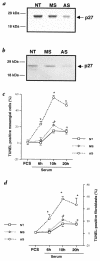
(a and b) Western blot analysis for p27. Transfecting rat mesangial cells (a) and rat fibroblasts (b) with antisense oligonucleotides to p27 (AS), but not mismatch oligonucleotides (MS), lowered p27 protein levels compared with nontransfected cells (NT). (c and d) Quantitation of TUNEL staining in rat cells. TUNEL staining was detected at 6 h after growth factor deprivation (serum free) in AS-transfected cells in rat mesangial cells (c) and rat fibroblasts (d), but not in controls, and was increased at each time point compared with controls.
Figure 5
Figure 5
CDK2 activity and protein levels. Total cell protein was immunoprecipitated with an antibody to CDK2, and CDK2 activity was detected by H1 kinase assay. CDK2 protein levels were measured by Western blot analysis. In the presence of survival and mitogenic growth factors (FCS), CDK2 activity was increased in p27–/– mesangial cells (a) and p27–/– fibroblasts (c) compared with p27+/+ cells. CDK2 activity remained elevated at 6 h and 18 h after growth factor deprivation (serum free) in p27–/– cells compared with p27+/+ cells. The protein levels for CDK2 did not change in p27–/– and p27+/+ mesangial cells (b) and fibroblasts (d) in the presence or absence of growth factors.
Figure 6
Figure 6
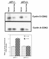
Cyclin E–CDK2 and cyclin A–CDK2 activity in p27–/– and p27+/+ mesangial cells. Total protein was extracted from p27–/– and p27+/+ mesangial cells and immunoprecipitated with antibodies to cyclin E (top) and cyclin A (middle) for histone H1 kinase. Quantitation of CDK2 kinase activity is shown (bottom). In the presence of growth factors (serum), there was an increase in cyclin E–CDK2 and cyclin A–CDK2 activity in p27–/– and p27+/+ cells, which decreased in p27+/+ cells after 6 h of growth factor deprivation (serum free). In p27–/– mesangial cells, cyclin A–CDK2 activity, but not cyclin E–CDK2 activity, remained increased after growth factor deprivation.
Figure 7
Figure 7
Effect of suppressing CDK2 activity on apoptosis in p27–/– fibroblasts in response to serum starvation. (a) p27–/– cells transfected with a dominant–negative mutant CDK2 (dnk2) plasmid were identified by cotransfection with a GFP plasmid (green nuclei). (b) Apoptosis (measured by Hoechst staining) was not detected in dnk2-transfected cells (thick arrow), but was detected in nontransfected cells (thin arrow). (c) p27–/– cells cotransfected with wild-type CDK2 (wtk2) plasmid and GFP were identified as green. (d) wtk2 did not protect p27–/– fibroblasts from apoptosis (arrow). (e) Quantitation of apoptosis. The percentage of apoptotic cells was evaluated in 200 cells. *P < 0.001 vs. nontransfected p27–/– fibroblasts (p27–/– alone) and wtk2-transfected cells. Similar results were obtained in p27–/– mesangial cells.
Figure 8
Figure 8
Proposed schema showing coordination of cell-cycle events by the CDK inhibitor p27. When grown in the presence of growth factors, reduced or absent p27 levels are associated with a coordinated and synchronous increase in cyclin E–CDK2 and cyclin A–CDK2 activity. This favors cell-cycle progression and proliferation. Under stress states such as growth factor deprivation, the loss of p27 is associated with an unconstrained increase in cyclin A–CDK2 activity, but not cyclin E–CDK2 activity. This unscheduled increase in CDK2 activity causes cell-cycle exit by apoptosis.

Similar articles

Cited by

References

    1. Sherr CJ, Roberts JM. Inhibitors of mammalian G1 cyclin-dependent kinases. Genes Dev. 1995;9:1149–1163. - PubMed
    1. Durand B, Gao F-B, Raff M. Accumulation of the cyclin-dependent kinase inhibitor p27/Kip1 and the timing of oligodendrocyte differentiation. EMBO J. 1997;16:306–317. - PMC - PubMed
    1. Zabludoff S, Csete M, Wagner R, Yu X, Wold BJ. p27Kip1 is expressed transiently in developing myotomes and enhances myogenesis. Cell Growth Differ. 1998;9:1–11. - PubMed
    1. Nourse J, et al. Interleukin-2-mediated elimination of the p27kip1 cyclin-dependent kinase inhibitor prevented by rapamycin. Nature. 1994;372:570–573. - PubMed
    1. Polyak K, et al. Cloning of p27Kip1, a cyclin-dependent kinase inhibitor and potential mediator of extracellular antimitogenic signals. Cell. 1994;78:59–66. - PubMed

Publication types

MeSH terms