. 1999 Mar;103(5):675-82.
doi: 10.1172/JCI4552.

Impairment of skeletal muscle adenosine triphosphate-sensitive K+ channels in patients with hypokalemic periodic paralysis

Affiliations

Impairment of skeletal muscle adenosine triphosphate-sensitive K+ channels in patients with hypokalemic periodic paralysis

D Tricarico et al. J Clin Invest. 1999 Mar.

Abstract

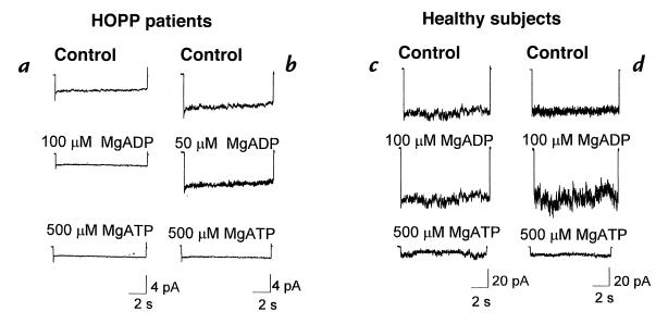
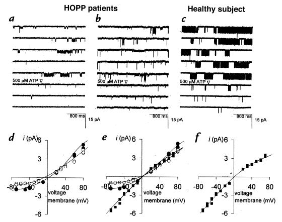
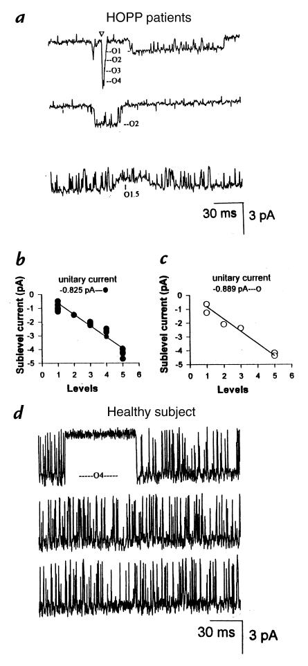
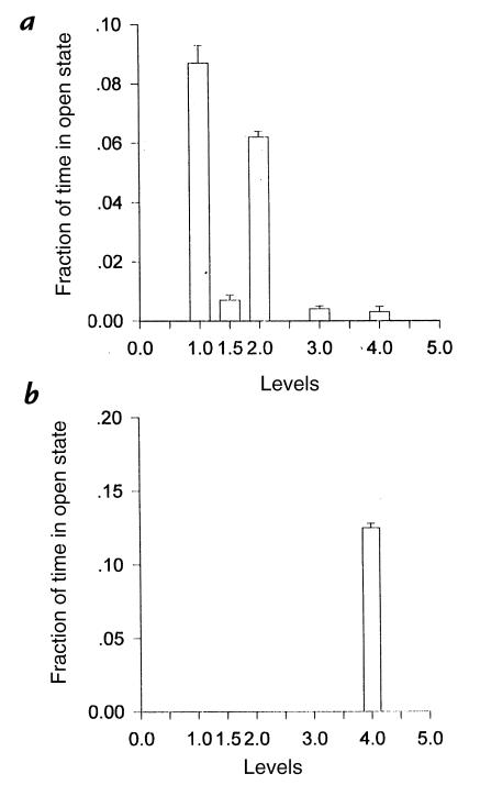
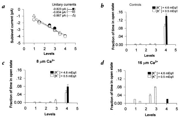
The adenosine triphosphate (ATP)-sensitive K+ (KATP) channel is the most abundant K+ channel active in the skeletal muscle fibers of humans and animals. In the present work, we demonstrate the involvement of the muscular KATP channel in a skeletal muscle disorder known as hypokalemic periodic paralysis (HOPP), which is caused by mutations of the dihydropyridine receptor of the Ca2+ channel. Muscle biopsies excised from three patients with HOPP carrying the R528H mutation of the dihydropyridine receptor showed a reduced sarcolemma KATP current that was not stimulated by magnesium adenosine diphosphate (MgADP; 50-100 microM) and was partially restored by cromakalim. In contrast, large KATP currents stimulated by MgADP were recorded in the healthy subjects. At channel level, an abnormal KATP channel showing several subconductance states was detected in the patients with HOPP. None of these were surveyed in the healthy subjects. Transitions of the KATP channel between subconductance states were also observed after in vitro incubation of the rat muscle with low-K+ solution. The lack of the sarcolemma KATP current observed in these patients explains the symptoms of the disease, i.e., hypokalemia, depolarization of the fibers, and possibly the paralysis following insulin administration.


Figures

Figure 1
Figure 1
Digital average of the sarcolemma KATP currents of patients with HOPP and healthy subjects. KATP currents of the 18-year-old boy with HOPP (seven macropatches) (a) and of the 40-year-old woman with HOPP (14 macropatches) are shown (b). MgADP at 50 μM concentration failed to stimulate the current, whereas at 100 μM, current was reduced. In both patients with HOPP, MgATP (500 μM) completely reduced the currents. KATP currents of the 17-year-old healthy boy (eight macropatches) (c) and 40-year-old healthy woman (seven macropatches) are shown (d). In contrast to the patients with HOPP, in the healthy subjects large KATP currents were recorded that were stimulated by MgADP (100 μM) and completely reduced by MgATP (500 μM). HOPP, hypokalemic periodic paralysis; KATP, adenosine triphosphate–sensitive K+ channel; MgADP, magnesium adenosine diphosphate; MgATP, magnesium adenosine triphosphate.
Figure 2
Figure 2
Sample traces of single sarcolemma KATP channel from two patients with HOPP and from a healthy subject. The KATP channel of the 40-year-old woman with HOPP (a) and of the 18-year-old boy with HOPP (b) transits through subconductance states of low open probability, all blocked by ATP. In the 17-year-old healthy boy, only one open level blocked by ATP is clearly visible (c). In the 40-year-old woman with HOPP, the O1 (open circles) and O2 (closed circles) levels had slope conductances of 25 pS and 32 pS at negative potentials, and 40 pS and 47 pS at positive potentials, respectively (d). In the 18-year-old boy with HOPP, the slope conductances of O1 (open circles), O2 (closed circles), and O4 (closed squares) levels were 27 pS, 35 pS, and 72 pS at negative potentials, and 40 pS, 47 pS, and 46 pS at positive potentials, respectively (e). In the 18-year-old healthy boy, the O4 level (closed squares) had a slope conductances of 72 pS and 45 pS at the negative and positive potentials, respectively (f).
Figure 3
Figure 3
Conductance levels of the sarcolemma KATP channels in the patients with HOPP and in a healthy subject. Five subconductance levels are clearly visible in these sample traces (a). They are labeled on the basis of their specific amplitudes. The arrow marks the simultaneous opening from the closed state to the O4 level. Linear relationships between the number of levels vs. the amplitude counted in the fibers of the 40-year-old woman with HOPP (b) and of the 18-year-old boy with HOPP (c). In the 40-year-old healthy woman (d), only one open conductance state, O4, was clearly visible.
Figure 4
Figure 4
Fraction of time spent in open state by each subconductance level counted in the fibers of the 40-year-old woman with HOPP (a) and in the 40-year-old healthy woman (b). In the patient with HOPP, the KATP channel resides mainly in the subconductance levels, whereas in the healthy subject, the KATP channel resides only in the O4 level.
Figure 5
Figure 5
In vitro effects of low-K+ solution and different concentrations of free Ca2+ ion on KATP channel. The muscle fibers were preincubated for 80 min with low-K+ solution (open bars) or with normokalemic solution (closed bars) before recordings. A linear relationship between the number of levels of the KATP channels vs. the corresponding amplitude in the absence (closed circles), or in the presence, of 8 μM (open circles) and 16 μM (open triangle) concentrations of free Ca2+ ion (a) was found. Fraction of time spent in open state by each subconductance level in the absence (b) or in the presence of 8 μM (c) and 16 μM (d) concentrations of free Ca2+ ion (a). Only after incubation of the muscle with the low-K+ solution, the Ca2+ ion forced the channel into the subconductance states, reducing the open probability of the O4 level and increasing the open probability of the sublevels.

Similar articles

Cited by

References

    1. Noma A. ATP-regulated K+ channels in cardiac muscle. Nature. 1983;305:147–148. - PubMed
    1. Sakura H, Ämmälä C, Smith PA, Gribble FM, Ashcroft FM. Cloning and functional expression of the cDNA encoding a novel ATP-sensitive potassium channel subunit expressed in pancreatic beta-cells, brain, heart and skeletal muscle. FEBS Lett. 1995;377:338–344. - PubMed
    1. Tricarico D, Conte Camerino D. ATP-sensitive K+ channels of skeletal muscle fibers from young adult and aged rats: possible involvement of thiol-dependent redox mechanisms in the age-dependent modifications of their biophysical and pharmacological properties. Mol Pharmacol. 1994;46:754–761. - PubMed
    1. Tricarico D, Petruzzi R, Conte Camerino D. Different sulfonylurea and ATP sensitivity characterizes the juvenile and the adult form of KATP channel complex of rat skeletal muscle. Eur J Pharmacol. 1997;321:369–378. - PubMed
    1. Tricarico D, et al. The biophysical and pharmacological characteristics of skeletal muscle KATP channels are modified in K+ depleted rat, an animal model of hypokalemic periodic paralysis. Mol Pharmacol. 1998;54:197–206. - PubMed

Publication types

Grants and funding