. 1999 Mar;103(5):683-9.
doi: 10.1172/JCI4445.

Hypertension in mice lacking 11beta-hydroxysteroid dehydrogenase type 2

Affiliations

Hypertension in mice lacking 11beta-hydroxysteroid dehydrogenase type 2

Y Kotelevtsev et al. J Clin Invest. 1999 Mar.

Abstract

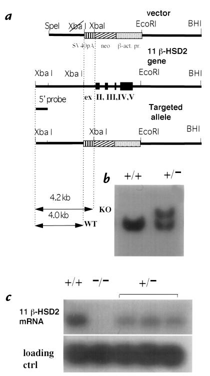
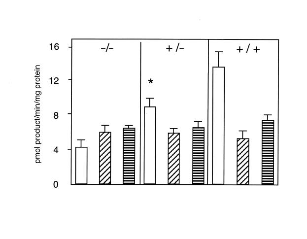
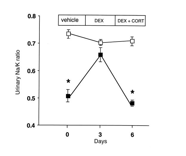
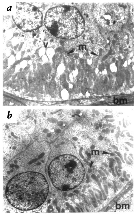
Deficiency of 11beta-hydroxysteroid dehydrogenase type 2 (11beta-HSD2) in humans leads to the syndrome of apparent mineralocorticoid excess (SAME), in which cortisol illicitly occupies mineralocorticoid receptors, causing sodium retention, hypokalemia, and hypertension. However, the disorder is usually incompletely corrected by suppression of cortisol, suggesting additional and irreversible changes, perhaps in the kidney. To examine this further, we produced mice with targeted disruption of the 11beta-HSD2 gene. Homozygous mutant mice (11beta-HSD2(-/-)) appear normal at birth, but approximately 50% show motor weakness and die within 48 hours. Both male and female survivors are fertile but exhibit hypokalemia, hypotonic polyuria, and apparent mineralocorticoid activity of corticosterone. Young adult 11beta-HSD2(-/-) mice are markedly hypertensive, with a mean arterial blood pressure of 146 +/- 2 mmHg, compared with 121 +/- 2 mmHg in wild-type controls and 114 +/- 4 mmHg in heterozygotes. The epithelium of the distal tubule of the nephron shows striking hypertrophy and hyperplasia. These histological changes do not readily reverse with mineralocorticoid receptor antagonism in adulthood. Thus, 11beta-HSD2(-/-) mice demonstrate the major features of SAME, providing a unique rodent model to study the molecular mechanisms of kidney resetting leading to hypertension.


Figures

Figure 1
Figure 1
Targeted inactivation of 11β-HSD2 gene. (a) Structure of the targeting vector, the 11β-HSD2 locus, and the targeted allele. (b) Southern blot hybridization analysis of XbaI-digested genomic DNA from neomycin-resistant nontargeted (+/+) and targeted (+/–) ES clones. (c) Northern blot analysis of 11β-HSD2 expression. RNA (10 μg) from kidney of wild-type (+/+), homozygous (–/–) mutants and three heterozygous (+/–) littermates was hybridized with mouse 11β-HSD2 and control (ctrl) U-1 snRNA cDNA probes. 11β-HSD2, 11β-hydroxysteroid dehydrogenase type 2; ES, embryonic stem cells.
Figure 2
Figure 2
Variation in 11β-HSD2 dehydrogenase and 11β-HSD1 oxidoreductase activities in placentas with different 11β-HSD2 genotypes. Activities (mean ± SEM) expressed as conversion of nmol [3H]-steroid substrate (either corticosterone for 11β-dehydrogenase or 11-dehydrocorticosterone for 11β-reductase) to pmol product/mg protein/min. (Open bars) NAD-dependent 11β-corticosterone dehydrogenase activity. (Diagonally striped bars) NADP-dependent 11β-corticosterone dehydrogenase activity. (Horizontally striped bars) NADPH-dependent 11-dehydrocorticosterone reductase activity. Note that activity due to 11β-HSD1 does not vary with genotype. In contrast, NAD-dependent predominantly 11β-HSD2 activity varies with genotype, resulting in overall NADP-preferring 11β-HSD activity in 11β-HSD2–/– mice. The residual NAD-dependent activity in 11β-HSD2–/– placentas presumably reflects activity of 11β-HSD1 enzyme. *Significantly greater than corresponding 11β-HSD2–/– value (P < 0.001) and significantly smaller than corresponding wild-type (+/+) values (P < 0.001). NAD, nicotinamide adenine dinucleotide; NADP, nicotinamide adenine dinucleotide phosphate.
Figure 3
Figure 3
Corticosterone acts as a mineralocorticoid in 11β-HSD2–/– mice. Wild-type (open squares; n = 4) and 11β-HSD2–/– (closed squares; n = 4) mice were injected with vehicle (days –3 to 0), followed by dexamethasone (DEX; 100 μg/kg, days 0–3) to suppress endogenous corticosterone, and finally dexamethasone with replacement corticosterone (DEX + CORT; 10 mg/kg, days 3–6). Urine was collected for the last 24 h of each treatment and electrolytes measured by automated analyzer. The abnormally low urinary Na/K ratio in 11β-HSD2–/– mice is reversed by dexamethasone and recurs with corticosterone replacement, whereas the treatments have no effect in wild-type mice. *Significantly different from corresponding wild-type value (P < 0.0005).
Figure 4
Figure 4
Hyperplasia and hypertrophy of the distal renal tubular epithelium in 11β-HSD2–/– mice. (a) In adult 11β-HSD2–/– mice, the distal tubules (d) show considerable enlargement: the tubular diameter is increased threefold compared with the distal tubule in the wild-type mice (b). Note that the proximal tubules (p) and glomeruli (g) are unaffected in the 11β-HSD2–/– mice. (c) Hyperplasia is apparent in the 11β-HSD2–/– mouse distal tubule (d), which shows over 20 cells in the cross-sectional profile compared with (d) the four to six cells observed in controls. (e) The distal tubular enlargement (d) is evident in 11β-HSD2–/– mice at the anatomical origin of the distal tubule at the juxtaglomerular apparatus (j) adjacent to the glomerulus (g), compared with (f) the nonenlarged wild-type distal tubule. md, macula densa. (g) Proximal tubule (p) is normal in 11β-HSD2–/– kidneys. 11β-HSD2–/– proximal tubules are indistinguishable from (h) wild-type proximal tubules. The tubules are of similar diameter and cell number in cross section. They are lined by cuboidal cells with granular cytoplasm and a brush border in both animals. The same magnification was used in 11β-HSD2–/– and corresponding wild-type control photographs. In a and b, H&E ×60; in c and d:, H&E ×120; in eh, H&E ×180. H&E, hematoxylin and eosin.
Figure 5
Figure 5
Electron microscopy of the distal tubular epithelial cells of 11β-HSD2–/– (a) and wild-type controls (b). Distal tubular epithelial cells from 11β-HSD2–/– animals show an increase in size. The subnuclear, or basal, pole is expanded with apical displacement of the nucleus. The number of mitochondria (m) in each distal tubular cell is increased, although the mitochondria remain located at the expanded basal pole (bm, basal membrane). Occasional membrane-enclosed vacuoles (v) are seen in the cytoplasm of the distal tubular epithelial cells from the 11β-HSD2–/– animals. ×3,800.

References

    1. Guyton AC. Blood pressure control—special role of the kidneys and body fluids. Science. 1991;252:1813–1816. - PubMed
    1. Shimkets RA, et al. Liddle's syndrome: heritable human hypertension caused by mutations in the beta subunit of the epithelial sodium channel. Cell. 1994;79:407–414. - PubMed
    1. Simon DB, et al. Mutations in the chloride channel gene, CLCNKB, cause Bartter's syndrome type III. Nat Genet. 1997;17:171–178. - PubMed
    1. White PC, Vitec A, Dupont B, New MI. Characterization of frequent deletions causing steroid 21-hydroxylase deficiency. Proc Natl Acad Sci USA. 1988;85:4436–4440. - PMC - PubMed
    1. Lifton RP, et al. A chimaeric 11β-hydroxylase/aldosterone synthase gene causes glucocorticoid-remediable aldosteronism and human hypertension. Nature. 1992;355:262–265. - PubMed

Publication types

Substances