. 1999 Apr;37(4):1084-91.
doi: 10.1128/JCM.37.4.1084-1091.1999.

DNA banding pattern polymorphism in vancomycin-resistant Enterococcus faecium and criteria for defining strains

Affiliations

DNA banding pattern polymorphism in vancomycin-resistant Enterococcus faecium and criteria for defining strains

D Morrison et al. J Clin Microbiol. 1999 Apr.

Abstract

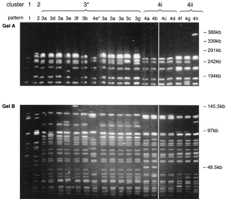
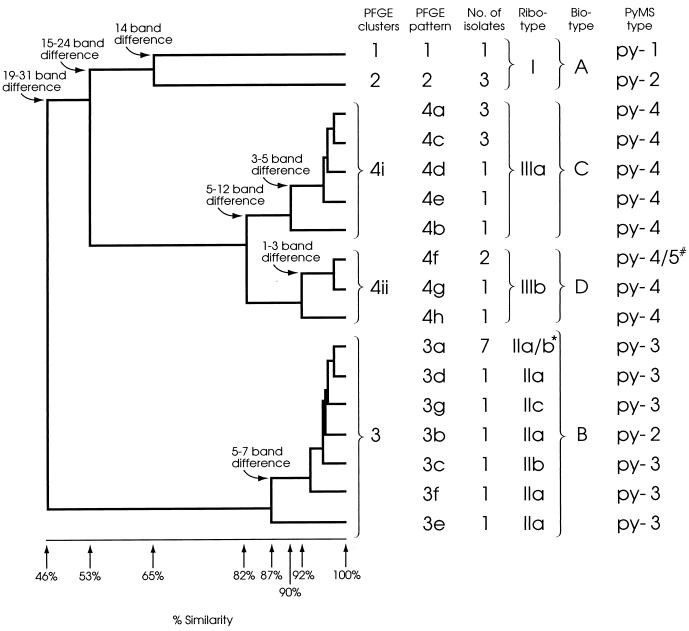
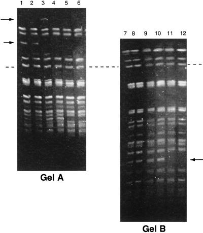
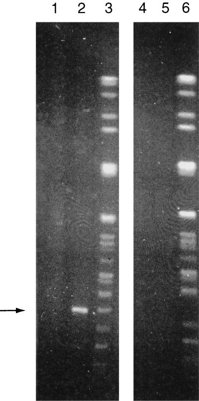
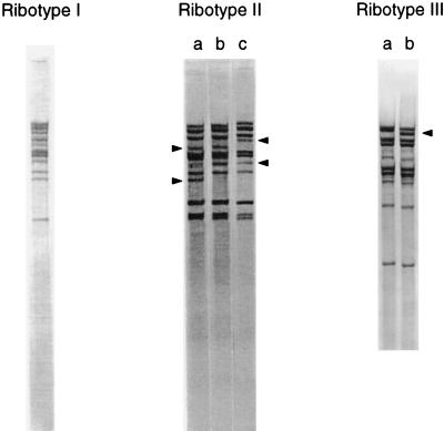
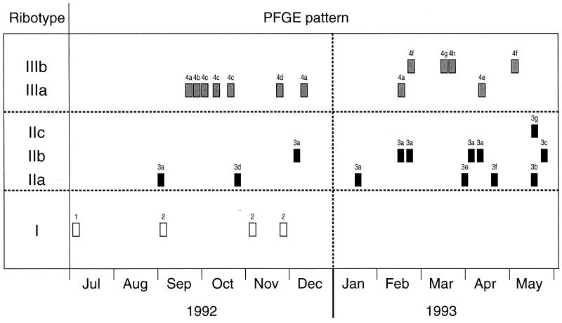
The degree of DNA banding pattern polymorphism exhibited by vancomycin-resistant Enterococcus faecium (VREM) strains isolated on a renal unit over an 11-month period was investigated. Thirty VREM strains from different patients were analyzed by pulsed-field gel electrophoresis (PFGE; with extended run and optimal pulse times), ribotyping, plasmid profile analysis, biotyping, pyrolysis mass spectrometry, and antibiogram analysis. PFGE resolved 17 banding patterns which formed four distinct clusters at the 82% similarity level. Intercluster band differences ranged from 14 to 31 bands. The strains in one cluster, which contained seven patterns that differed from each other by one to seven bands and from the common pattern by five bands, were confirmed to be a single strain by four of the five other typing methods. The strains in a second cluster with eight patterns, which differed from each other by 1 to 12 bands, contained two subclusters. This subdivision was supported by ribotyping and biotyping. However, it was unclear whether these subclusters represented distinct strains. In one strain, marked polymorphism (patterns that differed from each other by up to four bands) was observed in the ribotype pattern. This study demonstrates the high degree of DNA banding pattern polymorphism found for some strains of VREM and illustrates the complexity involved in defining such strains.


Figures

FIG. 1
FIG. 1
PFGE of SmaI-digested DNA from VREM showing representative pattern of all 17 patterns that were resolved. (Gel A) Linear ramped pulse time of 10 to 40 s with a run time of 40 h. Only well-separated bands above 145 kb are shown. (Gel B) Linear ramped pulse time of 1 to 10 s with a run time of 40 h. Only well-separated bands below 145 kb are shown. *, pattern 4e is included among the cluster 3 patterns on this gel.
FIG. 2
FIG. 2
Dendrogram produced following Dice and UPGMA analysis of the PFGE patterns of SmaI-digested DNA. The percent similarities and band differences between and within clusters are shown. The corresponding ribotype, biotype, and PyMS type of each isolate are also included. *, two of the PFGE pattern 3a isolates were ribotype IIa and five were ribotype IIb; #, one of the PFGE pattern 4f isolates was PyMS type py-4 and the other was PyMS type py-5.
FIG. 3
FIG. 3
PFGE banding patterns of the small colonial variant of an isolate of VREM following 20 serial single colony subcultures. (Gel A) Linear ramped pulse time of 10 to 40 s with a run time of 40 h. (Gel B) Linear ramped pulse time of 1 to 10 s with a run time of 40 h. Lanes 1 and 7, original culture; lanes 2 and 8, 1st subculture; lanes 3 and 9, 5th subculture; lanes 4 and 10, 10th subculture; lanes 5 and 11, 15th subculture; lanes 6 and 12, 20th subculture. Arrows, bands which appeared and/or disappeared during subculture; dashed line, the positions of the same band on both gels.
FIG. 4
FIG. 4
PFGE of total DNA and plasmid preparation by using ramped pulse times of 1 to 10 s for 30 h followed by 10 to 30 s for 15 h. Lanes 1 to 3, isolate with PFGE pattern 4a; lanes 4 to 6, isolate with PFGE pattern 4c; lanes 1 and 4, undigested plasmid preparation; lanes 2 and 5, plasmid preparation digested with SmaI; lanes 3 and 6, total DNA digested with SmaI. Arrow, plasmid band.
FIG. 5
FIG. 5
Ribotyping patterns of the VREM strains. Arrowheads, alteration in bands responsible for the subtypes (a, b, and c) of ribotype II and the subtypes (a and b) of ribotype III.
FIG. 6
FIG. 6
Time chart showing the date when each of the 30 VREM isolates was isolated on the renal unit and the corresponding ribotype and PFGE pattern for each strain.

References

    1. Arpin C, Coze C, Rogues A M, Gachie J P, Bebear C, Quentin C. Epidemiological study of an outbreak due to multidrug-resistant Enterobacter aerogenes in a medical intensive care unit. J Clin Microbiol. 1996;34:2163–2169. - PMC - PubMed
    1. Bonten M J M, Hayden M K, Nathan C, Rice T W, Weinstein R A. Stability of vancomycin-resistant enterococcal genotypes isolated from long-term-colonized patients. J Infect Dis. 1998;177:378–382. - PubMed
    1. Chetoui H, Melin P, Struelens M J, Delhalle E, Mutro Nigro M, de Ryck R, de Mol P. Comparison of biotyping, ribotyping, and pulsed-field gel electrophoresis for investigation of a common-source outbreak of Burkholderia pickettii bacteremia. J Clin Microbiol. 1997;35:1398–1403. - PMC - PubMed
    1. Chow J W, Perri M B, Thal L A, Zervos M J. Mobilization of the penicillinase gene in Enterococcus faecalis. Antimicrob Agents Chemother. 1993;37:1187–1189. - PMC - PubMed
    1. Clermont D, Delbos F, de Cespedes G, Horaud T. Old and new (Tn3708) mobile chromosomal elements in streptococci and enterococci. In: Ferretti J J, Gilmore M S, Klaenhammer T R, Brown F, editors. Genetics of streptococci, enterococci and lactococci. Basel, Switzerland: Krager; 1995. pp. 55–61. - PubMed

MeSH terms

LinkOut - more resources