Comparative Study
. 1999 Apr;37(4):1137-43.
doi: 10.1128/JCM.37.4.1137-1143.1999.

Genetic diversity of the 28-kilodalton outer membrane protein gene in human isolates of Ehrlichia chaffeensis

Affiliations
Comparative Study

Genetic diversity of the 28-kilodalton outer membrane protein gene in human isolates of Ehrlichia chaffeensis

X J Yu et al. J Clin Microbiol. 1999 Apr.

Abstract

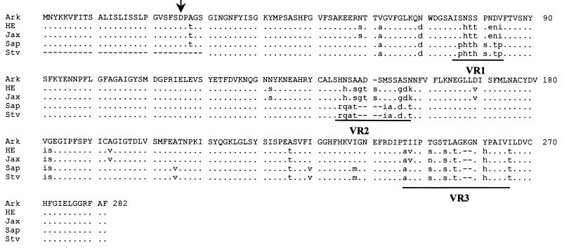
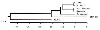
The Ehrlichia chaffeensis 28-kDa outer membrane protein (p28) gene was sequenced completely by genomic walking with adapter PCR. The DNA sequence of the p28 gene was nearly identical to the previously reported sequence (N. Ohashi, N. Zhi, Y. Zhang, and Y. Rikihisa, Infect. Immun. 66:132-139, 1998), but analysis of a further 75 bp on the 5' end of the gene revealed DNA that encoded a 25-amino-acid signal sequence. The leader sequence was removed from the N terminus of a 30-kDa precursor to generate the mature p28 protein. A monoclonal antibody (MAb), 1A9, recognizing four outer membrane proteins of E. chaffeensis (Arkansas strain) including the 25-, 26-, 27-, and 29-kDa proteins (X.-J. Yu, P. Brouqui, J. S. Dumler, and D. Raoult, J. Clin. Microbiol. 31:3284-3288, 1993) reacted with the recombinant p28 protein. This result indicated that the four proteins recognized by MAb 1A9 were encoded by the multiple genes of the 28-kDa protein family. DNA sequence alignment analysis revealed divergence of p28 among all five human isolates of E. chaffeensis. The E. chaffeensis strains could be divided into three genetic groups on the basis of the p28 gene. The first group consisted of the Sapulpa and St. Vincent strains. They had predicted amino acid sequences identical to each other. The second group contained strain 91HE17 and strain Jax, which only showed 0.4% divergence from each other. The third group contained the Arkansas strain only. The amino acid sequences of p28 differed by 11% between the first two groups, by 13.3% between the first and third groups, and by 13.1% between the second and third groups. The presence of antigenic variants of p28 among the strains of E. chaffeensis and the presence of multiple copies of heterogeneous genes suggest a possible mechanism by which E. chaffeensis might evade the host immune defenses. Whether or not immunization with the p28 of one strain of E. chaffeensis would confer cross-protection against other strains needs to be investigated.


Figures

FIG. 1
FIG. 1
Amino acid sequence alignment of the p28 proteins of five E. chaffeensis human isolates. The complete amino acid sequence of the Arkansas strain is presented as the consensus sequence. Differences from the consensus sequence are presented in lowercase. Dots represent the amino acids identical to those of the Arkansas strain, and dashes indicate gaps which were introduced for optimal alignment of the amino acid sequences. The variable regions (VR1, VR2, and VR3) are underlined. The arrow indicates the cleavage site of the signal peptide. The N-terminal portion of the St. Vincent strain sequence is incomplete. Ark, Arkansas; HE, 91HE17; Sap, Sapulpa; Stv, St. Vincent.
FIG. 2
FIG. 2
Protein immunoblotting of MAb 1A9 reacted with the p28 recombinant protein. Lanes: 1, heat-denatured E. chaffeensis (Arkansas strain) antigen; 2, GST fusion protein with p28; 3, thrombin-cleaved GST fusion protein (the arrow indicates the thrombin-cleaved recombinant p28); 4, GST protein only. The multiple bands in lanes 2 and 3 were apparently degradation products of the GST fusion protein.
FIG. 3
FIG. 3
Diagram of PCR amplification of the p28 gene. The dark boxes represent noncoding DNA sequences bordering the p28 gene. The shaded box and the open box represent the DNA sequence encoding the leader peptide and the sequence encoding the mature p28, respectively. The numbers on the top of the gene indicate the nucleotide positions in base pairs. Arrows indicate the directions of primers. The start point of each primer corresponds to the number at the end of the arrow and on the top of the p28 gene.
FIG. 4
FIG. 4
Alignment of the p28 gene coding DNA sequences. The complete DNA sequence of the Arkansas strain is presented as a consensus sequence. Differences from the consensus sequence are presented in lowercase. Dots represent the nucleotides of other strains of E. chaffeensis identical to those of the Arkansas strain, and dashes indicate gaps which were introduced for optimal alignment of the DNA sequences. The sequence of the St. Vincent strain is incomplete at the 5′ end. Ark, Arkansas; HE, 91HE17; Sap, Sapulpa; Stv, St. Vincent.
FIG. 5
FIG. 5
Phylogenetic tree constructed on the basis of the predicted amino acid sequences from the p28 genes of the E. chaffeensis strains. The predicted amino acid sequences of E. chaffeensis OMP-1F (19) and C. ruminantium MAP-1 (25) were included in the analysis to build the root of the tree. The Megalign program of Lasergene software was used to construct the tree. The length of each pair of branches represents the distance between sequence pairs. The scale beneath the tree measures the distance between the sequences.
FIG. 6
FIG. 6
Comparison of the predicted protein characteristics from the amino acids of the p28 proteins of the Arkansas and Jax strains. Surface probability predicts the surface residues by using a window consisting of a hexapeptide. A surface residue is any residue with >2.0 nm2 of water-accessible surface area. A hexapeptide with a value of greater than 1 was considered as surface region. The antigenic index predicts potential antigenic determinants. The regions with a value above 0 are potential antigenic determinants. The T-cell motif locates the potential T-cell antigenic determinants by using a motif of five amino acids with residue 1 glycine or polar, residue 2 hydrophobic, residue 3 hydrophobic, residue 4 hydrophobic or proline, and residue 5 polar or glycine. The scale indicates the amino acid positions.

Similar articles

Cited by

References

    1. Anderson B E, Dawson J E, Jones D C, Wilson K H. Ehrlichia chaffeensis, a new species associated with human ehrlichiosis. J Clin Microbiol. 1991;29:2838–2842. - PMC - PubMed
    1. Anderson B E, Sims K G, Olson J G, Childs J E, Piesman J F, Happ C M, Maupin G O, Johnson B J. Amblyomma americanum: a potential vector of human ehrlichiosis. Am J Trop Med Hyg. 1993;49:239–244. - PubMed
    1. Brouqui P, Dumler J S, Raoult D, Walker D H. Antigenic characterization of ehrlichiae: protein immunoblotting of Ehrlichia canis, Ehrlichia sennetsu, and Ehrlichia risticii. J Clin Microbiol. 1992;30:1062–1066. - PMC - PubMed
    1. Chen S-M, Cullman L C, Walker D H. Western immunoblotting analysis of the antibody responses of patients with human monocytotropic ehrlichiosis to different strains of Ehrlichia chaffeensis and Ehrlichia canis. Clin Diagn Lab Immunol. 1997;4:731–735. - PMC - PubMed
    1. Chen S M, Dumler J S, Feng H M, Walker D H. Identification of the antigenic constituents of Ehrlichia chaffeensis. Am J Trop Med Hyg. 1994;50:52–58. - PubMed

Publication types

MeSH terms

LinkOut - more resources