. 1999 Apr;44(4):476-82.
doi: 10.1136/gut.44.4.476.

The effect of hiatus hernia on gastro-oesophageal junction pressure

Affiliations

The effect of hiatus hernia on gastro-oesophageal junction pressure

P J Kahrilas et al. Gut. 1999 Apr.

Abstract

Background: Hiatus hernia and lower oesophageal sphincter hypotension are often viewed as opposing hypotheses for gastro-oesophageal junction incompetence.

Aims: To examine the interaction between hiatus hernia and lower oesophageal sphincter hypotension.

Methods: In seven normal subjects and seven patients with hiatus hernia, the squamocolumnar junction and intragastric margin of the gastro-oesophageal junction were marked with endoscopically placed clips. Axial and radial characteristics of the gastro-oesophageal junction high pressure zone were mapped relative to the hiatus and clips during concurrent fluoroscopy and manometry. Responses to inspiration and abdominal compression were also analysed.

Results: In normal individuals the squamocolumnar junction was 0.5 cm below the hiatus and the gastro-oesophageal junction high pressure zone extended 1.1 cm distal to that. In those with hiatus hernia, the gastro-oesophageal junction high pressure zone had two discrete segments, one proximal to the squamocolumnar junction and one distal, attributable to the extrinsic compression within the hiatal canal. Inspiration and abdominal compression mainly augmented the distal one. Simulation of hernia reduction by algebraically summing the proximal segment pressures with the hiatal canal pressures restored normal maximal pressure, radial asymmetry, and dynamic responses of the gastro-oesophageal junction.

Conclusions: Hiatus hernia reduces lower oesophageal sphincter pressure and alters its dynamic responsiveness by spatially separating pressure components derived from the intrinsic lower oesophageal sphincter and the extrinsic compression of the oesophagus within the hiatal canal.


Figures

Figure 1
Figure 1
Positioning of mucosal clips in normal subjects and patients with hiatus hernia. The central panel is a tracing from a fluoroscopic image of a normal subject illustrating the position of the mucosal clips relative to fluoroscopic landmarks. The opposing open arrows indicate the position of the centre of the hiatal canal as determined by the impression into the barium column during a barium swallow.
Figure 2
Figure 2
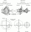
Examples of manometric pull through tracings with single (top) and double (bottom) peak axial pressure profiles. The median axial position and the pressure of the maximum (double vertical lines), minimum (single vertical lines), and three equispaced intermediate positions (vertical dotted lines) of each upward and downward slope were used to summarise group data. The open clips show the position of the squamocolumnar junction, the closed clips indicate the position of the intragastric clip, and the open squares indicate the centre of the hiatal canal.
Figure 3
Figure 3
Axial (top) and radial (bottom) pressure topography of the gastro-oesophageal junction of normal subjects and patients with hiatus hernia. The radial pressure profiles at the peak of each high pressure zone are diagrammed underneath. Position zero on the axial scale is the midpoint of the diaphragmatic hiatus. The wireframe representations are rotated such that the right anterior pressure is at the top and left posterior pressure is at the bottom, thereby accentuating the radial pressure asymmetry. The proximal clip indicates the median position of the squamocolumnar junction (SCJ) and the distal clip marks the median position of the intragastric aspect to the gastro-oesophageal junction as imaged endoscopically. All values of length and pressure are the medians of the subject groups.
Figure 4
Figure 4
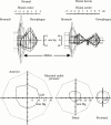
Change in the pressure topography of the gastro-oesophageal junction resulting from abdominal compression with an abdominal binder. The solid lines represent the condition at rest (represented as in fig 3) and the broken lines show movement and pressure augmentation associated with abdominal compression. Note that abdominal compression shifts the position of the squamocolumnar junction proximally by about 0.5 cm, increases the intragastric pressure, and causes a substantial increase in pressure within the hiatal canal of the hernia subjects. All values of length and pressure are the medians of the subject groups.
Figure 5
Figure 5
Change in the pressure topography of the gastro-oesophageal junction resulting from a Müller manoeuvre. The solid lines represent the condition at rest (represented as in fig 3) and the broken lines show movement and pressure augmentation associated with inspiration against a closed glottis. Note that the Müller manoeuvre shifts the position of the squamocolumnar junction distally by more than 1 cm in the normals but by less than 0.5 cm in the patients with hiatus hernia. In both subject groups, the Müller manoeuvre increases the intragastric pressure, and causes a substantial increase in pressure within the hiatal canal. All values of length and pressure are the medians of the subject groups.
Figure 6
Figure 6
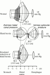
Simulation of reducing the hiatus hernia by algebraically repositioning the pressure values of the intrinsic LOS (pressure peak proximal to the squamocolumnar junction (SCJ)) to within the extrinsically determined pressure of the hiatal canal. For each subject the positioning of the proximal high pressure zone was such that the squamocolumnar junction mucosal clip attained the median normal position, 0.5 cm distal to the hiatus. The shaded area indicates the portion of the sphincter segment distal to the squamocolumnar junction in the normals and in the transposed panels. All values of length and pressure are the medians of the subject groups.

Comment in

References

    1. Physiol Rev. 1958 Oct;38(4):533-84 - PubMed
    1. Gut. 1997 Feb;40(2):167-74 - PubMed
    1. Surg Gynecol Obstet. 1964 Jun;118:1291-4 - PubMed
    1. Gastroenterology. 1962 Nov;43:521-31 - PubMed
    1. Gastrointest Endosc. 1996 Nov;44(5):541-7 - PubMed

Publication types