. 1999 Apr;44(4):504-10.
doi: 10.1136/gut.44.4.504.

Bacteroides fragilis toxin 2 damages human colonic mucosa in vitro

Affiliations

Bacteroides fragilis toxin 2 damages human colonic mucosa in vitro

M Riegler et al. Gut. 1999 Apr.

Abstract

Background: Strains of Bacteroides fragilis producing a 20 kDa protein toxin (B fragilis toxin (BFT) or fragilysin) are associated with diarrhoea in animals and humans. Although in vitro results indicate that BFT damages intestinal epithelial cells in culture, the effects of BFT on native human colon are not known.

Aims: To examine the electrophysiological and morphological effects of purified BFT-2 on human colonic mucosa in vitro.

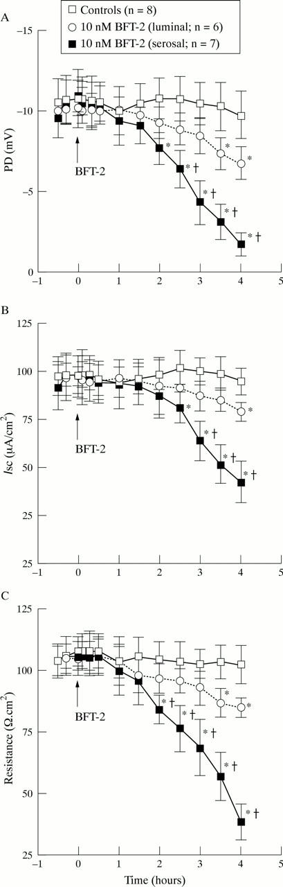
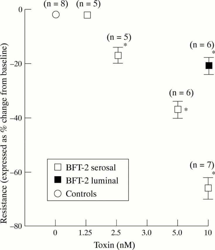
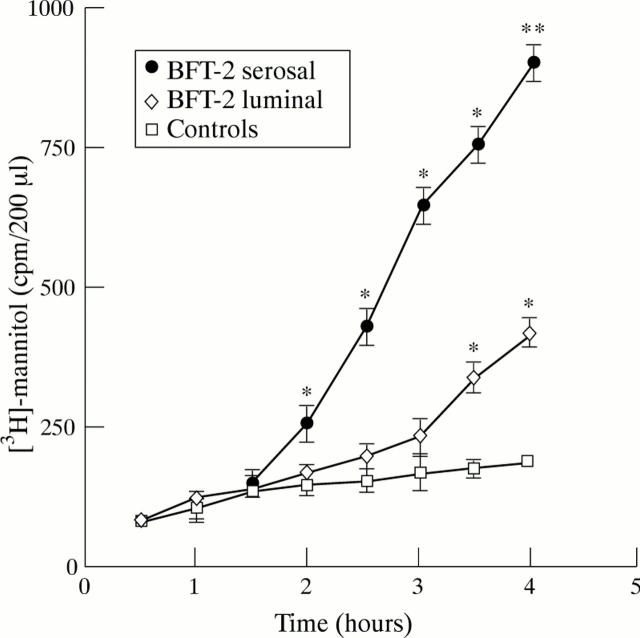
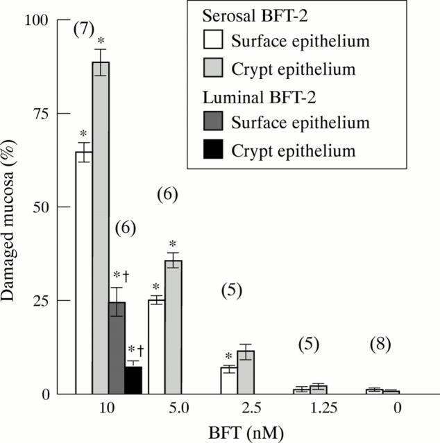
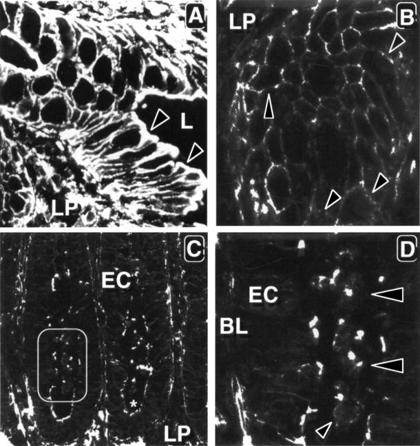
Methods: For resistance (R) measurements, colonic mucosa mounted in Ussing chambers was exposed to luminal or serosal BFT-2 (1.25-10 nM) and after four hours morphological damage was measured on haematoxylin and eosin stained sections using morphometry. F actin distribution was assessed using confocal microscopy.

Results: Serosal BFT-2 for four hours was four-, two-, seven-, and threefold more potent than luminal BFT-2 in decreasing resistance, increasing epithelial 3H-mannitol permeability, and damaging crypt and surface colonocytes, respectively (p<0.05). Confocal microscopy showed reduced colonocyte F actin staining intensity after exposure to BFT-2.

Conclusions: BFT-2 increases human colonic permeability and damages human colonic epithelial cells in vitro. These effects may be important in the development of diarrhoea and intestinal inflammation caused by B fragilis in vivo.


Figures

Figure 1
Figure 1
Time course of the effect of BFT-2 on (A) colonic potential difference (PD), (B) short circuit current (Isc), and (C) resistance. Values represent mean (SEM); n = number of experiments. *p<0.05 versus controls; †p<0.05 versus luminal BFT.
Figure 2
Figure 2
Dose dependent effect of BFT-2 on colonic resistance. Values represent mean (SEM); n = number of experiments. *p<0.05 versus controls.
Figure 3
Figure 3
Effect of BFT-2 on colonic 3H-mannitol permeability. Values are mean (SEM) of five experiments performed in triplicate. *p<0.05, **p<0.01 versus controls.
Figure 4
Figure 4
Morphological effect of BFT-2 on human colonic mucosa. Human colonic mucosal sheets were incubated with either buffer alone for four hours (A) or serosal buffer containing 10 nM BFT-2 for 2.5 hours (B) or four hours (C, D). At the end of the incubation period sheets were fixed in formalin and processed for light microscopy. (A) Control tissue appears normal and epithelial cells form a continuous sheet over the lamina propria. (B) The mucosal sheet shows disruption of crypt epithelium whereas the surface epithelium remains intact. (C) The mucosal sheet shows disruption of surface and crypt epithelium. The arrowhead marks an area shown at higher magnification in (D) which shows rounding and detachment of crypt enterocytes into the lumen (long arrows). Surface colonocytes show cytoplasmic elongation and apical localisation of the pyknotic nucleus (arrowheads). Note loosening of the basal lamina (broad arrows). Original magnification × 140 (A, B); × 80 (C); × 420 (D).
Figure 5
Figure 5
Morphometric analysis of colonic epithelial cell damage caused by BFT-2. Values are mean (SEM). The number of experiments is given in brackets. *p<0.05 versus controls; †p<0.05 versus serosal 10 nM BFT-2.
Figure 6
Figure 6
Effect of BFT-2 on cellular F actin. Fluorescent photomicrograph of human colonic mucosal sheet incubated with buffer alone (A) or serosal buffer containing 10 nM BFT-2 for two hours (B) or four hours (C, D). Samples were fixed and fresh frozen sections cut perpendicularly (B) or vertically (A, C, D) to the long axis of surface cells. (A) Control cells had a polygonal shape with F actin distributed in the peripheral actin ring. Arrowheads mark intense F actin staining of the apical brush border. L, lumen; LP, lamina propria. (B) There was an overall reduction in F actin staining intensity and disruption of the F actin ring (arrowheads). (C) There was an overall reduction in F actin staining in epithelial cells (EC). The rectangle marks a crypt shown at higher magnification in (D), and the asterisk marks the basal lumen of the crypt. (D) Staining of the enterocyte F actin ring was nearly abolished. Some F actin staining remained in the brush border of damaged enterocytes (EC) or in exfoliated enterocytes (arrowheads). BL, basal lamina. Original magnification × 400 (A, B); × 200 (C); ×480 (D).

References

    1. Nature. 1995 Jun 8;375(6531):500-3 - PubMed
    1. J Clin Invest. 1995 May;95(5):2004-11 - PubMed
    1. Infect Immun. 1995 Oct;63(10):3820-6 - PubMed
    1. Clin Infect Dis. 1995 Jun;20 Suppl 2:S142-8 - PubMed
    1. Infect Immun. 1996 Jan;64(1):113-9 - PubMed

Publication types