

Clinical spectral characterisation of colonic mucosal lesions using autofluorescence and delta aminolevulinic acid sensitisation
- PMID: 10075958
- PMCID: PMC1727450
- DOI: 10.1136/gut.44.4.511
Clinical spectral characterisation of colonic mucosal lesions using autofluorescence and delta aminolevulinic acid sensitisation
Abstract
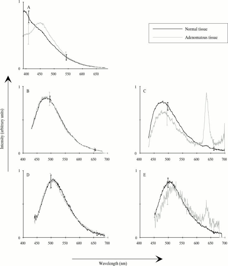
Background and aims: Laser induced fluorescence (LIF) from colonic mucosa was measured in vivo with and without delta aminolevulinic acid (ALA) in an attempt to differentiate between neoplasia and non-neoplasia in real time during colonoscopy.
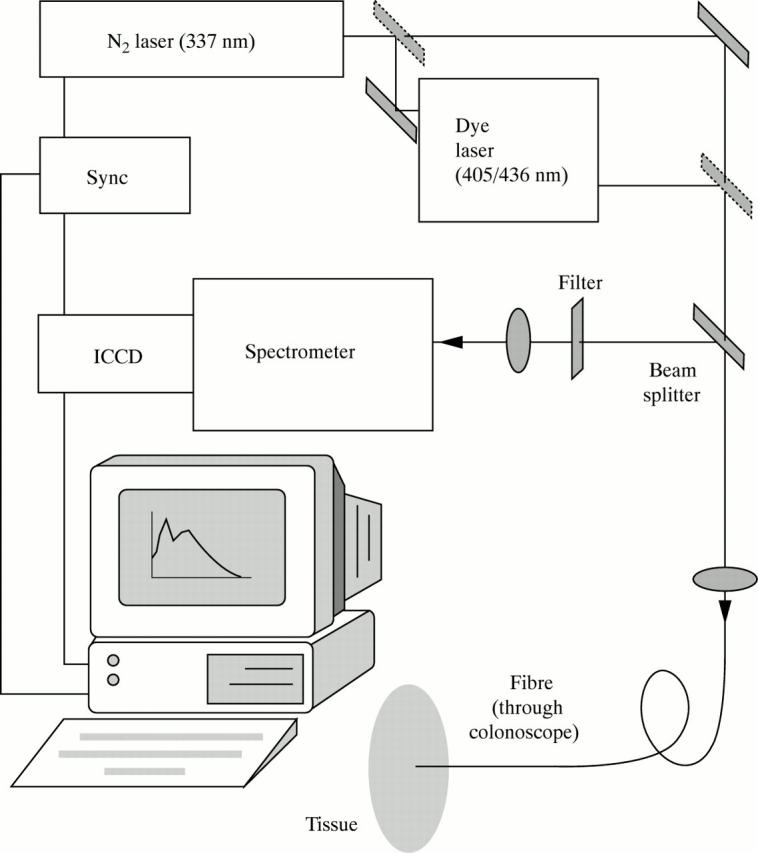
Methods: Spectra from 32 adenomas, 68 normal sites, and 14 hyperplastic polyps in 41 patients were obtained with a point monitoring system. Twenty one of the patients had been given a low dose of ALA as a photosensitiser before the examination. Light of 337, 405, or 436 nm wavelength was used as excitation. Stepwise multivariate linear regression analysis was performed.
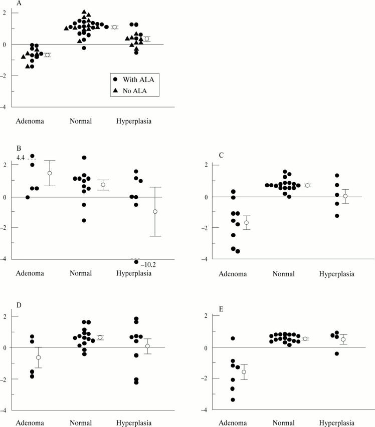
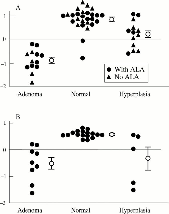
Results: With 337 nm excitation, 100% sensitivity and 96% specificity was obtained between normal mucosa and adenomas. Seventy seven per cent of the hyperplastic polyps were classified as non-neoplastic. When exciting with 405 and 436 nm, the possibility of distinguishing different types of tissue was considerably better in the ALA patients than in the non-ALA patients.
Conclusions: The in vivo point measurements imply that a good discrimination between normal tissue and adenomatous polyps can be obtained using the LIF technique. Excitation at 337 nm and at 405 nm or 436 nm using ALA gives good results. LIF also shows potential for distinguishing adenomatous from hyperplastic polyps. The number of detection wavelengths could be reduced if chosen properly.
Figures
Similar articles
-
Ultraviolet laser-induced fluorescence of colonic tissue: basic biology and diagnostic potential.Lasers Surg Med. 1992;12(1):63-78. doi: 10.1002/lsm.1900120111. Lasers Surg Med. 1992. PMID: 1614265
-
Autofluorescence characterisation of isolated whole crypts and primary cultured human epithelial cells from normal, hyperplastic, and adenomatous colonic mucosa.J Clin Pathol. 2005 Jul;58(7):766-74. doi: 10.1136/jcp.2004.023804. J Clin Pathol. 2005. PMID: 15976349 Free PMC article.
-
Evaluation of autofluorescence colonoscopy for the detection and diagnosis of colonic polyps.Gastrointest Endosc. 2008 Aug;68(2):283-90. doi: 10.1016/j.gie.2007.10.039. Epub 2008 Mar 10. Gastrointest Endosc. 2008. PMID: 18329642
-
Narrow-band imaging in the colon: limitations and potentials.J Gastroenterol Hepatol. 2011 Nov;26(11):1589-96. doi: 10.1111/j.1440-1746.2011.06877.x. J Gastroenterol Hepatol. 2011. PMID: 21793916 Review.
-
From colonic polyps to colon cancer: pathophysiology, clinical presentation, screening and colonoscopic therapy.Minerva Gastroenterol Dietol. 2007 Dec;53(4):351-73. Minerva Gastroenterol Dietol. 2007. PMID: 18043553 Review.
Cited by
-
Improving endoscopic resolution and sampling: fluorescence techniques.Gut. 2003 Jun;52 Suppl 4(Suppl 4):iv30-3. doi: 10.1136/gut.52.suppl_4.iv30. Gut. 2003. PMID: 12746266 Free PMC article. Review.
-
Methotrexate used in combination with aminolaevulinic acid for photodynamic killing of prostate cancer cells.Br J Cancer. 2006 Aug 21;95(4):485-95. doi: 10.1038/sj.bjc.6603273. Epub 2006 Jul 25. Br J Cancer. 2006. PMID: 16868543 Free PMC article.
-
Evolutionary Trend Analysis of Research on 5-ALA Delivery and Theranostic Applications Based on a Scientometrics Study.Pharmaceutics. 2022 Jul 15;14(7):1477. doi: 10.3390/pharmaceutics14071477. Pharmaceutics. 2022. PMID: 35890373 Free PMC article.
-
Fluorescence spectroscopy of neoplastic and non-neoplastic tissues.Neoplasia. 2000 Jan-Apr;2(1-2):89-117. doi: 10.1038/sj.neo.7900077. Neoplasia. 2000. PMID: 10933071 Free PMC article. Review.
-
Evaluation of wavelength ranges and tissue depth probed by diffuse reflectance spectroscopy for colorectal cancer detection.Sci Rep. 2021 Jan 12;11(1):798. doi: 10.1038/s41598-020-79517-2. Sci Rep. 2021. PMID: 33436684 Free PMC article.
References
Publication types
MeSH terms
Substances
LinkOut - more resources
Full Text Sources
Other Literature Sources