. 1999 Mar 15;189(6):957-68.
doi: 10.1084/jem.189.6.957.

The cyclin-dependent kinase Cdk2 regulates thymocyte apoptosis

Affiliations

The cyclin-dependent kinase Cdk2 regulates thymocyte apoptosis

A Hakem et al. J Exp Med. .

Abstract

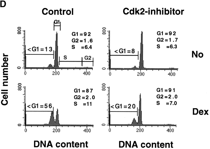
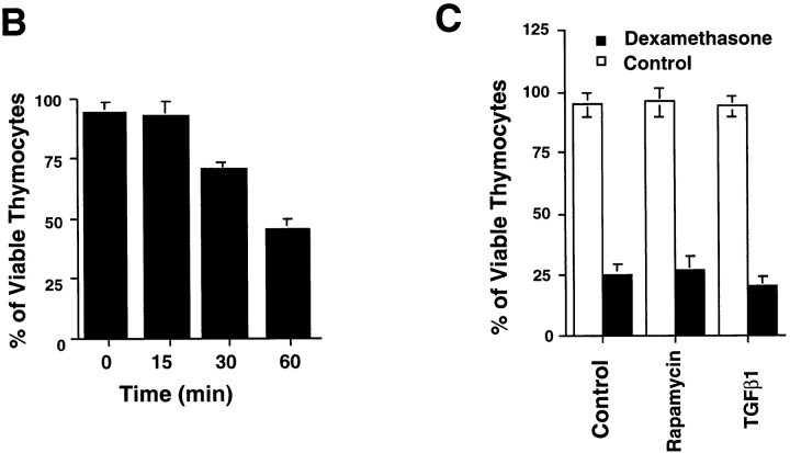
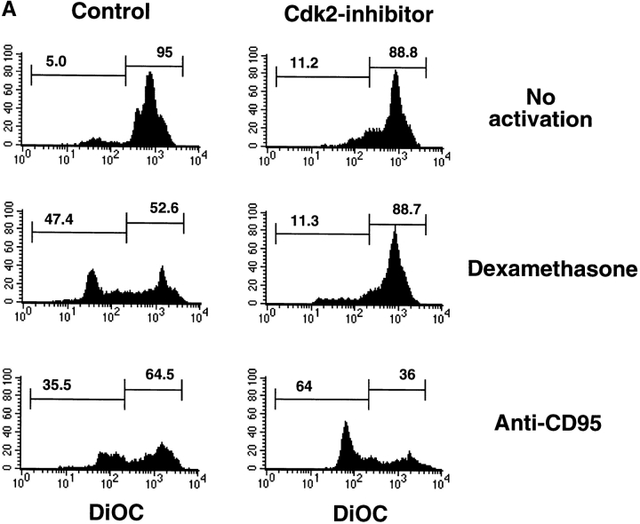
Aberrant activation of cell cycle molecules has been postulated to play a role in apoptosis ("catastrophic cell cycle"). Here we show that in noncycling developing thymocytes, the cyclin- dependent kinase Cdk2 is activated in response to all specific and nonspecific apoptotic stimuli tested, including peptide-specific thymocyte apoptosis. Cdk2 was found to function upstream of the tumor suppressor p53, transactivation of the death promoter Bax, alterations of mitochondrial permeability, Bcl-2, caspase activation, and caspase-dependent proteolytic cleavage of the retinoblastoma protein. Inhibition of Cdk2 completely protected thymocytes from apoptosis, mitochondrial changes, and caspase activation. These data provide the first evidence that Cdk2 activity is crucial for the induction of thymocyte apoptosis.


Figures

Figure 1
Figure 1
Induction of Cdk2 kinase activity in response to apoptotic stimuli. (A) Kinetics of Cdk2 and Cdc2 activity after dexamethasone treatment. Freshly isolated thymocytes were cultured in the absence or presence of dexamethasone (1 μM), and Cdk2 and Cdc2 activities were assessed. After the indicated periods of stimulation, cells were harvested and lysed, and proteins were immunoprecipitated using Abs against Cdk2 and Cdc2. Cdk2 and Cdc2 kinase activities in immunoprecipitates were assayed using [γ-32P]ATP and histone H1 as the substrate. One result representative of four independent experiments is shown. (B) Induction of Cdk2 activity in thymocytes in response to dexamethasone (1 μM), heat shock (42°C for 1 h), anti-CD95 (1 μg/ml), and γ-irradiation (500 rads). Thymocytes were harvested 5 h after stimulation. Top panel, histone H1 phosphorylation; middle panel, level of Cdk2 protein; bottom panel, kinase activity measured in arbitrary units normalized to background protein and kinase levels. One result representative of five independent experiments is shown. (C) Activity of Cdc2 and Cdk4 after induction of apoptosis. Thymocytes were cultured with dexamethasone (Dex) to induce apoptosis. After 5 h, cells were lysed and Cdk4 and Cdc2 were immunoprecipitated. Cdc2 and Cdk4 kinase activities were determined using histone H1 and GST-Rb as substrates, respectively. (D) Blockage of Cdk2 activity in thymocytes by specific inhibitor roscovitine (50 μM) after dexamethasone (Dex) treatment. Similar results were obtained using the Cdk2 blocker olomoucine. Note the background kinase activity of Cdk2 in A, B, and D which is due to the spontaneous apoptosis of thymocytes in culture (∼20% of thymocytes die within 24 h by default). Blockage of Cdk2 inhibits this form of default death and even after 1 wk in culture, >90% of thymocytes were viable in the presence of roscovitine or olomoucine (not shown). (E) Activity of Cdk2–cyclin A or Cdk2–cyclin E complexes on histone H1 after dexamethasone (1 μM) treatment. Cyclin A or cyclin E was immunoprecipitated using specific Abs, and kinase assays were performed as in A.
Figure 2
Figure 2
Inhibition of apoptosis by Cdk2 blockers. (A) CD4+CD8+ thymocyte apoptosis in response to various death stimuli. Thymocytes (2 × 10⁶/well) were cultured in the absence of stimulation (Control), or with dexamethasone (1 μM), heat shock (42°C for 1 h), γ-irradiation (500 rads), anti-CD95 (1 μg/ml), plate-bound anti-CD3ε (10 μg/ml), etoposide (2.5 μg/ml), or PMA (12.5 ng/ml) in the absence (black bars, Control) or presence of olomoucine (100 μM; white bars, Cdk2-inhibitor). After 22 h, thymocytes were harvested and apoptosis was determined. Similar results were obtained using roscovitine (not shown). 1 result representative of 15 independent experiments is shown. (B) Time course for the action of the Cdk2 inhibitors. Thymocytes were cultured in the presence of dexamethasone (1 μM), and olomoucine (100 μM) was added at the different indicated times. The “point of no return” is between 15 and 30 min. (C) Thymocytes were cultured in the absence of stimulation (Control) or with dexamethasone (1 μM) in the absence (Control) or presence of rapamycin (2 ng/ml) or TGF-β1 (1 nM). Thymocyte apoptosis was determined 24 h after addition of dexamethasone. (D) Cell cycle status and apoptosis of thymocytes. Thymocytes were activated with the indicated stimuli (as described for A) in the absence (Control) or presence of olomoucine (Cdk2-inhibitor). No (activation) indicates a culture not treated with death-inducing stimuli. After 5 h, cells were harvested and stained with PI. The subdiploid (<G1) peak represents the number of apoptotic cells among total cells. Cell cycle phases (G1, S, G2/M) are indicated. Numbers indicate the percentages of viable cells in the G1, S, and G2/M populations. 1 result representative of 10 independent experiments is shown.
Figure 2
Figure 2
Inhibition of apoptosis by Cdk2 blockers. (A) CD4+CD8+ thymocyte apoptosis in response to various death stimuli. Thymocytes (2 × 10⁶/well) were cultured in the absence of stimulation (Control), or with dexamethasone (1 μM), heat shock (42°C for 1 h), γ-irradiation (500 rads), anti-CD95 (1 μg/ml), plate-bound anti-CD3ε (10 μg/ml), etoposide (2.5 μg/ml), or PMA (12.5 ng/ml) in the absence (black bars, Control) or presence of olomoucine (100 μM; white bars, Cdk2-inhibitor). After 22 h, thymocytes were harvested and apoptosis was determined. Similar results were obtained using roscovitine (not shown). 1 result representative of 15 independent experiments is shown. (B) Time course for the action of the Cdk2 inhibitors. Thymocytes were cultured in the presence of dexamethasone (1 μM), and olomoucine (100 μM) was added at the different indicated times. The “point of no return” is between 15 and 30 min. (C) Thymocytes were cultured in the absence of stimulation (Control) or with dexamethasone (1 μM) in the absence (Control) or presence of rapamycin (2 ng/ml) or TGF-β1 (1 nM). Thymocyte apoptosis was determined 24 h after addition of dexamethasone. (D) Cell cycle status and apoptosis of thymocytes. Thymocytes were activated with the indicated stimuli (as described for A) in the absence (Control) or presence of olomoucine (Cdk2-inhibitor). No (activation) indicates a culture not treated with death-inducing stimuli. After 5 h, cells were harvested and stained with PI. The subdiploid (<G1) peak represents the number of apoptotic cells among total cells. Cell cycle phases (G1, S, G2/M) are indicated. Numbers indicate the percentages of viable cells in the G1, S, and G2/M populations. 1 result representative of 10 independent experiments is shown.
Figure 3
Figure 3
Cdk2 kinase is activated in peptide-specific thymocyte apoptosis. (A) Inhibition of Cdk2 blocks induction of peptide-specific apoptosis of P14 Tg thymocytes, which express an α/β TCR (TCRVα2Vβ8) specific for p33 peptide of LCMV. Purified P14 Tg thymocytes were cultured on p33 glycoprotein peptide– pulsed MC57/L fibroblasts in the absence (Peptide) or presence of roscovitine (50 μM; Peptide+Cdk2-inhibitor). Thymocytes were harvested after 22 h incubation and stained with anti-CD4–PE and anti-CD8–FITC. Percent survival was calculated as follows: (total number of viable CD4+ CD8+ thymocytes cultured at a given concentration of p33 peptide)/(total number of viable CD4+CD8+ thymocytes cultured with MC57/L cells at 37°C in the absence of peptide) × 100. Molarity of peptide concentrations is shown on the x-axis. Cells were harvested after 22 h of culture, and cell death was determined. Numbers are the percentages of viable CD4+CD8+ thymocytes cultured at various p33 peptide concentrations compared with control thymocytes cultured in the absence of p33 peptide (Control). Apoptosis of P14 thymocytes in the presence of roscovitine alone is also shown (Cdk2-inhibitor). Similar results were obtained using olomoucine (not shown). One result representative of five independent experiments is shown. (B) Induction of Cdk2 kinase activity by peptide-specific negative selection. P14 Tg thymocytes (106/well) were cultured on a monolayer of adherent MC57/L fibroblasts (H-2b/b) in the presence or absence of the deleting p33 peptide. After 5 h of culture, thymocytes were harvested, Cdk2 was immunoprecipitated, and Cdk2 activity was assessed using histone H1 as substrate. (C) Cdk2 inhibitors prevent cOVA protein (Ova-protein)–induced apoptosis of OVA-specific TCR Tg thymocytes in FTOCs. Thymi were removed at embyronic day 16 and cultured with olomoucine (100 μM) and cOVA protein (1 mg/ml). Percentages of viable thymocytes were determined.
Figure 3
Figure 3
Cdk2 kinase is activated in peptide-specific thymocyte apoptosis. (A) Inhibition of Cdk2 blocks induction of peptide-specific apoptosis of P14 Tg thymocytes, which express an α/β TCR (TCRVα2Vβ8) specific for p33 peptide of LCMV. Purified P14 Tg thymocytes were cultured on p33 glycoprotein peptide– pulsed MC57/L fibroblasts in the absence (Peptide) or presence of roscovitine (50 μM; Peptide+Cdk2-inhibitor). Thymocytes were harvested after 22 h incubation and stained with anti-CD4–PE and anti-CD8–FITC. Percent survival was calculated as follows: (total number of viable CD4+ CD8+ thymocytes cultured at a given concentration of p33 peptide)/(total number of viable CD4+CD8+ thymocytes cultured with MC57/L cells at 37°C in the absence of peptide) × 100. Molarity of peptide concentrations is shown on the x-axis. Cells were harvested after 22 h of culture, and cell death was determined. Numbers are the percentages of viable CD4+CD8+ thymocytes cultured at various p33 peptide concentrations compared with control thymocytes cultured in the absence of p33 peptide (Control). Apoptosis of P14 thymocytes in the presence of roscovitine alone is also shown (Cdk2-inhibitor). Similar results were obtained using olomoucine (not shown). One result representative of five independent experiments is shown. (B) Induction of Cdk2 kinase activity by peptide-specific negative selection. P14 Tg thymocytes (106/well) were cultured on a monolayer of adherent MC57/L fibroblasts (H-2b/b) in the presence or absence of the deleting p33 peptide. After 5 h of culture, thymocytes were harvested, Cdk2 was immunoprecipitated, and Cdk2 activity was assessed using histone H1 as substrate. (C) Cdk2 inhibitors prevent cOVA protein (Ova-protein)–induced apoptosis of OVA-specific TCR Tg thymocytes in FTOCs. Thymi were removed at embyronic day 16 and cultured with olomoucine (100 μM) and cOVA protein (1 mg/ml). Percentages of viable thymocytes were determined.
Figure 4
Figure 4
Cdk2 acts upstream of mitochondrial permeability transition, caspase activation, and Rb cleavage. (A) Mitochondrial permeability transition (ΔΨm disruption). Thymocytes were cultured for 5 h in medium alone, or in medium containing dexamethasone (1 μM) or anti-CD95 (1 μg/ml) in the presence or absence of roscovitine (50 μM). Cells induced to undergo apoptosis manifest an early reduction in the incorporation of ΔΨm-sensitive dyes, indicating a disruption of ΔΨm. For DiOC6(3) staining, 106 thymocytes were incubated with DiOC6(3) (final concentration 20 nM in PBS) for 20 min at 37°C. DiOC6(3) staining was analyzed immediately using a FACSCalibur™ flow cytometer. One result representative of three independent experiments is shown. (B) Thymocytes were stimulated with the indicated death stimuli for 5 h, and proteolytic activation of caspase 3 (Cpp32) was assessed by Western blotting. The anti–caspase 3 Ab recognizes both the intact caspase 3 molecule (procaspase 3) and its cleaved 17-kD active form (p17). Addition of roscovitine inhibits caspase 3 cleavage in response to dexamethasone (right) and in response to all other death stimuli tested, except for anti-CD95. (C) Thymocytes were stimulated with the indicated stimuli, and the proteolytic activation of caspase 2 (Nedd2) was assessed by Western blotting. The addition of roscovitine (Rosco) after irradiation blocked caspase 2 cleavage into a p14 fragment. (D) The retinoblastoma protein Rb is proteolytically processed in response to all cell death stimuli. Inhibition of Cdk2 blocked Rb cleavage. Thymocytes were stimulated with dexamethasone and γ-irradiation in the presence or absence of roscovitine (Rosco, 50 μM). After 5 h, cells were harvested and lysed, and the status of Rb was assessed by Western blotting. Rb-reactive Ab recognizes aa 300–380 of Rb. As previously described in cell lines (reference 53), Rb was cleaved of the COOH-terminal end in apoptotic thymocytes in response to all apoptotic stimuli.
Figure 4
Figure 4
Cdk2 acts upstream of mitochondrial permeability transition, caspase activation, and Rb cleavage. (A) Mitochondrial permeability transition (ΔΨm disruption). Thymocytes were cultured for 5 h in medium alone, or in medium containing dexamethasone (1 μM) or anti-CD95 (1 μg/ml) in the presence or absence of roscovitine (50 μM). Cells induced to undergo apoptosis manifest an early reduction in the incorporation of ΔΨm-sensitive dyes, indicating a disruption of ΔΨm. For DiOC6(3) staining, 106 thymocytes were incubated with DiOC6(3) (final concentration 20 nM in PBS) for 20 min at 37°C. DiOC6(3) staining was analyzed immediately using a FACSCalibur™ flow cytometer. One result representative of three independent experiments is shown. (B) Thymocytes were stimulated with the indicated death stimuli for 5 h, and proteolytic activation of caspase 3 (Cpp32) was assessed by Western blotting. The anti–caspase 3 Ab recognizes both the intact caspase 3 molecule (procaspase 3) and its cleaved 17-kD active form (p17). Addition of roscovitine inhibits caspase 3 cleavage in response to dexamethasone (right) and in response to all other death stimuli tested, except for anti-CD95. (C) Thymocytes were stimulated with the indicated stimuli, and the proteolytic activation of caspase 2 (Nedd2) was assessed by Western blotting. The addition of roscovitine (Rosco) after irradiation blocked caspase 2 cleavage into a p14 fragment. (D) The retinoblastoma protein Rb is proteolytically processed in response to all cell death stimuli. Inhibition of Cdk2 blocked Rb cleavage. Thymocytes were stimulated with dexamethasone and γ-irradiation in the presence or absence of roscovitine (Rosco, 50 μM). After 5 h, cells were harvested and lysed, and the status of Rb was assessed by Western blotting. Rb-reactive Ab recognizes aa 300–380 of Rb. As previously described in cell lines (reference 53), Rb was cleaved of the COOH-terminal end in apoptotic thymocytes in response to all apoptotic stimuli.
Figure 5
Figure 5
P53 is a substrate for Cdk2 activity. (A) Phosphorylation of p53 by Cdk2. p53 phosphorylation was determined after γ-irradiation (500 rads), dexamethasone treatment (1 μM), or no treatment (Control). Thymocytes were cultured and harvested as described in the legend to Fig. 1. Cdk2 was immunoprecipitated, and Cdk2 activity was determined using recombinant human p53 as the substrate. The levels of phosphorylated p53 (top), p53 protein (middle), and Cdk2 protein (bottom) are shown. (B) Coimmunoprecipitation of p53 and Cdk2. Thymocytes were harvested 2 h after γ-irradiation (500 rads) or after 2 h culture without a death stimulus (Control). Western blot analysis was performed after immunoprecipitation (IP) using an anti-Cdk2 Ab and blotting with anti-p53 and anti-Cdk2. Similar results were obtained using p53 immunoprecipitations. One result representative of three independent experiments is shown. (C) Levels of p53 protein. Thymocytes were cultured for 2 h after γ-irradiation (500 rads) or the absence of a death stimulus (Control) in the presence or absence of roscovitine (50 μM, Rosco). Cells were lysed, and extracts were analyzed by Western blotting using anti-p53 and anti–Bcl-XL Abs. One result representative of three independent experiments is shown. (D) Induction of Bax mRNA. Thymocytes were cultured as described in B and harvested after 3 h. 10 μg total RNA was Northern blotted and hybridized to probes for both Bax and β-actin (loading control). Hybridization with the Bax probe shows two alternatively spliced RNA transcripts of 1.5 and 1.0 kb. The increase in Bax mRNA induced by γ-irradiation is abolished in the presence of roscovitine (50 μM).
Figure 5
Figure 5
P53 is a substrate for Cdk2 activity. (A) Phosphorylation of p53 by Cdk2. p53 phosphorylation was determined after γ-irradiation (500 rads), dexamethasone treatment (1 μM), or no treatment (Control). Thymocytes were cultured and harvested as described in the legend to Fig. 1. Cdk2 was immunoprecipitated, and Cdk2 activity was determined using recombinant human p53 as the substrate. The levels of phosphorylated p53 (top), p53 protein (middle), and Cdk2 protein (bottom) are shown. (B) Coimmunoprecipitation of p53 and Cdk2. Thymocytes were harvested 2 h after γ-irradiation (500 rads) or after 2 h culture without a death stimulus (Control). Western blot analysis was performed after immunoprecipitation (IP) using an anti-Cdk2 Ab and blotting with anti-p53 and anti-Cdk2. Similar results were obtained using p53 immunoprecipitations. One result representative of three independent experiments is shown. (C) Levels of p53 protein. Thymocytes were cultured for 2 h after γ-irradiation (500 rads) or the absence of a death stimulus (Control) in the presence or absence of roscovitine (50 μM, Rosco). Cells were lysed, and extracts were analyzed by Western blotting using anti-p53 and anti–Bcl-XL Abs. One result representative of three independent experiments is shown. (D) Induction of Bax mRNA. Thymocytes were cultured as described in B and harvested after 3 h. 10 μg total RNA was Northern blotted and hybridized to probes for both Bax and β-actin (loading control). Hybridization with the Bax probe shows two alternatively spliced RNA transcripts of 1.5 and 1.0 kb. The increase in Bax mRNA induced by γ-irradiation is abolished in the presence of roscovitine (50 μM).

Similar articles

Cited by

References

    1. White E. Death-defying acts: a meeting review on apoptosis. Genes Dev. 1993;7:2277–2284. - PubMed
    1. Russell JH, White CL, Loh DY, Meleedy R-P. Receptor-stimulated death pathway is opened by antigen in mature T cells. Proc Natl Acad Sci USA. 1991;88:2151–2155. - PMC - PubMed
    1. Green DR, Scott DW. Activation-induced apoptosis in lymphocytes. Curr Opin Immunol. 1994;6:476–487. - PubMed
    1. King LB, Ashwell JD. Signaling for death of lymphoid cells. Curr Opin Immunol. 1993;5:368–373. - PubMed
    1. Martin JS, Green RD. Protease activation during apoptosis: death by thousand cuts? . Cell. 1995;82:349–352. - PubMed

Publication types

MeSH terms