. 1999 Mar 15;189(6):979-90.
doi: 10.1084/jem.189.6.979.

Characterization of the 4C8 antigen involved in transendothelial migration of CD26(hi) T cells after tight adhesion to human umbilical vein endothelial cell monolayers

Affiliations

Characterization of the 4C8 antigen involved in transendothelial migration of CD26(hi) T cells after tight adhesion to human umbilical vein endothelial cell monolayers

J Masuyama et al. J Exp Med. .

Abstract

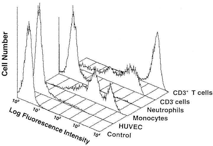
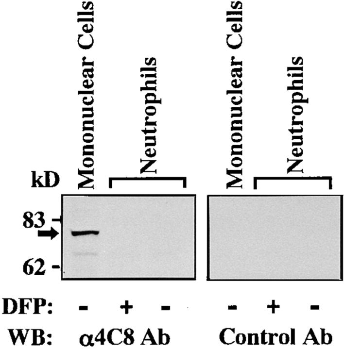
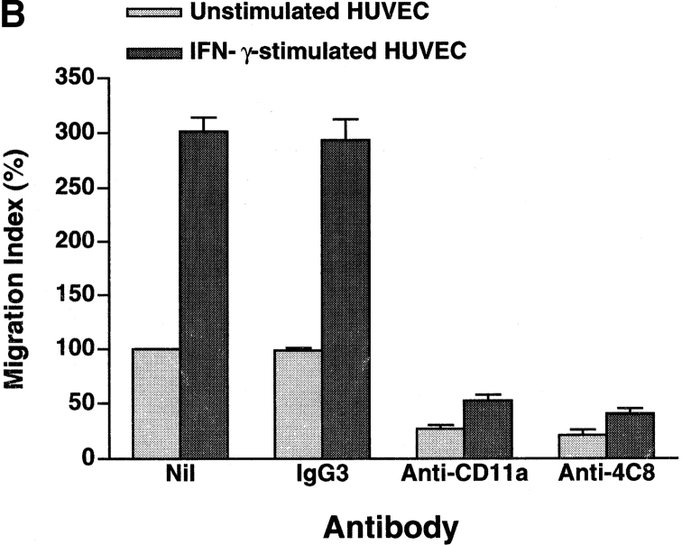
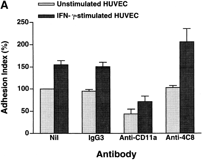
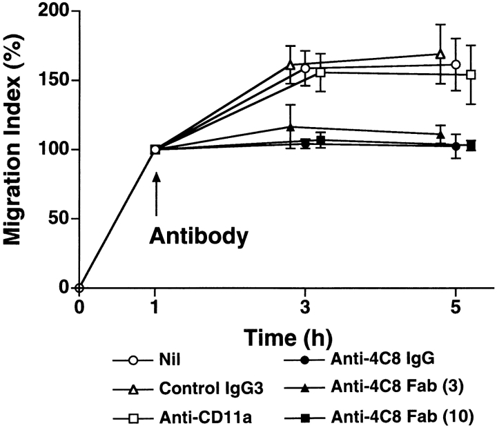
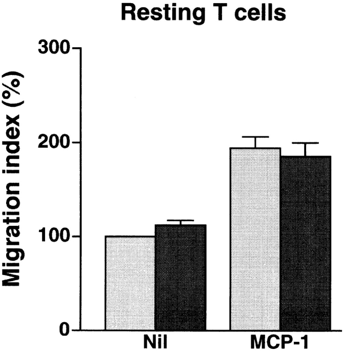
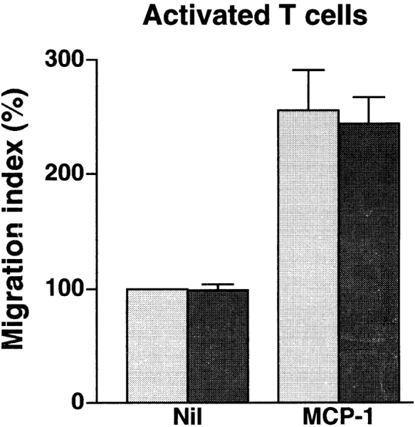
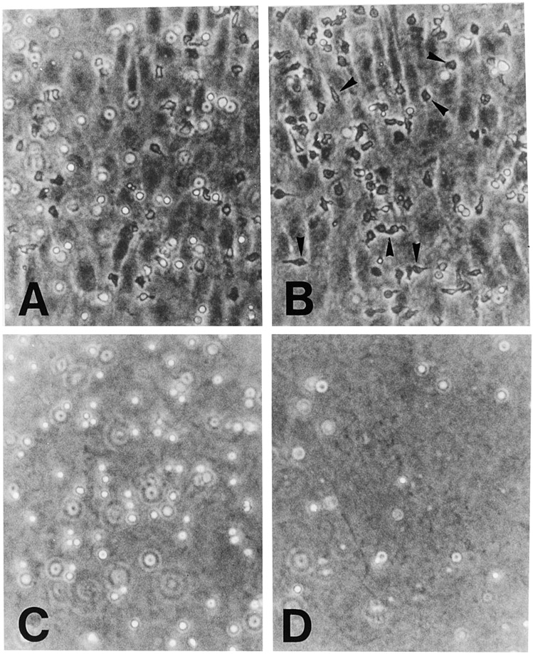
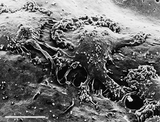
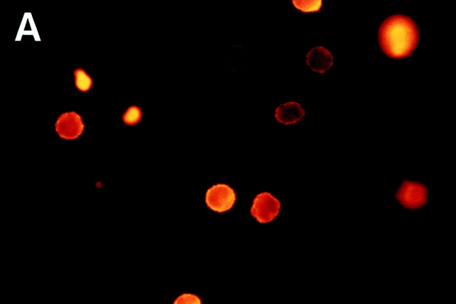
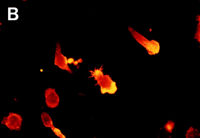
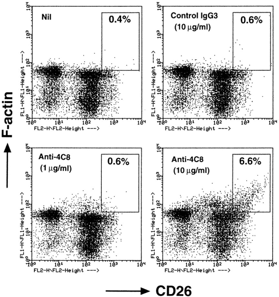
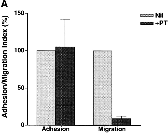
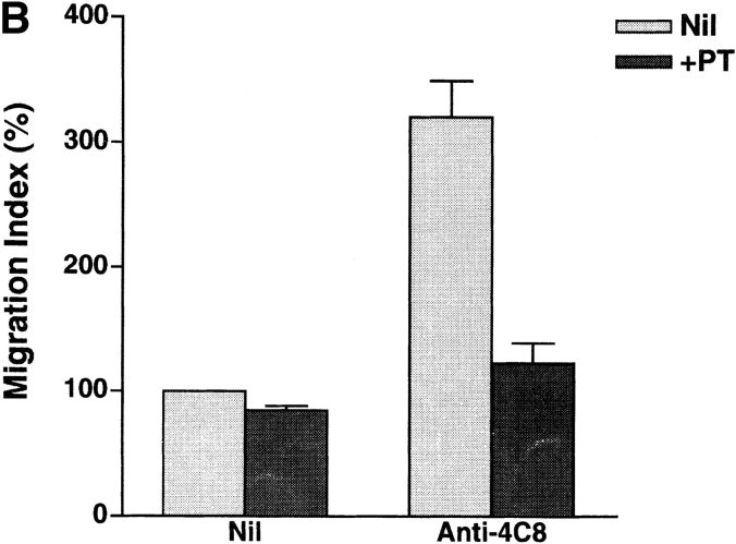
In extravasation of T cells, little is known about the mechanisms of transendothelial migration subsequent to the T cells' tight adhesion to endothelium. To investigate these mechanisms, we developed a monoclonal antibody (mAb), termed anti-4C8, that blocks transmigration but not adhesion in a culture system in which high CD26-expressing (CD26(hi)) T cells preferentially migrate through human umbilical vein endothelial cell (HUVEC) monolayers cultured on collagen gels. Anti-4C8 reacted with all CD3(+) T cells and monocytes but not neutrophils or HUVECs. The structure defined by this antibody was an 80-kD molecule. The mAb at 1 mug/ml inhibited 80-90% of migration of CD3(+) T cells through unstimulated and interferon gamma-stimulated HUVEC monolayers without interfering with adhesion and cell motility. When added to the cultures after the adhesion, anti-4C8 completely blocked subsequent transmigration of adherent T cells. Phase-contrast and electron microscopy revealed that T cells are arrested at the intercellular junctions of HUVECs in the presence of anti-4C8. Anti-4C8 exhibited agonistic effects on resting T cells without other stimuli under culture conditions in which anti-4C8 can stimulate T cells. First, in the checkerboard assay using collagen gels, the antibody promoted chemokinetic migration of the cells in a dose-dependent manner from 0.1 to 10 mug/ml. The predominant population of T cells that migrated into collagen gels with impregnated anti-4C8 were CD26(hi). Second, solid-phase-immobilized anti-4C8 induced adhesion of T cells to the substrate, often with polarizations in cell shape and large pseudopods rich in filamentous (F-) actin. Third, soluble anti-4C8 augmented F-actin content preferentially in CD26(hi) T cells when added to T cells at a high dose of 10 mug/ml. Finally, both anti-4C8-induced chemokinetic migration and transendothelial migration were inhibited by pretreatment of T cells with pertussis toxin. These findings suggest that stimulation via the 4C8 antigen increases cell motility of CD26(hi) cells with profound cytoskeletal changes through signaling pathways including G proteins. The 4C8 antigen may be involved in preferential transmigration of CD26(hi) cells adherent to HUVECs.


Figures

Figure 1
Figure 1
Immunofluorescence profiles of the 4C8 antigen on human PBLs and HUVECs. PBMC and neutrophils from a healthy donor were stained with FITC-conjugated anti-4C8 and phycoerythrin-Cy5–conjugated anti-CD3 mAb or control FITC-conjugated IgG3. The stained leukocytes as well as HUVECs (10,000 cells) were analyzed by a FACScan® (Becton Dickinson) flow cytometer with gating on the lymphocyte, monocyte, and neutrophil populations.
Figure 2
Figure 2
Western blot analysis of the 4C8 antigen. SDS–gel electrophoresis of lysates from PBMC and neutrophils (5.5 × 10⁶ cells/lane) was performed under reducing conditions. DFP, a serine protease inhibitor, was used to prevent proteolytic activity in the neutrophil lysates. Control Ab was purified mouse IgG3 of the same isotype as anti-4C8. See Materials and Methods for details.
Figure 3
Figure 3
Anti-4C8 mAb inhibits T cell migration through, but not adhesion to, HUVEC monolayers. Freshly isolated CD3+ T cells were incubated with unstimulated and IFN-γ–stimulated (500 U/ml, 48 h) HUVEC monolayers cultured on collagen gels in the continuous presence of anti-4C8 (1 μg/ml), anti-CD11a (10 μg/ml), or control IgG3 (10 μg/ml). The numbers of adherent cells and migrated cells were determined as described in Materials and Methods. Results of T cell adhesion (A) and migration (B) are expressed as the adhesion and the migration index, respectively, and represent the mean ± SD of five independent experiments. The index (%) is calculated as follows: the number of adherent or migrated T cells with antibody/the number of adherent or migrated T cells without antibody × 100. The numbers of the background adhesion and migration with unstimulated HUVEC are 376 ± 84 and 131 ± 4, respectively.
Figure 3
Figure 3
Anti-4C8 mAb inhibits T cell migration through, but not adhesion to, HUVEC monolayers. Freshly isolated CD3+ T cells were incubated with unstimulated and IFN-γ–stimulated (500 U/ml, 48 h) HUVEC monolayers cultured on collagen gels in the continuous presence of anti-4C8 (1 μg/ml), anti-CD11a (10 μg/ml), or control IgG3 (10 μg/ml). The numbers of adherent cells and migrated cells were determined as described in Materials and Methods. Results of T cell adhesion (A) and migration (B) are expressed as the adhesion and the migration index, respectively, and represent the mean ± SD of five independent experiments. The index (%) is calculated as follows: the number of adherent or migrated T cells with antibody/the number of adherent or migrated T cells without antibody × 100. The numbers of the background adhesion and migration with unstimulated HUVEC are 376 ± 84 and 131 ± 4, respectively.
Figure 4
Figure 4
Anti-4C8 mAb inhibits postadhesive transmigration of T cells. T cells were incubated for 1 h with IFN-γ–stimulated HUVEC monolayers. After nonadherent T cells were washed out, the cultures were further incubated with anti-4C8 IgG (1 μg/ml), anti-4C8 Fab fragments (3 and 10 μg/ml), anti-CD11a (10 μg/ml), or control IgG3 (10 μg/ml). Results are expressed as the migration index calculated as described in the Fig. 3 legend and represent the mean ± SD of four independent experiments.
Figure 5
Figure 5
Anti-4C8 mAb has no effect on spontaneous or chemotactic migration of resting or activated T cells into collagen gels. Resting and activated T cells were prepared by incubation for 2 d without stimuli and for 6 d on anti-CD3–coated dishes with IL-2, respectively. The cells were placed on collagen gels with or without 100 ng/ml of impregnated MCP-1 in 96-multiwell plates and incubated for 1.5–2 h in the presence or absence of 1 μg/ml of anti-4C8 in medium. After incubation, T cells that had migrated into the gels were counted. Results are expressed as the migration index (calculated as described in the Fig. 3 legend), and represent the mean ± SD of three independent experiments. Gray bar, Nil; black bar, +Anti-4C8.
Figure 5
Figure 5
Anti-4C8 mAb has no effect on spontaneous or chemotactic migration of resting or activated T cells into collagen gels. Resting and activated T cells were prepared by incubation for 2 d without stimuli and for 6 d on anti-CD3–coated dishes with IL-2, respectively. The cells were placed on collagen gels with or without 100 ng/ml of impregnated MCP-1 in 96-multiwell plates and incubated for 1.5–2 h in the presence or absence of 1 μg/ml of anti-4C8 in medium. After incubation, T cells that had migrated into the gels were counted. Results are expressed as the migration index (calculated as described in the Fig. 3 legend), and represent the mean ± SD of three independent experiments. Gray bar, Nil; black bar, +Anti-4C8.
Figure 6
Figure 6
Anti-4C8 mAb inhibits T cell transmigration at the intercellular junctions of HUVECs. T cells were incubated on IFN-γ–stimulated (500 U/ml, 48 h) HUVEC monolayers cultured on collagen gels in the presence of anti-4C8 (1 μg/ml; B and D) or control IgG3 (1 μg/ml; A and C). After 4 h, monolayers were washed to remove nonadherent cells and fixed with 1% paraformaldehyde in PBS or further treated with 0.4% EDTA to remove monolayers from the surface of collagen gels. Cells were photographed under a phase–contrast microscope (×100). In the control Ab sample, although a number of T cells still adhered to the apical surface of the HUVEC monolayer (A), numerous cells that migrated into the collagen gel below could be seen (C). However, in the anti-4C8– treated sample, migration was strongly inhibited (D), whereas the number of adherent cells was increased (B) compared to the control. Most adherent cells appear to be arrested at the EC junctions (B). The black arrows indicate these T cells.
Figure 7
Figure 7
Anti-4C8 mAb blocks transmigration of T cells at the junctional level. T cells were incubated for 4 h on an IFN-γ–stimulated HUVEC monolayer in the presence of anti-4C8 (1 μg/ml). The culture was washed, fixed, and prepared for scanning electron microscopy. Tightly adherent cells extending pseudopods into the junction could be seen. Bar, 5 μm.
Figure 8
Figure 8
The profile of CD26 expression on T cells that migrated through HUVEC monolayers (A) and into collagen gels with impregnated anti-4C8 mAb (B). (A) After T cells were incubated for 5 h with resting HUVEC monolayers, unbound, adherent (but not migrating), and migrated T cells were isolated as described in Materials and Methods. (B) T cells were incubated for 5 h on collagen gels with impregnated anti-4C8 (10 μg/ml) or control IgG3 (10 μg/ml). After unbound cells and cells adhering to the apical surface of collagen gels were removed with EDTA treatment, migrated cells were released from the gels by treatment with collagenase. The isolated cells were stained with fluorescein-conjugated anti-CD26 and anti-CD3 mAbs. The CD26 expression of the cells was analyzed by flow cytometry with gating on CD3+ cells.
Figure 9
Figure 9
Morphologic changes of T cells and redistribution of F-actin induced by immobilized anti-4C8 mAb. T cells were incubated for 2 h on glass slides precoated with anti-4C8 (10 μg/ml) or control IgG3 (10 μg/ml). After staining with TRITC-conjugated phalloidin, microscopic observation was performed (×1,000). Extreme cell polarization and large pseudopods rich in F-actin were noted with immobilized anti-4C8 (B) but not IgG3 (A).
Figure 9
Figure 9
Morphologic changes of T cells and redistribution of F-actin induced by immobilized anti-4C8 mAb. T cells were incubated for 2 h on glass slides precoated with anti-4C8 (10 μg/ml) or control IgG3 (10 μg/ml). After staining with TRITC-conjugated phalloidin, microscopic observation was performed (×1,000). Extreme cell polarization and large pseudopods rich in F-actin were noted with immobilized anti-4C8 (B) but not IgG3 (A).
Figure 10
Figure 10
Soluble anti-4C8 mAb augments F-actin content in CD26hi T cells. T cells were incubated for 3 h with anti-4C8 or control IgG3. The cells were fixed, permeabilized, and stained with FITC-conjugated phalloidin and rhodamine-conjugated anti-CD26 mAb. The stained cells were analyzed by a flow cytometer. Increased F-actin content was observed in CD26hi T cells at 10 μg/ml, but not 1 μg/ml, of anti-4C8. The staining shown is representative of three independent experiments.
Figure 11
Figure 11
Inhibitory effect of PT on T cell transmigration through HUVEC monolayers and migration into collagen gels impregnated with anti-4C8 mAb. T cells were pretreated for 1 h with PT (100 ng/ml), washed three times, and cultured for 2 h with IFN-γ–stimulated HUVEC monolayers (A) or for 3 h on collagen gels with or without impregnated anti-4C8 (10 μg/ml; B). Adhesion and migration are expressed as the adhesion and the migration index, respectively. The data are presented as the mean ± SD of three independent experiments.
Figure 11
Figure 11
Inhibitory effect of PT on T cell transmigration through HUVEC monolayers and migration into collagen gels impregnated with anti-4C8 mAb. T cells were pretreated for 1 h with PT (100 ng/ml), washed three times, and cultured for 2 h with IFN-γ–stimulated HUVEC monolayers (A) or for 3 h on collagen gels with or without impregnated anti-4C8 (10 μg/ml; B). Adhesion and migration are expressed as the adhesion and the migration index, respectively. The data are presented as the mean ± SD of three independent experiments.

Similar articles

Cited by

References

    1. Springer TA. Traffic signals for lymphocyte recirculation and leukocyte emigration: the multistep paradigm. Cell. 1994;76:301–314. - PubMed
    1. Butcher EC, Picker LJ. Lymphocyte homing and homeostasis. Science. 1996;272:60–66. - PubMed
    1. Bianchi E, Bender JR, Blasi F, Pardi R. Through and beyond the wall: late steps in leukocyte transendothelial migration. Immunol Today. 1997;18:586–591. - PubMed
    1. Muller WA. The role of PECAM-1 (CD31) in leukocyte emigration: studies in vitro and in vivo. J Leukoc Biol. 1995;57:523–528. - PubMed
    1. Allport JR, Ding H, Collins T, Gerristen ME, Luscinskas FW. Endothelial-dependent mechanisms regulate leukocyte transmigration: a process involving the proteasome and disruption of the vascular endothelial-cadherin complex at endothelial cell-to-cell junctions. J Exp Med. 1997;186:517–527. - PMC - PubMed

Publication types