. 1999 Mar 15;189(6):1005-10.
doi: 10.1084/jem.189.6.1005.

Transgenic interleukin 10 prevents induction of experimental autoimmune encephalomyelitis

Affiliations

Transgenic interleukin 10 prevents induction of experimental autoimmune encephalomyelitis

D J Cua et al. J Exp Med. .

Abstract

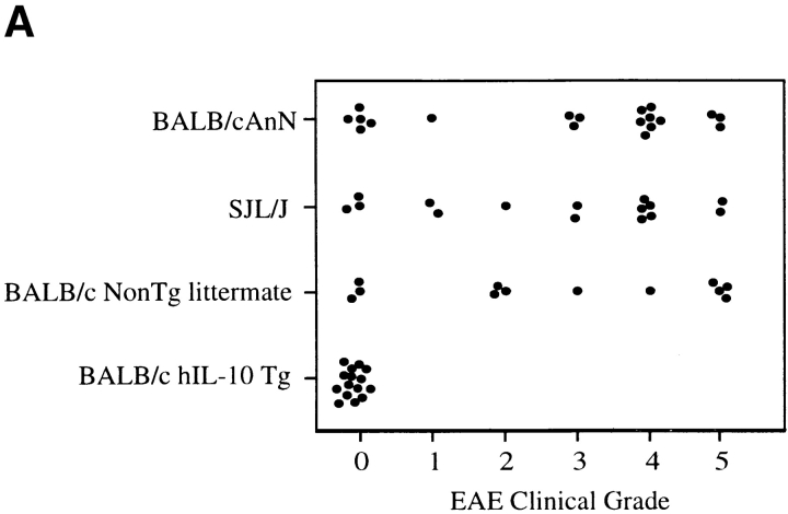
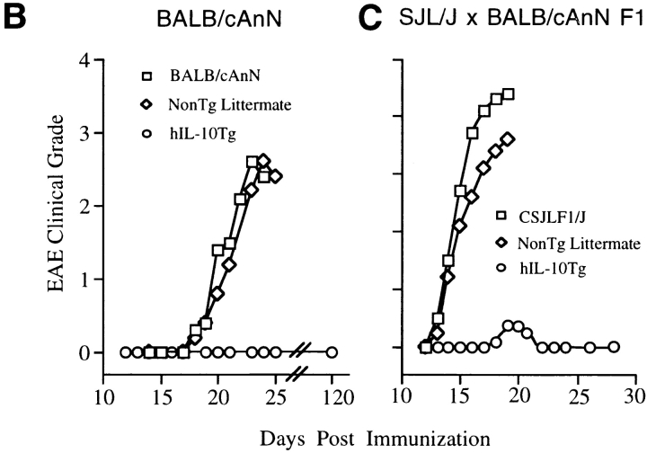
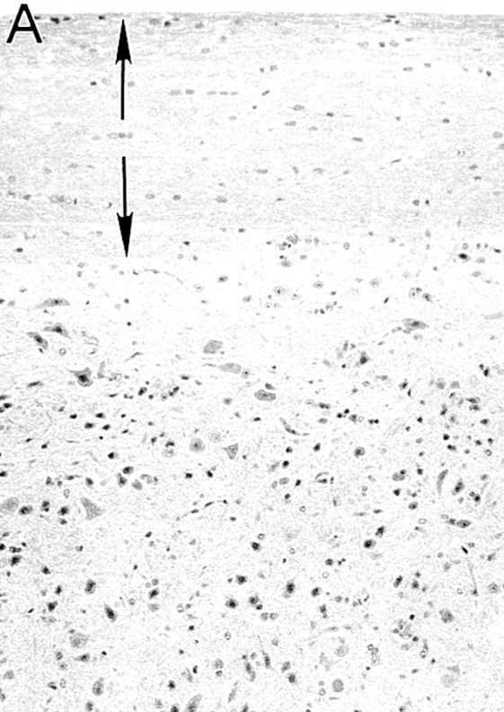
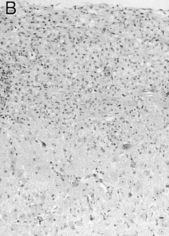
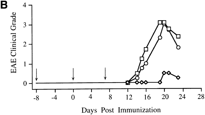
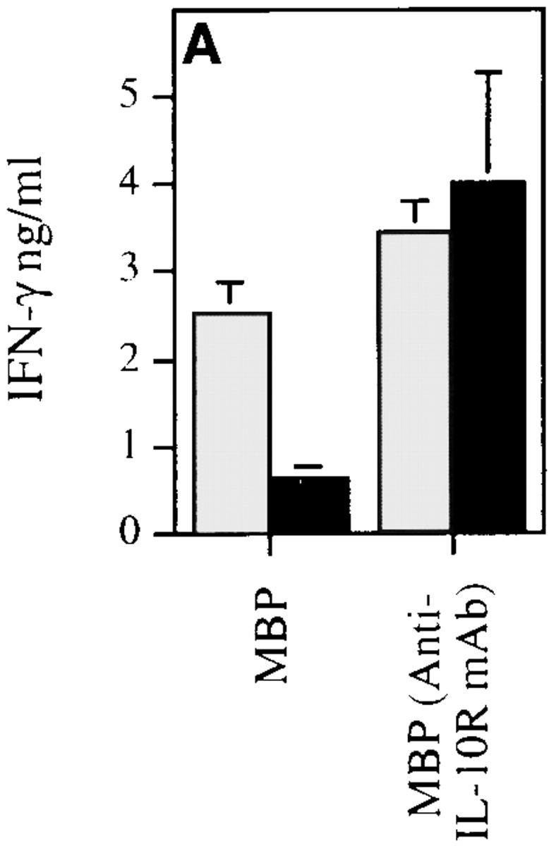
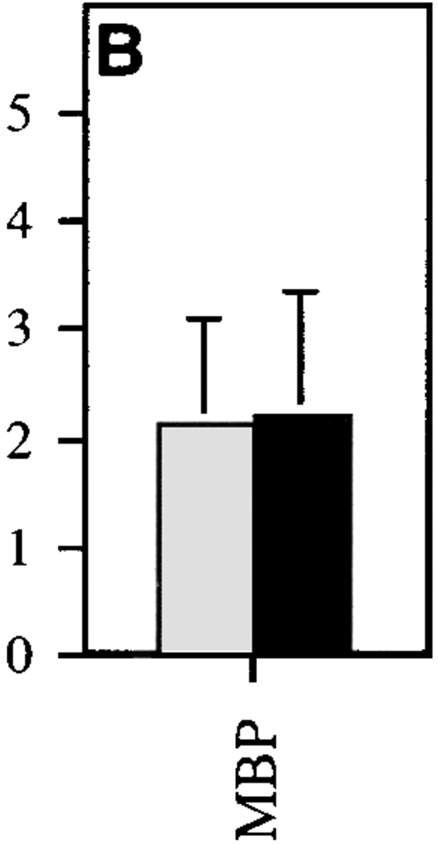
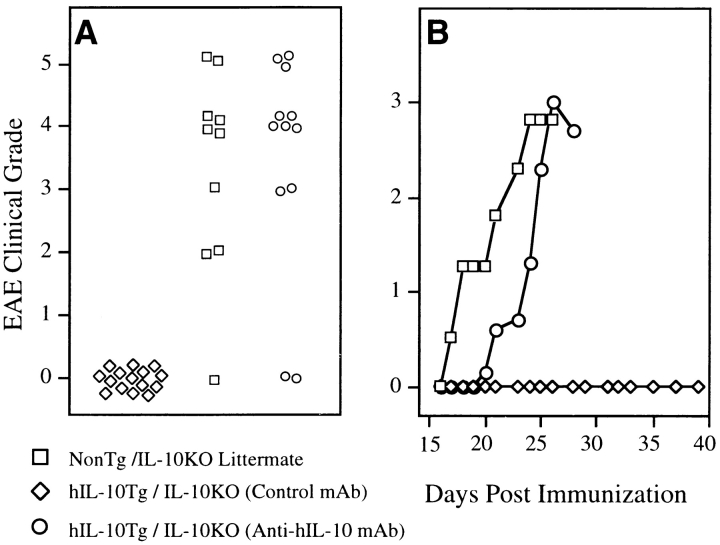
The effectiveness of interleukin 10 (IL-10) in the treatment of autoimmune-mediated central nervous system inflammation is controversial. Studies of the model system, experimental autoimmune encephalomyelitis (EAE), using various routes, regimens, and delivery methods of IL-10 suggest that these variables may affect its immunoregulatory function. To study the influence of these factors on IL-10 regulation of EAE pathogenesis, we have analyzed transgenic mice expressing human IL-10 (hIL-10) transgene under the control of a class II major histocompatibility complex (MHC) promoter. The hIL-10 transgenic mice are highly resistant to EAE induced by active immunization, and this resistance appears to be mediated by suppression of autoreactive T cell function. Myelin-reactive T helper 1 cells are induced but nonpathogenic in the IL-10 transgenic mice. Antibody depletion confirmed that EAE resistance is dependent on the presence of the transgenic IL-10. Mice expressing the hIL-10 transgene but not the endogenous murine IL-10 gene demonstrated that transgenic IL-10 from MHC class II-expressing cells is sufficient to block induction of EAE. This study demonstrates that IL-10 can prevent EAE completely if present at appropriate levels and times during disease induction.


Figures

Figure 1
Figure 1
hIL-10Tg mice are EAE resistant. (A) Circles represent peak severity of clinical disease for individual mice on the indicated genetic backgrounds. Data compiled from three separate experiments. (B) Average severity score for hIL-10Tg mice on the BALB/cAnN background. One representative experiment of five is shown. (C) Average severity score for hIL-10Tg mice on SJL/J × BALB/cAnN F1 background. One representative experiment of four is shown.
Figure 1
Figure 1
hIL-10Tg mice are EAE resistant. (A) Circles represent peak severity of clinical disease for individual mice on the indicated genetic backgrounds. Data compiled from three separate experiments. (B) Average severity score for hIL-10Tg mice on the BALB/cAnN background. One representative experiment of five is shown. (C) Average severity score for hIL-10Tg mice on SJL/J × BALB/cAnN F1 background. One representative experiment of four is shown.
Figure 2
Figure 2
Histology of spinal cords of hIL-10Tg mice and nonTg littermates immunized to induce EAE. (A) hIL-10Tg mice were EAE resistant and spinal cords showed no inflammatory infiltrates or plaques of demyelination in the white matter (normal white matter outlined by arrows). (B) NonTg littermates were highly EAE susceptible and showed inflammatory infiltrates and extensive demyelination in the white matter. Luxol fast blue stain with hematoxylin counterstain. ×125.
Figure 2
Figure 2
Histology of spinal cords of hIL-10Tg mice and nonTg littermates immunized to induce EAE. (A) hIL-10Tg mice were EAE resistant and spinal cords showed no inflammatory infiltrates or plaques of demyelination in the white matter (normal white matter outlined by arrows). (B) NonTg littermates were highly EAE susceptible and showed inflammatory infiltrates and extensive demyelination in the white matter. Luxol fast blue stain with hematoxylin counterstain. ×125.
Figure 3
Figure 3
Induction of EAE in hIL-10Tg mice treated with anti– hIL-10 mAb. Days of mAb treatment are indicated by arrows. (A) Tg mice were injected with anti–hIL-10 mAb (1 mg/dose) or isotype-matched mAb starting at the day of MSCH immunization. One representative experiment out of five is shown. (B) Mice were injected with the mAb starting 8 d before MSCH immunization. One representative experiment out of three is shown.
Figure 3
Figure 3
Induction of EAE in hIL-10Tg mice treated with anti– hIL-10 mAb. Days of mAb treatment are indicated by arrows. (A) Tg mice were injected with anti–hIL-10 mAb (1 mg/dose) or isotype-matched mAb starting at the day of MSCH immunization. One representative experiment out of five is shown. (B) Mice were injected with the mAb starting 8 d before MSCH immunization. One representative experiment out of three is shown.
Figure 4
Figure 4
MBP-specific Th1 cells are induced in hIL-10Tg mice. hIL-10Tg mice (black bar) and control littermate mice (gray bar) were immunized twice at 1-wk intervals with MSCH emulsified in CFA. (A) 3 d after the last immunization, DLN cells from hIL-10Tg or control littermate mice were stimulated with MBP or MBP plus anti–IL-10 receptor mAb. One representative experiment out of three is shown. (B) CD4+ T cells were purified and stimulated in vitro with MBP plus irradiated T cell–depleted splenocytes from control CSJLF1/J mice for 60 h. The amount of IFN-γ in the supernatants was measured by ELISA. One representative experiment out of two is shown. The results are the mean of three or four individual mice/group ± SEM.
Figure 4
Figure 4
MBP-specific Th1 cells are induced in hIL-10Tg mice. hIL-10Tg mice (black bar) and control littermate mice (gray bar) were immunized twice at 1-wk intervals with MSCH emulsified in CFA. (A) 3 d after the last immunization, DLN cells from hIL-10Tg or control littermate mice were stimulated with MBP or MBP plus anti–IL-10 receptor mAb. One representative experiment out of three is shown. (B) CD4+ T cells were purified and stimulated in vitro with MBP plus irradiated T cell–depleted splenocytes from control CSJLF1/J mice for 60 h. The amount of IFN-γ in the supernatants was measured by ELISA. One representative experiment out of two is shown. The results are the mean of three or four individual mice/group ± SEM.
Figure 5
Figure 5
hIL-10Tg mice on the IL-10KO genetic background are EAE resistant. (A) Peak severity scores of individual mice treated with isotype control mAb or anti–hIL-10 mAb (1 mg/dose) on days 0, 7, and 14. Results shown are compiled from two separate experiments. (B) Average severity score over time for the three groups of mice shown in A.
Figure 6
Figure 6
Histology of spinal cords of hIL-10Tg mice on the IL-10KO background shown in Fig. 5. (A) NonTg IL-10KO mice were EAE susceptible and showed inflammatory infiltrates and plaques of demyelination in spinal cords (arrowheads). (B) hIL-10Tg IL-10KO mice were EAE resistant and their spinal cords showed no inflammatory infiltrates or demyelination. Luxol fast blue stain with hematoxylin counterstain. ×125.
Figure 6
Figure 6
Histology of spinal cords of hIL-10Tg mice on the IL-10KO background shown in Fig. 5. (A) NonTg IL-10KO mice were EAE susceptible and showed inflammatory infiltrates and plaques of demyelination in spinal cords (arrowheads). (B) hIL-10Tg IL-10KO mice were EAE resistant and their spinal cords showed no inflammatory infiltrates or demyelination. Luxol fast blue stain with hematoxylin counterstain. ×125.

References

    1. Moore KW, O'Garra A, de Waal R, Malefyt, Vieira P, Mosmann TR. Interleukin-10. Annu Rev Immunol. 1993;11:165–190. - PubMed
    1. Pennline KJ, Roque-Gaffney E, Monahan M. Recombinant human IL-10 prevents the onset of diabetes in the nonobese diabetic mouse. Clin Immunol Immunopathol. 1994;71:169–175. - PubMed
    1. Balasa B, Sarvetnick N. The paradoxical effects of interleukin 10 in the immunoregulation of autoimmune diabetes. J Autoimmun. 1996;9:283–286. - PubMed
    1. Tanaka Y, Otsuka T, Hotokebuchi T, Miyahara H, Nakashima H, Kuga S, Nemoto Y, Niiro H, Niho Y. Effect of IL-10 on collagen-induced arthritis in mice. Inflamm Res. 1996;45:283–288. - PubMed
    1. Persson S, Mikulowska A, Narula S, O'Garra A, Holmdahl R. Interleukin-10 suppresses the development of collagen type II-induced arthritis and ameliorates sustained arthritis in rats. Scand J Immunol. 1996;44:607–614. - PubMed

Publication types