

doi: 10.1073/pnas.96.6.2627.
Modulation of CREB binding protein function by the promyelocytic (PML) oncoprotein suggests a role for nuclear bodies in hormone signaling
Affiliations
- PMID: 10077561
- PMCID: PMC15819
- DOI: 10.1073/pnas.96.6.2627
Item in Clipboard
Modulation of CREB binding protein function by the promyelocytic (PML) oncoprotein suggests a role for nuclear bodies in hormone signaling
Proc Natl Acad Sci U S A.
.
Abstract
Disaggregation of the spherical nuclear bodies termed promyelocytic (PML) oncogenic domains (PODs) is a characteristic of acute promyelocytic leukemia. Here, we demonstrate that the cAMP enhancer binding protein (CREB)-binding protein (CBP) associates with PML in vitro and is recruited to the PODs in vivo. Through its association with CBP, wild-type PML dramatically stimulates nuclear receptor transcriptional activity. These results demonstrate that a fraction of CBP is compartmentalized to the POD through its association with PML and thus suggest that PML and other POD-associated proteins may play an unexpectedly broad role in aspects of transcriptional regulation and human disease.
Figures
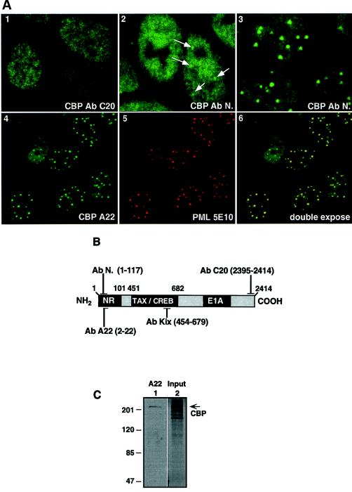
Differential localization of CBP in the nucleus. (A) Immunohistochemistry of CV1 cells, fixed at 80–90% confluence and analyzed in confocal microscopy. Green corresponds to the CBP staining revealed with the FITC-conjugated secondary Ab, red corresponds to the PML staining revealed with the Texas red-conjugated secondary Ab, and the yellow color in the double-exposure image indicates the sites where PML and CBP colocalize. Primary Abs are used as indicated. (2) The arrow shows the CBP speckled-like structures that colocalize with PML protein. 2 and 3 show two independent, randomly selected, fields of asynchronous cells populations. 1-5 are single-exposure photographs, and 6 is a double exposure. (B) Schematic representation of CBP primary structure. The location of the epitopes for the corresponding CBP Abs is indicated. (C) immunoblotting of total reticulocyte extracts expressing the full-length CBP protein. Proteins were analyzed in a 7.5% SDS gel and were probed with the A22 CBP Ab. The arrow shows the 270-kDa CBP.
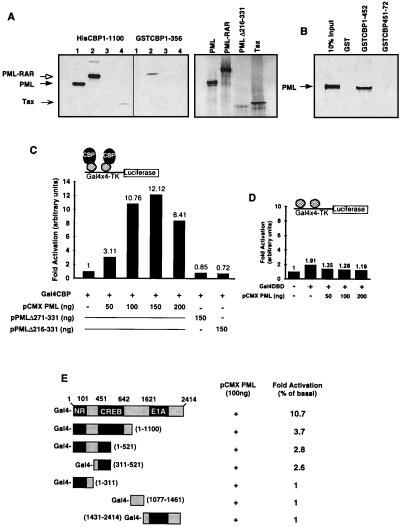
PML and CBP proteins interact in vivo and in vitro. (A) In vitro association of bacterial expressed CBP and S35 radiolabeled PML wild-type and mutant PMLΔ216-331, PML-RAR, and Tax proteins. In the pull-down experiment, the loaded proteins were used at 5:1 ratio compared with the input proteins represented on the right part of the figure. All samples were analyzed in a 4–20% SDS gradient gel, as indicated. (B) In vitro mapping of the PML-CBP interaction domain. GST fusion proteins containing the indicated residues of CBP were tested for binding to radiolabeled PML. CBP451-722 contains the Tax/CREB interaction domain (i.e., amino acids 451–661). (C) PML-CBP interaction in a one-hybrid analysis. CV1 cells (48-well plates) were transiently cotransfected with 100 ng of Gal4-tk-Luc reporter construct, 90 ng of CMXβgal, 60 ng of Gal4CBP, and the expression vectors for PML wild type and mutant, as indicated, per transfection point. All of the transfection points were equalized for the total amount of expression vectors, i.e., CMX empty vector, at a 200-ng final concentration. All points were performed in triplicate and varied by <10%. The presented values correspond to a representative experiment of at least four independent assays. (D) Transfections using the Gal4 DNA-binding domain as a control. (E) Mapping the PML-CBP interaction domain in vivo. CV1 cells were transfected and analyzed as in C. Expressions vectors at 100 ng were used as indicated.
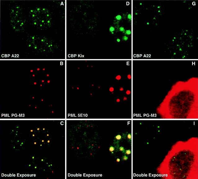
PML recruits CBP to PODs. (A–I) Double immunofluorescence of CV1 cells (6-well dishes) analyzed in confocal microscopy. Cells were transfected at 70% confluence with 2.5 μg pCMXPML expression vector (A–C); 1 μg of pCMXPML and 2.5 μg pCMXCBPm (mouse) (D–F); and 2.5 μg pSVPMLΔ216-331 (G–I). Primary Abs were used as indicated. Green corresponds to the CBP staining revealed with the FITC-conjugated secondary Ab, red corresponds to the PML staining revealed with the Texas red-conjugated secondary Ab, and the yellow color in the double-exposure image indicates the sites where PML and CBP colocalize.
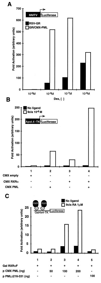
PML activates NR transcription. (A) PML potentiates GR-mediated transactivation of the mouse mammary tumor virus (MMTV) promoter. CV1 cells (48-well plates) were transiently transfected with 120 ng of reporter construct and 90 ng of CMXβgal. Transient transfection of GR at 60 ng induces MMTV-Luc transcription from 30- to 200-fold in a dexamethasone concentration-dependent manner. Coexpression of PML at 120 ng synergizes with GR and activates the MMTV transcription (600-fold) at 10 nM of ligand. The synergism of PML and GR is more dramatic in conditions at which lower concentrations of ligand and GR protein are tested. The open columns represent the points of PML and GR cotransfection, and the black ones are single transfected with GR expression vector, as indicated. (B) PML potentiates RXR-mediated transactivation of the Apol4-tk-Luc synthetic promoter. CV1 cells (48-well plates) were transiently transfected with 120 ng of reporter construct, 90 ng of CMXβgal, 120 ng of CMX-RXRα and CMXPML, as indicated. All of the transfection points were equalized with CMX empty vector at 240 ng. Transfected cells were treated with 9-cis retinoic acid at 1 μM for 8 h before the assay. (C) PML potentiates Gal-4 RXRαF-dependent transcription. Cells were transfected as before. Gal-4 RXRαF was transfected at 120 ng, and pCMXPML and pSVPMLΔ216-331 were used as indicated. Transfected cells were treated with 9-cis retinoic acid at 1 μM for 8 h before the assay. All points were performed in triplicate and varied by <10%. The presented values correspond to a representative experiment of at least four independent assays.
References
Publication types
MeSH terms
Substances
Grants and funding
LinkOut - more resources
Full Text Sources
Other Literature Sources
Molecular Biology Databases
