. 1999 Mar 16;96(6):2805-10.
doi: 10.1073/pnas.96.6.2805.

Syndecan-4 signals cooperatively with integrins in a Rho-dependent manner in the assembly of focal adhesions and actin stress fibers

Affiliations

Syndecan-4 signals cooperatively with integrins in a Rho-dependent manner in the assembly of focal adhesions and actin stress fibers

S Saoncella et al. Proc Natl Acad Sci U S A. .

Abstract

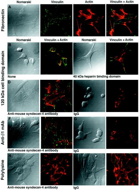
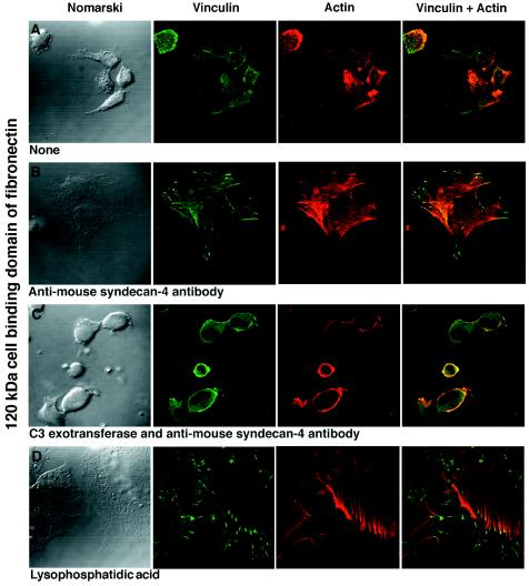
The assembly of focal adhesions and actin stress fibers by cells plated on fibronectin depends on adhesion-mediated signals involving both integrins and cell-surface heparan sulfate proteoglycans. These two cell-surface receptors interact with different domains of fibronectin. To attempt to identify the heparan sulfate proteoglycans involved, we used fibronectin-null (FN-/-) mouse fibroblasts to eliminate the contribution of endogenous fibronectin during the analysis. FN-/- fibroblasts plated on the cell-binding domain of fibronectin or on antibodies directed against mouse beta1 integrin chains attach but fail to spread and do not form focal adhesions or actin stress fibers. When such cells are treated with antibodies directed against the ectodomain of mouse syndecan-4, they spread fully and assemble focal adhesions and actin stress fibers indistinguishable from those seen in cells plated on intact fibronectin. These results identify syndecan-4 as a heparan sulfate proteoglycan involved in the assembly process. The antibody-stimulated assembly of focal adhesions and actin stress fibers in cells plated on the cell-binding domain of fibronectin can be blocked with C3 exotransferase, an inhibitor of the small GTP-binding protein Rho. Treatment of cells with lysophosphatidic acid, which activates Rho, results in full spreading and assembly of focal adhesions and actin stress fibers in fibroblasts plated on the cell-binding domain of fibronectin. We conclude that syndecan-4 and integrins can act cooperatively in generating signals for cell spreading and for the assembly of focal adhesions and actin stress fibers. We conclude further that these joint signals are regulated in a Rho-dependent manner.


Figures

Figure 1
Figure 1
Representative PCR analysis of DNA from ES cells generated from blastocysts of a FN/129 het × het mating. Shown are the 900-bp product from the wild-type allele and the 1,060-bp product from the targeted allele. Lanes: MW, 1-kb DNA ladder; 1, 3, and 5, heterozygous ES cell clones; 2 and 6, wild-type ES cell clones; and 4, the homozygous null ES cell clone used for the derivation of FN null fibroblasts.
Figure 2
Figure 2
Fibroblasts differentiated from ES cells heterozygous (+/−) or homozygous (−/−) for the fibronectin mutation were cultured on glass coverslips for 2 days in DMEM containing 10% fetal calf serum without (A and C) or with 10 mg/ml human plasma fibronectin (B and D). The cells were washed, fixed in 4% paraformaldehyde, and stained with rabbit anti-human fibronectin that crossreacted with mouse and bovine fibronectins (20).
Figure 3
Figure 3
(A) PCR analysis of FN−/− and 3T3 fibroblasts. The 388-bp band (arrowhead) indicates the presence of mRNA for syndecan-4 in 3T3 (lane 1) and FN−/− (lane 2) fibroblasts. No band is visible in PCR reactions where the cDNA is replaced by nonreverse-transcribed RNA from FN−/− cells (lane 3) or H2O (lane 4). MW, 1-kb DNA ladder. (B) Surface expression of syndecan-4 and β1 integrins analyzed by flow cytometry after staining with affinity-purified rabbit antibody against mouse syndecan-4 or with a rat monoclonal antibody against mouse β1 integrin. (C) Bacterially expressed mouse syndecan-4 ectodomain (M) or chicken full-length syndecan-4 core protein (C) was analyzed by SDS/PAGE and Western blotting with preimmune (IgG), anti-mouse syndecan-4 antibodies (MS-4-E), or anti-chicken syndecan-4 antibodies (CS-4-E). The lower bands (arrowheads) in lane C of the right panel are degradation products of the full-length fusion protein (arrow).
Figure 4
Figure 4
Analysis of the spreading and assembly of focal adhesions and actin stress fibers by FN−/− fibroblasts. The degree of spreading is seen from the Nomarski micrographs. Focal adhesions are visualized with the vinculin staining and actin stress fibers by means of tetramethyl rhodamine isothiocyanate–phalloidin staining. (A) FN−/− fibroblasts plated on fibronectin. The cells are fully spread. They have assembled focal adhesions and actin stress fibers. (B–E) FN−/− fibroblasts plated on the 120-kDa cell-binding domain of fibronectin. Cell spreading and assembly of focal adhesions and actin stress fibers are present in cells supplemented with the 40-kDa heparin-binding domain of fibronectin (C) or supplemented with antibodies directed against the ectodomain of mouse syndecan-4 (D) but absent in unsupplemented cells (B) or in cells supplemented with IgG (E). (F and G) FN−/− fibroblasts plated on rat monoclonal antibody directed against mouse β1 integrin chains. Cell spreading and assembly of focal adhesions and actin stress fibers are present in cells supplemented with antibodies directed against the ectodomain of mouse syndecan-4 (F) but not in those supplemented with IgG (G). (H and I) FN−/− fibroblasts plated on polylysine. No assembly of focal adhesions and actin stress fibers is evident in cells supplemented either with antibodies directed against the ectodomain of mouse syndecan-4 (H) or with IgG (I).
Figure 5
Figure 5
Assembly of focal adhesions and actin stress fibers is absent in unsupplemented FN−/− fibroblasts plated on the 120-kDa cell-binding domain of fibronectin (A). The cell spreading and assembly of focal adhesions and actin stress fibers as a result of supplementation with antibodies directed against the ectodomain of mouse syndecan-4 (B) is inhibited by the C3 exotransferase treatment that inhibits Rho (C). Supplementation with LPA leads to full spreading and assembly of focal adhesions and actin stress fibers in cells plated on the 120-kDa cell-binding domain of fibronectin alone (D).

References

    1. Adams J C, Watt F M. Development. 1993;117:1183–1198. - PubMed
    1. Ashkenas J, Muschler J, Bissell M J. Dev Biol. 1996;180:433–444. - PMC - PubMed
    1. Gumbiner B M. Cell. 1996;84:345–357. - PubMed
    1. Giancotti F G. Curr Opin Cell Biol. 1997;9:691–700. - PubMed
    1. Howe A, Aplin A E, Alahari S K, Juliano R L. Curr Opin Cell Biol. 1998;10:220–231. - PubMed

Publication types

LinkOut - more resources