. 1999 Mar 16;96(6):2811-6.
doi: 10.1073/pnas.96.6.2811.

Angiostatin binds ATP synthase on the surface of human endothelial cells

Affiliations

Angiostatin binds ATP synthase on the surface of human endothelial cells

T L Moser et al. Proc Natl Acad Sci U S A. .

Abstract

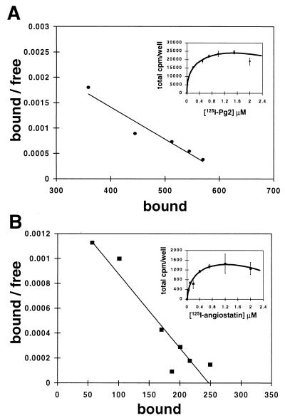
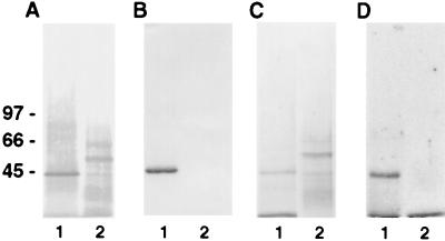
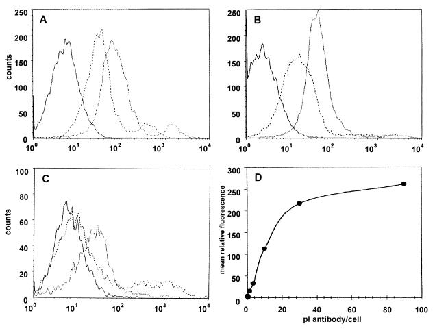
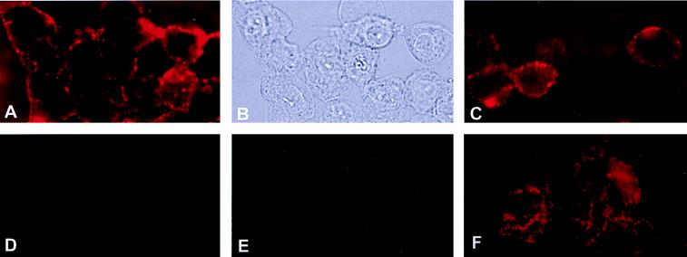
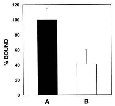
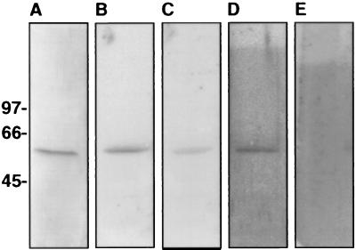
Angiostatin, a proteolytic fragment of plasminogen, is a potent antagonist of angiogenesis and an inhibitor of endothelial cell migration and proliferation. To determine whether the mechanism by which angiostatin inhibits endothelial cell migration and/or proliferation involves binding to cell surface plasminogen receptors, we isolated the binding proteins for plasminogen and angiostatin from human umbilical vein endothelial cells. Binding studies demonstrated that plasminogen and angiostatin bound in a concentration-dependent, saturable manner. Plasminogen binding was unaffected by a 100-fold molar excess of angiostatin, indicating the presence of a distinct angiostatin binding site. This finding was confirmed by ligand blot analysis of isolated human umbilical vein endothelial cell plasma membrane fractions, which demonstrated that plasminogen bound to a 44-kDa protein, whereas angiostatin bound to a 55-kDa species. Amino-terminal sequencing coupled with peptide mass fingerprinting and immunologic analyses identified the plasminogen binding protein as annexin II and the angiostatin binding protein as the alpha/beta-subunits of ATP synthase. The presence of this protein on the cell surface was confirmed by flow cytometry and immunofluorescence analysis. Angiostatin also bound to the recombinant alpha-subunit of human ATP synthase, and this binding was not inhibited by a 2,500-fold molar excess of plasminogen. Angiostatin's antiproliferative effect on endothelial cells was inhibited by as much as 90% in the presence of anti-alpha-subunit ATP synthase antibody. Binding of angiostatin to the alpha/beta-subunits of ATP synthase on the cell surface may mediate its antiangiogenic effects and the down-regulation of endothelial cell proliferation and migration.


Figures

Figure 1
Figure 1
Direct binding assay and Scatchard analysis of plasminogen and angiostatin with endothelial cells. HUVEC were plated at a density of 10,000 cells/well and incubated with increasing concentrations of 125I-labeled-plasminogen or -angiostatin as described in Materials and Methods. (A) 125I-labeled plasminogen binding was concentration dependent and saturable with an apparent dissociation constant (Kd) of 158 nM and 870,000 sites/cell. (B) Binding to HUVEC with 125I-labeled angiostatin was concentration dependent and saturable with a Kd of 245 nM and 38,000 sites/cell. Error bars represent SD.
Figure 2
Figure 2
Competition binding assay between plasminogen and angiostatin. HUVEC were plated at a density of 10,000 cells/well and incubated with 1.0 μM 125I-labeled plasminogen in the presence of 100-fold molar excess of unlabeled angiostatin for 1 h at 4°C. Cells were washed, and the remaining radioactivity was quantified by γ-counting. (A) Total binding of 1.0 μM 125I-labeled plasminogen was designated as 100%. (B) Plasminogen binding is inhibited by ≈80% in the presence of a 25-fold molar excess of unlabeled plasminogen. (C) Plasminogen binding was not inhibited in the presence of a 100-fold molar excess of unlabeled angiostatin, suggesting distinct binding sites for each on the cells. Similar experiments by using 125I-labeled angiostatin (D) showed no inhibition of binding in the presence of a 2-fold molar excess unlabeled plasminogen (E). Error bars represent SD.
Figure 3
Figure 3
Affinity purification of plasminogen and angiostatin binding sites. Plasma membranes were prepared as described in Materials and Methods. SDS/PAGE containing membrane proteins then were analyzed by Western blotting. Membranes were incubated in 10 mM Tris⋅HCl, 0.15 M NaCl, 0.05% Nonidet P-40, pH 7.5 containing (A) streptavidin-alkaline phosphatase conjugate antibody or (B) anti-annexin II antibody and developed by using 5-bromo-4-chloroindol-3-yl-phosphate nitro blue tetrazolium. Membrane stained with Coomassie brilliant blue (C), showing affinity-purified membrane proteins. Membrane incubated with 125I-labeled plasminogen (D), showing binding to the plasminogen-purified membrane and not the angiostatin. Lane 1 represents protein eluted from the plasminogen-Sepharose column. Lane 2 represents protein eluted from the angiostatin-Sepharose column. The relative molecular masses of α-ATP synthase and β-ATP synthase are ≈55 and ≈50 kDa, respectively.
Figure 4
Figure 4
Binding of antibody directed against the α-subunit of ATP synthase on the surface of HUVEC by flow cytometry. HUVEC were analyzed by FACScan flow cytometry as described in Materials and Methods. Histogram plots are shown for HUVEC (A) and A549 (B) where dotted lines represent cells incubated with antibody directed against the α-subunit of ATP synthase, dashed lines preimmune serum, and solid lines secondary antibody only. Histogram plot of A549 shown in C are similar with dotted lines representing antibody incubated with a 5-fold molar excess α ATP synthase protein. (D) HUVEC demonstrate specific, saturable binding of antibodies directed against the α-subunit of ATP synthase. The mean relative fluorescence of HUVEC incubated with preimmune rabbit serum subtracted from the mean relative fluorescence of HUVEC incubated with the same volume of anti-α ATP synthase gave the mean relative fluorescence resulting from the specific binding of antibodies directed against the α-subunit of ATP synthase on the HUVEC surface.
Figure 5
Figure 5
Immunofluorescence microscopy of ATP-synthase on HUVEC surface. HUVEC were incubated with rabbit polyclonal antiserum raised against the α-subunit of ATP synthase from E. coli as described in Materials and Methods. (A) HUVEC under epi-illumination showing immunofluorescent surface staining for the α-subunit of ATP synthase. (B) Same field of HUVEC under visible light. (C) Human dermal microvascular endothelial cells also showed immunofluorescent surface staining for the α-subunit of ATP synthase. Control experiments were performed with (D) preimmune serum and (E) secondary antibody alone. (F) HUVEC were permeabilized by acetone fixation before adding antibodies for the α-subunit of ATP synthase.
Figure 6
Figure 6
Competition binding assay between angiostatin and the antibody against the α-subunit of ATP synthase from E. coli. HUVEC were plated at a constant density of 10,000 cells/well and incubated with 0.5 μM 125I-labeled angiostatin in the presence of 1:10 dilution of antibody against the α-subunit of ATP synthase from E. coli for 1 h at 4°C. Cells were washed and remaining bound radioactivity was quantified by γ-counting. Nonspecific binding was measured in the presence of excess unlabeled angiostatin and was subtracted from total binding. (A) Total binding of 0.5 μM 125I-labeled angiostatin was designated as 100%. (B) Angiostatin binding is inhibited by 59% in the presence of a 1:10 dilution of anti-α-subunit ATP synthase antibody. Competition studies also were performed simultaneously by using rabbit preimmune serum to account for nonspecific inhibition. Error bars represent SD. A one-tailed homoscedastic t test was used for statistical analysis; P < 0.10.
Figure 7
Figure 7
Angiostatin binding to the recombinant α-subunit of human ATP synthase. The α-subunit of human ATP synthase was cloned and expressed in E. coli and purified by using Qiagen’s nickel-Sepharose protein purification system before dialyzing in PBS, pH 7.0. Recombinant protein was electrophoresed on 5–15% SDS/PAGE, electroblotted onto Immobilon membrane, and incubated 18 h in 10 mM Tris⋅HCl/0.15 M NaCl/0.05% Nonidet P-40, pH 7.5 (TSN buffer) containing 125I-angiostatin. For competition studies unlabeled ligand was added 4 h before radiolabeled ligand. Blots were washed in TSN buffer containing 0.05% Tween80 and bound radioactivity was quantified on a Molecular Dynamics PhosphorImager. (A) Coomassie stain of Immobilon membrane containing the α-subunit of human ATP synthase. (B) Binding of 0.5 μM 125I-labeled angiostatin. (C) Binding of 0.5 μM 125I-labeled angiostatin in the presence of a 250-fold molar excess of unlabeled angiostatin. Binding of angiostatin is inhibited by ≈56%. (D) Binding of 0.5 μM 125I-labeled angiostatin in the presence of a 2,500-fold molar excess of unlabeled plasminogen. Binding of angiostatin is not inhibited. (E) Binding of 0.5 μM 125I-labeled plasminogen to the α-subunit of human ATP synthase. Plasminogen did not bind to the recombinant α-subunit of ATP synthase; however, it did bind the annexin II control (as shown in Fig. 3).

References

    1. O’Reilly M S, Holmgren L, Shing Y, Chen C, Rosenthal R A, Moses M, Lane W S, Cao Y, Sage E H, Folkman J. Cell. 1994;79:315–328. - PubMed
    1. Sottrup-Jensen L, Claeys H, Zajdel M, Petersen T E, Magnusson S. Prog Chem Fibrinolysis Thrombolysis. 1978;3:191–209.
    1. Gately S, Twardowski P, Stack M S, Patrick M, Boggio L, Cundiff D L, Schnaper H W, Madison L, Volpert O, Bouck N, et al. Cancer Res. 1996;36:4887–4890. - PubMed
    1. Dong Z, Kumar R, Yang X, Fidler I J. Cell. 1997;88:801–810. - PubMed
    1. Lijnen H R, Ugwu F, Collen D. Biochemistry. 1998;37:4699–4702. - PubMed

Publication types

MeSH terms