. 1999 Mar 16;96(6):2817-22.
doi: 10.1073/pnas.96.6.2817.

Cyclin-dependent kinase control of centrosome duplication

Affiliations

Cyclin-dependent kinase control of centrosome duplication

K R Lacey et al. Proc Natl Acad Sci U S A. .

Abstract

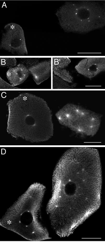
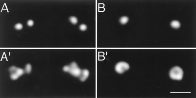
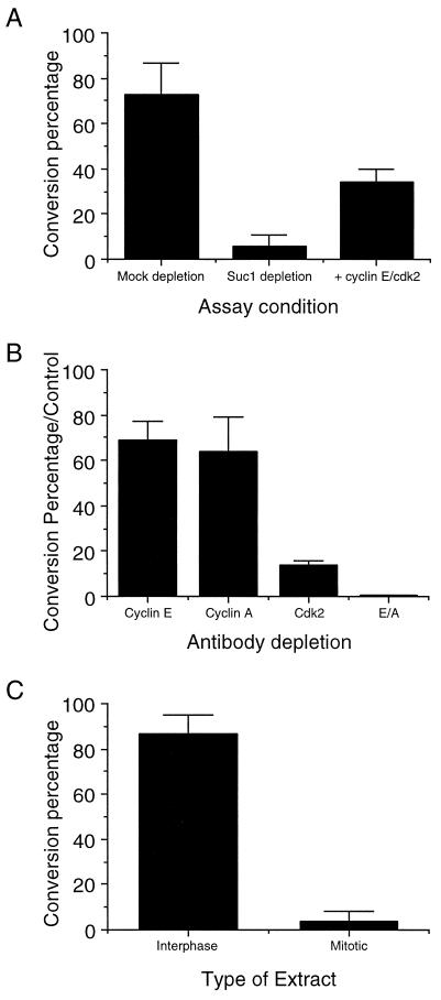
Centrosomes nucleate microtubules and duplicate once per cell cycle. This duplication and subsequent segregation in mitosis results in maintenance of the one centrosome/cell ratio. Centrosome duplication occurs during the G1/S transition in somatic cells and must be coupled to the events of the nuclear cell cycle; failure to coordinate duplication and mitosis results in abnormal numbers of centrosomes and aberrant mitoses. Using both in vivo and in vitro assays, we show that centrosome duplication in Xenopus laevis embryos requires cyclin/cdk2 kinase activity. Injection of the cdk (cyclin-dependent kinase) inhibitor p21 into one blastomere of a dividing embryo blocks centrosome duplication in that blastomere; the related cdk inhibitor p27 has a similar effect. An in vitro system using Xenopus extracts carries out separation of the paired centrioles within the centrosome. This centriole separation activity is dependent on cyclin/cdk2 activity; depletion of either cdk2 or of the two activating cyclins, cyclin A and cyclin E, eliminates centriole separation activity. In addition, centriole separation is inhibited by the mitotic state, suggesting a mechanism of linking the cell cycle to periodic duplication of the centrosome.


Figures

Figure 1
Figure 1
The cdk inhibitor p21 blocks centrosome duplication in frog embryos. Embryonic cells were injected with various inhibitors and treated with cycloheximide for 4 h to allow for centrosome overduplication. Asterisks mark the injected cells, as visualized by coinjection of a fluorescein-conjugated dextran. (A) p21. (B and B′) p21 and cyclin E (two different sections are shown to view all centrosomes). (C) p21 N terminus. (D) p21 C terminus. (A) γ-Tubulin staining. (B, B′, C, and D) α-Tubulin staining. The difference in cell size is because of the variation in cell stage at the time of injection. (Bar = 100 μm.)
Figure 2
Figure 2
An in vitro centriole separation assay. Deconvolution images of centriole doublets and singlets. (A and A′) These doublets represent the 0-h starting point of the assay. (B and B′) At the 1-h endpoint, the majority of centrosomes are singlets, as depicted. (A and B) α-Tubulin staining. (A′ and B′) γ-Tubulin staining. (Bar = 1 μm.)
Figure 3
Figure 3
The dependence of centriole separation on cyclin-dependent kinases. The graphs express centriole separation activity as conversion percentages, as described in Table 2. (A) Depletion and rescue of centriole separation activity with Suc1p and cyclin E/cdk2. (B) Depletion of both cyclin E- and cyclin A-dependent kinases inhibits centriole separation. The conversion percentage has been normalized to the control depletion (uncoupled protein A-Sepharose). (C) The mitotic state inhibits centriole separation.

References

    1. Kellogg D R, Moritz M, Alberts B M. Annu Rev Biochem. 1994;63:639–674. - PubMed
    1. Kochanski R S, Borisy G G. J Cell Biol. 1990;110:1599–1605. - PMC - PubMed
    1. Kuriyama R, Borisy G G. J Cell Biol. 1981;91:814–821. - PMC - PubMed
    1. Winey M, Byers B. Trends Genet. 1993;9:300–304. - PubMed
    1. McDonald H B, Byers B. J Cell Biol. 1997;137:539–553. - PMC - PubMed

Publication types

MeSH terms

LinkOut - more resources