. 1999 Mar 16;96(6):2852-7.
doi: 10.1073/pnas.96.6.2852.

The tail domain of lamin Dm0 binds histones H2A and H2B

Affiliations

The tail domain of lamin Dm0 binds histones H2A and H2B

M Goldberg et al. Proc Natl Acad Sci U S A. .

Abstract

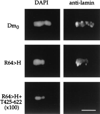
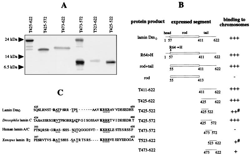
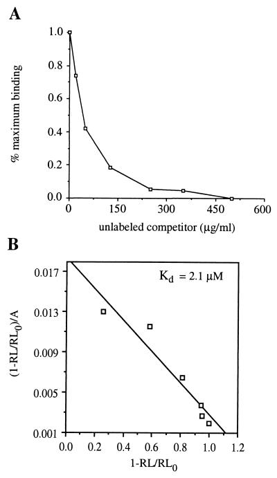
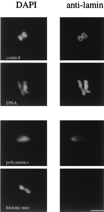
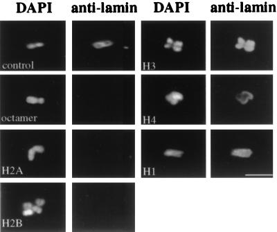
In multicellular organisms, the higher order organization of chromatin during interphase and the reassembly of the nuclear envelope during mitosis are thought to involve an interaction between the nuclear lamina and chromatin. The nuclear distribution of lamins and of peripheral chromatin is highly correlated in vivo, and lamins bind specifically to chromatin in vitro. Deletion mutants of Drosophila lamin Dm0 were expressed to map regions of the protein that are required for its binding to chromosomes. The binding activity requires two regions in the lamin Dm0 tail domain. The apparent Kd of binding of the lamin Dm0 tail domain was found to be approximately 1 microM. Chromatin subfractions were examined to search for possible target molecules for the binding of lamin Dm0. Isolated polynucleosomes, nucleosomes, histone octamer, histone H2A/H2B dimer, and histones H2A or H2B displaced the binding of lamin Dm0 tail to chromosomes. This displacement was specific, because polyamines or proteins such as histones H1, H3, or H4 did not displace the binding of the lamin Dm0 tail to chromosomes. In addition, DNA sequences, including M/SARs, did not interfere with the binding of lamin Dm0 tail domain to chromosomes. Taken together, these results suggest that the interaction between the tail domain of lamin Dm0 and histones H2A and H2B may mediate the attachment of the nuclear lamina to chromosomes in vivo.


Figures

Figure 1
Figure 1
The binding of lamin Dm0 to chromosomes is mediated by its tail domain. Binding of wild-type lamin Dm0 (Top) and lamin R64 → H, which is a mutant form of lamin Dm0 that cannot polymerize in vitro (Middle) (36) to mitotic CHO chromosomes was performed in the presence of 10% BSA. Staining was with affinity purified polyclonal antibodies directed against lamin Dm0 rod domain as primary antibodies and Cy3-conjugated anti rabbit antibodies as secondary antibodies. Staining was not observed when the chromatin was first incubated with a 100-fold molar excess of the tail domain (residues 425–622; Bottom). (Bar = 5 μm.)
Figure 2
Figure 2
The binding of lamin Dm0 to chromosomes requires amino acids 425–473 and 572–622. (A) SDS/PAGE (15%) analysis of different bacterially expressed and purified constructs of the lamin Dm0 tail domain. Proteins were stained with Comassie brillant blue. The names assigned to the different constructs are written above the lanes, and the positions of the size markers are indicated. (B) Summary of the binding of different lamin Dm0 constructs to chromosomes. Examples of the binding of lamin Dm0 and R64 → H to chromosomes are shown in Fig. 1, and an example of the binding of T425–622 to chromosomes is shown in Fig. 4. The position of the amino acid termini of each expressed segment are shown below the map of each construct. +++ indicates strong binding; ++ indicates medium binding; + indicates weak binding; − indicates lack of detectable binding; # indicates that the detection was performed with RGSHis antibody and the intensity of the binding was compared with that of T425–622 staining with RGSHis antibody. (C) Comparison between lamin Dm0, Drosophila lamin C, human lamin A/C, and Xenopus lamin B2 sequences in the amino-terminal part of the tail domains that bind chromosomes. Conserved amino acids are in boldface; similar amino acids are underlined. The position of the amino acids termini of each lamin segment are shown above the sequence; − indicates a gap.
Figure 3
Figure 3
Binding of the lamin Dm0 tail domain to immobilized chromatin with an apparent Kd of 2.1 μM. (A) 125I-labeled T425–622 protein (2 μg/ml) was incubated with immobilized polynucleosomes in the presence of increasing concentrations of unlabeled T425–622 protein. Correction for nonspecific binding was obtained by subtraction of values obtained with 250 μg/ml unlabeled T425–622. (B) Data for specific binding was analyzed as described (28). RL, amount of radioactive protein bound to chromatin at cold competitor concentration A; RL0, amount of radioactive protein bound to chromatin in the absence of unlabeled competitor. Assuming a single-site competitive interaction (47), the slope given by this plot equals −1/Kd.
Figure 4
Figure 4
The binding of lamin Dm0 to chromosomes is mediated by histones. T425–622 protein was used to bind chromosomes in the presence of 10% BSA (control). Mitotic chromosomes were added 5 min after incubation of the reaction mixture with a plasmid containing the Drosophila ftz M/SAR DNA (38, 39), a mixture of spermine and spermidine (polyamines), or the commercial mixture of histone proteins (histone-mix). (Bar = 5 μm.)
Figure 5
Figure 5
The binding of lamin Dm0 to chromosomes is mediated by histones H2A and H2B. T425–622 protein was used to bind chromosomes in the presence of 10% BSA (control). Chromosomes were added 5 min after incubation of the reaction mixture with histone octamers, purified histones H2A, H2B, H3, H4, or H1 (amino acids 1–142). (Bar = 6 μm.)

References

    1. Moir R D, Spann T P, Goldman R D. Int Rev Cytol. 1995;162:141–182. - PubMed
    1. Georgatos S D, Meier J, Simos G. Curr Opin Cell Biol. 1994;6:347–353. - PubMed
    1. Liu J, Lopez J M, Wolfner M F. Curr Top Dev Biol. 1997;35:47–70. - PubMed
    1. Hutchison C J, Bridger J M, Cox L S, Kill I R. J Cell Sci. 1994;107:3259–3269. - PubMed
    1. Benavente R, Krohne G. J Cell Biol. 1986;103:1847–1854. - PMC - PubMed

Publication types

LinkOut - more resources